- Oct 2022
-
fs2.american.edu fs2.american.edu
-
Not surprisingly, foes of the yellow press were more eager to disparage than define. Thus were the yellow journals accused of such malevolent effects as "corrupting the young and debauching the old, championing vice and lewdness, and defying respectability and decency."39 The practice of yellow journalism was likened, moreover, to a "contest of madmen for the primacy of the sewer."40
yellow journalism in this manner did not have good effects. It was spreading misinformation to audiences.
-
-
fs2.american.edu fs2.american.edu
-
The Journal gloated about its extravagant spending on newsgathering. Not atypical was this claim, in which the Journal disparaged its rivals, notably the New York Sun: "The reason the old journalism doesn't like the Journal is that the Journal gets the news, no matter what it costs. The Sun and its kind cannot afford to spend money since the Journal has taken their readers away from them, and the probability is they would not do so if they could afford it. They are still living in the Silurian age."16
This is what yellow journalism was- getting the information they needed at whatever cost, eve if it meant making up some facts in order to attract readers.
-
-
www.biorxiv.org www.biorxiv.org
-
Reviewer #3 (Public Review):
This manuscript aims to address whether age-related iron status influences differential effects of estrogen replacement on atherogenesis in postmenopausal females. Specifically, whether age-related iron accumulation reduces estrogen signaling through ERa receptor in relevant cell types (endothelial cells and macrophages). They test this fairly rigorously using in vitro, preclinical and clinical data, and the data is presented logically.
First, the authors demonstrate that in postmenopausal women, there is an inverse correlation between age-related increase in iron levels and age-related decrease in ERa levels in atherosclerotic plaques. This is consistent with the fact that, in ovariectomized (OVX) pro-atherogenic (ApoE KO) mice, there is differential effect of estrogen (E2) on ERa, atherosclerosis, lipid profiles and key biomarkers (ABCA1, eNOS, etc) based on age (early: 16 weeks, versus late: 40 weeks), with young mice responding favorably while older mice responding negatively. Consistent with this, in the older OVX ApoE KO mice, E2 treatment worsened atherosclerosis. Importantly, they point this differential effect of E2 to iron overload in macrophages using ApoE KO; LysM-macrophage-specific Fpn1 KO as E2 now has deleterious effects regardless of age. Next, using ferric ammonium citrate (FAC) to supplement iron and deferiprone (DFP) to chelate iron, they show that iron manipulation impacts the E2 response appropriately in the relevant cell types (endothelial cells and macrophage cells). Then they provide evidence for the MdM2-mediated post-translational regulation of ERa as a mechanism by which iron status impacts differential E2 has differential effects on ERa. Finally, they test the impact of systemic iron chelation on the OVX ApoE KO mice model of atherosclerosis and show that iron chelation attenuates the deleterious effects of E2 in late postmenopausal mice.
Overall the evidence is solid and logically laid out. Given that serum iron levels do not correlate with the rest of the story, inclusion of LysM-macrophage-specific Fpn1 KO provides the key evidence that iron loading in macrophages drives differential effects to E2 in postmenopausal mice. The paper provides evidence that iron levels influence how the relevant cells respond to E2 with clinical implications to hormonal replacement therapy in younger and older postmenopausal women. While this study is limited by a small clinical sample size, it provides an important framework for future studies on the impact of age-related iron status on the response to hormonal replacement therapy.
-
-
www.biorxiv.org www.biorxiv.org
-
Reviewer #3 (Public Review):
This paper demonstrates neural mechanisms important for the representation of moving stimuli. Specifically, using EEG, the authors investigated the temporal profiles of visual activities that correspond to changes in positions of moving stimuli.
Strengths:<br /> The authors examined an interesting question of how moving stimuli can be smoothly represented and perceived by using a neural recording modality with high temporal resolution. To my knowledge, the temporal dynamics of the neural correlates of successful motion perception are not well understood, and the study provides evidence for a plausible mechanism for this process. Additionally, the paper is well-written where the results are clearly communicated, and the figures are clearly presented.
Weaknesses:<br /> The findings reported are derived from a specific case of motion perception which may not reflect the general mechanisms optimized for motion perception. The limitations related to task designs and the neural readouts should be discussed as they affect the way that the reported results will be interpreted.
-
-
www.biorxiv.org www.biorxiv.org
-
Reviewer #3 (Public Review):
This study provides interesting new information on the cellular roles of different Arp2/3 complex isoforms. Previous studies have shown that T cell receptor signaling induces both cytoplasmic and nuclear actin filament assembly, which are dependent on the Arp2/3 complex. Moreover, nuclear actin filament assembly can be induced in response to DNA damage by the Arp2/3 complex. However, the possible roles of different Arp2/3 isoforms in these processes have not been reported. Here, Sadhu et al. demonstrate that the two isoforms of the ARPC5 subunit (ARPC5 and ARPC5L) have specific functions in these processes. By using knockdown and knockout cells, they provide evidence that cytoplasmic actin polymerization induced by T cell receptor activation is dependent on the ARPC5 isoform, whereas consequent nuclear actin filament assembly relies on the ARPC5L isoform. Interestingly, the nuclear actin polymerization induced by DNA replication stress is dependent on ARPC5. The authors also examined the upstream signaling pathways, and provide evidence that nuclear calcium-calmodulin signaling and N-WASP are specific activators of ARPC5L containing complexes.
Majority of the data presented in the manuscript appear of good technical quality, and the study provides interesting new insights into specific cellular roles of different Arp2/3 isoforms in T lymphocytes.
-
-
www.biorxiv.org www.biorxiv.org
-
Reviewer #3 (Public Review):
The article reports the functional analysis of one of the critical genetic determinants for bacterial infection, the RHIZOBIUM-DIRECTED POLAR GROWTH protein (RPG). The evolutionary pattern linking RPG to the Transcription Factor and the LysM receptor-like kinase, and the ability to form root nodule symbiosis makes it one of the prime candidates for engineering symbiotic nitrogen fixation in cereals. Therefore this analysis is timely and of huge importance. In a previous study, an EMS-induced mutant of M. truncatula (rpg-1 allele) was reported to have aberrant infection threads and poorly colonized nodules, the RPG expression was strongly associated with rhizobial infection, and the RPG protein showed nuclear localization when heterologously overexpressed in N. benthamiana (Arrighi et al., 2008), but its cellular and molecular function in M. truncatula have not been understood in detail.
In the present manuscript, the authors showed that RPG is a crucial component of the infectosome machinery. They conclude that RPG sustains polar growth of intracellular infection threads, being necessary to recruit via protein-protein interactions the VAPYRIN.<br /> In absence of rpg, the authors reported : (i) the absence of membrane polarization at the advancing tip of the infection thread using phosphoinositide reporters, (ii) the lack of connectivity between the infection thread tip and the nucleus via the microtubules cytoskeleton, (iii) the loss of polar secretion of the cell wall modifying enzyme NODULE PECTATE LYASE (NPL).
These results confirmed that RPG is part of the core host machinery required to support symbiont accommodation. Furthermore, this work shows that multimeric host factors work as a module committed to endosymbiosis to sustain the infection thread-mediated bacterial infection.
This paper is well written, with extremely beautiful cell biology and the conclusions are clearly connected to the data presented. However, in my view, some critical points need to be further addressed:
1. The author showed that RPG co-purified with EXO70H4 and VPY indicating that these proteins indeed belong to the same complex. Moreover, they showed that the major molecular determinant conferring functionality to RPG resides in its coiled-coil domain with structure-function analysis. Does the CC domain important for RPG interaction with "the multimeric host factors" described here? What is the localization of the truncated form of RPG (reported in this paper for rpg1 complementation analysis) in the WT plant?
2. It is not clear to me how RPG acts on the membrane polarity: Is there a direct role of RPG in the PIP2 polarization, and cytoskeleton arrangement in the exocytosis of cell wall modifying enzyme NODULE PECTATE LYASE (NPL)? Did the authors observe an increase in membrane polarity when RPG is overexpressed? The authors might consider adding a model for the different molecular components they studied in the context of this biotic interaction.
-
-
www.biorxiv.org www.biorxiv.org
-
Reviewer #3 (Public Review):
In this manuscript, the authors propose that there is a special, previously unrecognized, high-frequency population of a/b TCRs that are shared between people, have high generation probabilities, and react to many unrelated viral epitopes. Here is the main flow of the results, with comments on the strengths of the conclusions:
"Thymopoiesis selects a large and diverse set of clustered CDR3s with high generation probabilities" – this seems correct and has been noted in earlier work by Mora and Walczak and others. Selection leads to a focusing of the CDR3 length which likely increases the degree of clustering and increases Pgen.
"Clustered CDR3s are enriched for publicness" This also seems correct and again it makes sense: publicness is equivalent to having been independently rearranged (and sequenced) in another individual, which is determined by Pgen, and clustering is also determined to a large extent by Pgen (the factors that contribute to Pgen, shorter CDR3s for example, are largely shared between neighbor TCRs).
"Clustered public CDR3s are enriched in viral specificities" – This claim is not justified by the data, which comes from sequence matching against literature-derived databases. Rather, what is true is that "Clustered public CDR3s are enriched in public viral specificities". But this might be a simple consequence of the previous observation, that "clustered CDR3s are enriched for publicness". One would need experimental specificity data on the very same datasets to make a conclusion about viral specificities in general.
"Identification of polyspecific TCRs" – In this section, the authors report that some of the CDR3 clusters contain CDR3 sequences from literature-derived TCRs with multiple specificities. They conclude that these must represent polyspecific TCRs. The problem with this conclusion is that even having the same CDR3beta, let alone similar CDR3beta sequences, does not imply the same specificity. One can see the problem if one imagines a very deeply sequenced dataset, and focuses on a short CDR3 length with high frequency. WIth sufficient sampling, one will be able to navigate from nearly any single CDR3beta to any other CDR3beta of the same or similar length by jumping between single-mismatch variants. But this doesn't imply that all the TCRs from which these CDR3s were sampled, which likely have many different Vbeta genes and completely different TCRalpha sequences, must all bind the same thing.
"Binding properties of polyspecific TCRs" – Here the authors look to validate these results with paired TCR sequences. They analyze a public dataset made available by 10X genomics, featuring single-cell gene expression, TCR sequencing, and dextramer UMI counts for ~150,000 T cells. This is an amazing dataset with lots of interesting features, but, like any large high-throughput dataset, it needs to be analyzed with care. The authors claim to see evidence for large-scale cross-reactivity. This comes mainly from a set of dextramers for A*03 and A*11-restricted peptides. But these dextramers appear to be binding in a uniquely non-specific manner (by comparison with the other dextramers) and non-TCR-dependent manner in this experiment. One can see this, for example, by comparing the consistency of binding within expanded clonotypes: for a specific dextramer like A*02-GIL(Flu), positive binding for one cell in a clonotype greatly increases the likelihood of binding for other cells in the clonotype, suggesting that the binding is mediated by the TCR. This is not true for the A*03 and A*11 dextramers (except for a few expanded clonotypes in an A*11 donor). TCR sequence doesn't appear to be the determining factor for binding to these dextramers; rather it may be expression of KIR genes or other surface proteins that can interact with MHC.
"Polyspecific T cells are activated in vitro by multiple viral peptides" Here the authors explore polyspecificity experimentally. First they report that polyclonal populations of T cells, sorted for binding to one dextramer, can also produce IFNgamma upon stimulation with a distinct peptide, albeit more weakly than for the cognate peptide. But it's not clear that the concentrations of the peptides are appropriate for stringently detecting cross-reactivity. Then the authors actually synthesize and characterize individual TCRs. Here what is seen is consistent with expectation and does not seem to support the idea of substantial fuzzy cross-reactivity: binding to the cognate peptide is 3-4 orders of magnitude stronger than to the alternative peptides. The only exception is the GAD 114-122 TCR, where the different peptides appear to be closer in binding strength. But in this case, the authors state that they "analyzed their response to a set of peptides comprising their cognate peptide and peptides with no significant structural commonalities, selected by testing combinatorial peptide libraries". If the competitor peptides came from peptide library screening then the observation of strong binding to alternative peptides does not seem as surprising as a TCR that binds well to a Flu peptide, say, and also a CMV peptide, selected from a smallish set of possibilities.
It is pretty well established that TCRs are cross-reactive, both for nearby peptides and also for sequence-dissimilar peptides. The question is whether widespread, functionally relevant (not just dextramer binding at some concentration) poly-reactivity to diverse viral peptides is a defining feature of a large fraction of the TCR repertoire. The paper does not appear to present sufficiently strong evidence to support this claim.
-
-
www.biorxiv.org www.biorxiv.org
-
Reviewer #3 (Public Review):
This manuscript explores the concept of TCR convergence, defined here as the presence of TCRs with the same amino acid sequence but distinct nucleotide sequences. The central premise is that TCR convergence is a sign of antigen-driven selection. TCR convergence as a biomarker for immune checkpoint blockade (ICB) response is also investigated. Although both these ideas have been put forward in the literature, this manuscript provides some new analyses and a new perspective on these topics.
Main results:
- TCR convergence is different from publicity: The authors look at CDR3 sequence features of convergent TCRs in the large Emerson CMV cohort. Amino usage does not perfectly correlate with codon degeneracy, for example, arginine (which has 6 codons) is less common in convergent TCRs, whereas leucine and serine are elevated. It's argued that there's more to convergence than just recombination biases, which makes sense. (I wonder if the trends for charged amino acids could be explained by the enrichment of convergent TCRs in CD8 T cells, which tend to have more acidic CDR3 loops). There's also a claim that the overlap between convergent and public TCRs is lower in tumors with a high mutational burden (TMB), but this part is sketchy: the definition of public TCRs is murky and hard to interpret, and the correlation between TMB and convergence-publicity overlap is modest (two cohorts with low TMB have higher overlap, and the other three have lower, but there is no association over those three, if anything the trend is in the other direction). It's also not clear why the overlap between COVID19 cohort convergent TCRs and public TCRs defined by the pre-2019 Emerson cohort should be high. A confounder here is the potential association between convergence and clonal expansion since expanded clonotypes can spawn apparently convergent TCRs due to sequencing errors. The paper "TCR Convergence in Individuals Treated With Immune Checkpoint Inhibition for Cancer" (Ref#5 here) gives evidence that sequencing errors may be inflating convergence in this specific dataset.
- Convergent TCRs are more likely to be antigen-specific: This is nicely shown on two datasets: the large dextramer dataset from 10x genomics, and the COVID19 datasets from Adaptive biotech. But given previous work on TCR convergence, for example, the Pogorelyy ALICE paper, and many others, this is also not super-surprising.
- Convergent T cells exhibit a CD8+ cytotoxic gene signature: This is based on a nice analysis of mouse and human single-cell datasets. One striking finding is that convergent TCRs are WAY more common in CD8+ T cells than in CD4+ T cells. It would be interesting to know how much of this could be explained by greater clonal expansion of CD8+ T cells, together with sequencing errors. A subtle point here is that some of the P values are probably inflated by the presence of expanded clonotypes: a group of cells belonging to the same expanded clonotype will tend to have similar gene expression (and therefore similar cluster membership), and will necessarily all be either convergent or not convergent collectively since they share the same TCR. So it's probably not quite right to treat them as independent for the purposes of assessing associations between gene expression clusters and convergence (or any other TCR-defined feature). You can see evidence for clonal expansion in Figure 3C, where TRAV genes are among the most enriched, suggesting that Cluster 04 may contain expanded clones.
- TCR convergence is associated with the clinical outcome of ICB treatment: The associations for the first analysis are described as significant in the text, and they are, but just barely (0.045 and 0.047, but you have to check the figure to see that).
- Introduction/Discussion: Overall, the authors could do a better job citing previous work on convergence, for example, papers from Venturi on convergent recombination and the work from Mora and Walczak (ALICE, another recombination modeling). They also present the use of convergence as an ICB biomarker as a novel finding, but Ref 5 introduces this concept and validates it in another cohort. Ref 5 also has a careful analysis of the link between sequencing errors and convergence, which could have been more carefully considered here.
-
-
www.biorxiv.org www.biorxiv.org
-
Reviewer #3 (Public Review):
Gupte and colleagues develop an individual-based model to examine how the introduction of a novel pathogen influences the evolution of social cue use in a population of agents for which social cues can both facilitate more efficient foraging, but also expose individuals to infection. In their simulations, individuals move across a landscape in search of food, and their movements are guided by a combination of cues related to food patches, individuals that are currently handling food items, and individuals that are not actively handling food. The latter two cues can provide indirect information about the likely presence of food due to the patchiness of food across the landscape.
The authors find that prior to introducing the novel pathogen, selection favors strategies that home in on agents, regardless of whether those agents are currently handling food items. The overall contribution of these social cues to movement decisions, however, tends to be relatively small. After pathogen introduction, agents evolve to rely more heavily on social information and to either be more selective in their use of it (attending to other agents that are currently handling food and avoiding non-handlers) or avoiding other agents altogether. Gupte and colleagues further examine the ecological consequences of these shifts in social decision-making in terms of individuals' overall movement, food consumption, and infection risk. Relative to pre-introduction conditions, individuals move more, consume less food, and are less likely to be infected due to reduced contact with others. Epidemiological models on emergent social networks confirm that evolved behavioral changes generate networks that impede the spread of disease.
The introduction of novel pathogens into wild populations is expected to be increasingly common due to climate change and increasing global connectedness. The approach taken here by the authors is a potentially worthwhile avenue to explore the potential eco-evolutionary consequences of such introductions. A major strength of this study is how it couples ecological and evolutionary timescales. Dominant behavioral strategies evolve over time in response to changing environmental conditions and impact social, foraging, and epidemiological dynamics within generations. I imagine there are many further questions that could be fruitfully explored using the authors' framework. There are, however, important caveats that impact the interpretation of the authors' findings.
First, reproduction bears no cost in this model. Individuals produce offspring in proportion to their lifetime net energy intake, which is increased by consuming food and decreased by a set amount per turn once infected. However, prior to reproduction, net energy intake is normalized (0-1) according to the lowest individual value within the generation. This means that individuals need not maintain a positive energy balance nor even consume food at all to successfully reproduce, so long as they perform reasonably well relative to other members of the population. Since consuming food is not necessary to reproduce, declining per capita intake due to evolved social avoidance (Fig. 1d) likely decreases the importance of food to an individual's reproductive success relative to simply avoiding infection. This dynamic could explain the delayed emergence of the 'agent avoiding' strategy (Fig. 1a), as this strategy potentially is only viable once per capita intake reaches a sufficiently low level across the population (Fig. 1d). I am curious to know what the results would be if reproduction required some minimal positive net energy, such that individuals must risk food patches in order to reproduce. It would also be useful for the authors to provide information on how net energy intake changes across generations, as well as whether (and if so, how) attraction to the food itself may change over time.
A second important caveat is that the evolutionary responses observed in the model only appear when novel pathogen introductions are extremely frequent. The model assumes no pathogen co-evolution, but rather that the same (or a functionally identical) pathogen is re-introduced every generation (spillover rate = 1.0). When the authors considered whether evolutionary responses were robust to less frequent introductions, however, they found that even with a per-generation spillover rate of 0.5, there was no impact on social movement strategies. The authors do discuss this caveat, but it is worth highlighting here as it bears on how general the study's conclusions may be.
-
-
www.biorxiv.org www.biorxiv.org
-
Reviewer #3 (Public Review):
The authors have accomplished large amounts of work to prove the role of VPS9D1-AS1 in promoting immune escape from cytotoxic T cells, and the mechanistic exploration is valid enough to support the conclusions, as well as the translational significance of this target through in vivo experiments. However, the logicality of the diagram requires improvement, and several revisions are warranted.
-
-
www.biorxiv.org www.biorxiv.org
-
Reviewer #3 (Public Review):
The authors do an excellent job of producing relatively simple model neurons to characterize the key properties of two different cell classes (feedforward Somatostanin, SOM, interneurons and serotonergic, 5-HT, output neurons) in the dorsal raphe nucleus. They make a strong case for the role of an A-type potassium current and strong spike-threshold adaptation in the 5-HT neurons in reproducing the measured single-cell responses to stimuli and characterize well the interplay of these two currents in producing a transient measure of the derivative of inputs under some circumstances. While the demonstration of rapid adaptation leading to an output that resembles a derivative of the input under some circumstances is far from novel, the authors here have succeeded in matching such a computation to known properties of neurons in known circuitry. However, a little more care is needed in how the authors describe the output of serotonergic neurons being linear in the derivative of the inputs, because in general that is not the case. For example, the sustained response of the neurons depends on the net input current when the derivative of the input is zero in all cases. A neuron that really follows a derivative would, for example, respond in a manner independent of baseline current and produce a cosine output to a sinusoidal input. Rather, it is a transient output response that in some limited ranges of constant baseline input and ramping rate of the input, has a peak that is linearly dependent on the ramping rate. Also, only positive slopes were considered. Thus, it seems unlikely that the serotonergic output in the model is very close to the derivative of a general input signal, nor does it appear likely to operate on the long temporal timescale needed for reinforcement learning, as suggested.
-
-
www.biorxiv.org www.biorxiv.org
-
Reviewer #3 (Public Review):
In this work, the authors have assessed the bone phenotype of a mouse with targeted ablation of the vacuolar ATPase accessory protein ATP6AP2 in the osteoblast lineage. They observe a clear increase in cortical thickness, but the cortex is highly porous and contains remnant cartilage as well as extensive woven bone. They then follow this by suggesting that one cause of this phenotype may be a change in the surface expression of the protein MMP14, a matrix metalloproteinase, known to be involved in bone matrix degradation, at least in osteoclasts. They provide evidence that this protein may also regulate matrix degradation surrounding osteocytes and an increase in this protein in osteocytes lacking ATP6AP2 may be a cause of the initial phenotype described.
While the phenotype described is very dramatic, the interpretation that it reflects a defect in osteoblast to osteocyte transition is questioned by this reviewer. The phenotype appears to be an osteopetrosis, including a lack of remodelling of the cortex. Cartilage and woven bone are not replaced effectively by lamellar bone. The bone contains ample osteocytes, but they are the osteocytes typical of woven bone, with rounded cell bodies, disordered organisation, low sclerostin expression, and short dendritic processes. The defect in the ATP6AP2 mice is a lack of cortical remodelling during cortical consolidation (for review see PMID: 34196732). Cartilage and woven bone remnants, which are normally remodelled as cortical bone matures, remain in the cortex until adulthood. It is not clear whether this results from reduced or increased remodelling of the cortex, but it is not because the osteoblasts cannot form osteocytes.
Some of the data is very challenging to interpret because of low sample numbers (n=4 for much of the analysis), and lack of detail as to the sex of the animals. Regions used for imaging, histomorphometry, and dynamic histomorphometry all need to be defined throughout the work. Since the cortex differs dramatically by site, and by distance from the growth plate (due to the different stages of maturation) this is critical. Some methods are not defined, although they could be of great use to the field (e.g. the method for assessing bone degradation by MMP14).
-
-
www.biorxiv.org www.biorxiv.org
-
Reviewer #3 (Public Review):
The aim of Weber and colleagues' study was to generate arthropod environmental DNA extracted from a unique 30-year time series of deep-frozen leaf material sampled at 24 German sites, that represent four different land use types. Using this dataset, they explore how the arthropod community has changed through time in these sites, using both conventional metabarcoding to reconstruct the OTUs present, and a new qPCR assay developed to estimate the overall arthropod diversity on the collected material. Overall their results show that while no clear changes in alpha diversity are found, the β-diversity dropped significantly over time in many sites, most notable in the beech forests. Overall I believe their data supports these findings, and thus their conclusion that diversity is becoming homogenized through time is valid.
While overall I do not doubt the general findings, I have a number of comments. Firstly while I agree this is a very nice study on a unique dataset - other temporal datasets of insects that were used for eDNA studies do exist, and perhaps it would be relevant to put the findings into context (or even the study design) of other work that has been done on such datasets. One example that jumps to my mind is Thomsen et al. 2015 https://besjournals.onlinelibrary.wiley.com/doi/full/10.1111/1365-2656.12452 but I am sure there are others.
From a technical point of view, the conclusions of course rely on several assumptions, including (1) that the biomass assay is effective and (2) that the reconstructed levels of OTU diversity are accurate,
With regards to biomass although it is stated in the manuscript that "Relative eDNA copy number should be a predictor for relative biomass ", this is in fact only true if one assumes a number of things, e.g. there is a similar copy number of 18s rDNA per species, similar numbers of mtDNA per cell, a similar number of cells per individual species etc. In this regard, on the positive side, it is gratifying to see that the authors perform a validation assay on 7 mock controls, and these seem to indicate the assay works well. Given how critical this is, I recommend discussing the details of this a bit more, and why the authors are convinced the assay is effective in the main text so that the reader is able to fully decide if they are in agreement. However perhaps on the negative side, I am concerned about the strategy taken to perform the qPCR may have not been ideal. Specifically, the assay is based on nested PCR, where the authors first perform a 15cycle amplification, this product is purified, then put into a subsequent qPCR. Given how both PCR is notorious for introducing amplification biases in general (especially when performed on low levels of DNA), and the fact that nested PCRs are notoriously contamination prone - this approach seems to be asking for trouble. This raises the question - why not just do the qPCR directly on the extracts (one can still dilute the plant DNA 100x prior to qPCR if needed). Further, given the qPCRs were run in triplicate I think the full data (Ct values) for this should be released (as opposed to just stating in the paper that the average values were used). In this way, the readers will be able to judge how replicable the assay was - something I think is critical given how noisy the patterns in Fig S10 seem to be.
Next, with regards to the observation that the results reveal an overall decrease in arthropod biomass over time: The authors suggest one alternate to their theory, that the dropping DNA copy number may reflect taxonomic turnover of species with different eDNA shedding rates. Could there be another potential explanation - simply be that leaves are getting denser/larger? Can this be ruled out in some way, e.g. via data on leaf mass through time for these trees? (From this dataset or indeed any other place).
With regards to estimates of OTU/zOTU diversity. The authors state in the manuscript that zOTUs represent individual haplotypes, thus genetic variation within species. This is only true if they do not represent PCR and/or sequencing errors. Perhaps therefore they would be able to elaborate (for the non-computational/eDNA specialist reader) on why their sequence processing methods rule out this possibility? One very good bit of evidence would be that identical haplotypes for the individual species are found in the replicate PCRs. Or even between different extractions at single locations/timepoints.
With regards to the bigger picture, one thing I found very interesting from a technical point of view is that the authors explored how modifying the mass of plant material used in the extraction affects the overall results, and basically find that using more than 200mg provides no real advantage. In this regard, I draw the authors and readers attention to an excellent paper by Mata et al. (https://onlinelibrary.wiley.com/doi/full/10.1111/mec.14779) - where these authors compare the effect of increasing the amount of bat faeces used in a bat diet metabarcoding study, on the OTUs generated. Essentially Mata and colleagues report that as the amount of faeces increases, the rare taxa (e.g. those found at a low level in a single faeces) get lost - they are simply diluted out by the common taxa (e.g those in all faeces). In contrast, increasing biological replicates (in their case more individual faecal samples) increased diversity. I think these results are relevant in the context of the experiment described in this new manuscript, as they seem to show similar results - there is no benefit of considerably increasing the amount of leaf tissue used. And if so, this seems to point to a general principal of relevance to the design of metabarcoding studies, thus of likely wide interest.
-
-
www.medrxiv.org www.medrxiv.org
-
Reviewer #3 (Public Review):
Park and Bafna et al. applied a genetics-based epidemiological approach, the Mendelian randomization analysis (MR), to evaluate the potential causal roles of triglycerides across 2,600 disease traits (i.e., the phenome). In a typical two-sample MR framework, they utilized existing genome-wide association study (GWAS) summary statistics from two separate studies. They are Global Lipids Genetics Consortium (GLGC) and UK Biobank in the discovery analysis, and UK Biobank and FinnGen in the replication analysis. This replication design is a great strength of the study, enhancing the robustness and reproducibility of the results. For the candidate pairs of causal associations, the authors further perform multiple sensitivity analyses to evaluate the robustness of the results to possible violations of assumptions in MR. To disentangle the independent effects of triglycerides from other lipid fractions (i.e., LDL-cholesterol and HDL-cholesterol), the authors performed multivariable MR analysis. In the end, possible causal associations were revealed in three tiers, based on statistical significance in the two-stage analysis. The results support the causal effects of triglycerides in increasing the risk of atherosclerotic cardiovascular disease. They also reveal novel conditions, which are either new treatable conditions (e.g., leiomyoma, hypertension, calculus of kidney and ureter) for repurposing of triglycerides-lowering drug, or possible side effects (e.g., alcoholic liver disease) the triglyceride-lowering treatment should pay special attention to.
The analysis approaches in the paper are standard and solid. The discovery-replication study design is a great strength. Correction for multiple testing was implemented in a conservative way. The sensitivity analyses and MVMR strengthen the robustness of the results. The manuscript is very clearly written and pleasant to read. The limitations were well-presented. The conclusions and interpretations are mostly supported by the data, with one major concern as explained below. But overall, in addition to the specific findings, this study could be an exemplar study for the use of phenome-wide MR in identifying treatable conditions and side effects for most existing drugs.
1. My major concern is about reverse causation. For example, having atherosclerotic cardiovascular disease increases circulating triglycerides. Reverse causation can induce false positives in MR analysis. With the existing data in this study, the authors can perform a reverse MR to evaluate the effect of the 19 disease traits on triglycerides. Ruling out the presence of reserve causation is important to make sure that the current findings are not false positives.
-
-
www.biorxiv.org www.biorxiv.org
-
Reviewer #3 (Public Review):
This study examines behavior of humans and monkeys in a standard two-player game theory game called Bach or Stravinsky (also known as Battle of the Sexes) and, more technically, the iterated version of this game. This game is less well studied than the Prisoner's Dilemma or the Stag Hunt, but has an interesting twist relative to these - the optimal strategy in an iterated version is one of two options that provide a better reward to one player. For this reason, humans will typically show alternating behavior. This game then lets us ask whether and how well monkeys will also come to the same alternating behavioral pattern.
The study is unique in that it uses a novel format for interaction - the "transparent game" in which subjects press their fingers on a clear glass screen. Among other benefits, this feature allows the experimenters to study dynamics of choice, so that the decision takes on properties of continuousness. That in turn allows for reaction time biases.
In summary, this is an excellent and fascinating study. The authors have asked important and interesting questions, and have done a careful study that provides answers. I anticipate that their "transparent game" technique will become more popular due to its utility.
Another strength of this paper is the combination of human and macaque players (and the return of the macaque to the macaque-macaque group play). The result is exciting and surprising. This is a novel and remarkable element of the study and a source of great strength.
Limitation - lots of conceptual differences, including primary vs secondary reward, expectations, that may be due to learning/socialization. Having said that, these need to be acknowledged, but the study is good anyway. I will also note the authors already include a good and healthy limitation section in the Discussion.
-
-
www.biorxiv.org www.biorxiv.org
-
Reviewer #3 (Public review):
This papers builds on a previous publication from the same group that showed compartmentalisation model of beta-cell fuel metabolism in which plasma membrane-localized pyruvate kinase is sufficient to close KATP channels required for insulin secretion. In this current manuscript the authors identified the PK isoforms involved in this process using tissue specific KO mouse models. Using excised patch-clamp experiments, they demonstrated that although redundant in their function both the constitutively active PKm1 and allosterically PKm2 are associated with the PM and locally regulate KATP channel closure. Further, the authors showed that the mitochondrial PEP carboxylase (PCK2) is essential for amino acids to promote an increase in cytosolic ATP/ADP and closure of KATP channels. Therefore, this study very nicely demonstrates that he distinct response of PK isoforms to the mitochondrial and glycolytic sources of PEP impacts beta cell nutrient preference and affects the oscillatory cycle regulating secretion. These findings do provide new mechanistic information about the control of the regulated secretory pathway and will be of interest to broader audience.
Strength<br /> The major strength of the study is the use of tissue/isoform specific KO mouse models. Although limited by constitutive KOs with compensatory increase in other isoforms, the authors have achieved what they were set out to do i.e identify the PK isoform involved in the regulation of PM ATP generation and regulation of KATP channel closure. Their experimental rigorosity including the ability to perform the excised patch clamp experiments and use of PKa to show the specific effect of the allosterically regulated PKM2 are also strength.
Weakness<br /> It is not clear from the manuscript what the "littermate controls" are used in all the experiments. Given the limitations of the cre lox system, it is really important to clearly show what controls have been used and their phenotypes (and the rationale for pooling the different controls if that is what is done here).
The data adds to our understanding of the role of PM localised PK on the regulated exocytosis pathway however the claim that these findings question the canonical mitochondrial ATP coupled to KATP channel closure is not fully supported by the data especially given glucose induced insulin secretion is not affected by any of the KO models.
-
-
www.biorxiv.org www.biorxiv.org
-
Reviewer #3 (Public Review):
This manuscript presents a new tool, SIMMER, to predict bacterial enzyme-mediated transformations of compounds, an important and incompletely understood aspect of microbiome drug metabolism. The authors compare their resource to existing resources that allow users to generate hypotheses related to compound toxicity and putative routes of compound metabolism. The authors identify the key innovations of their resource as including full chemical representations of reactions and a novel method to predict an enzyme's EC number (a description of function) from its reaction.
Strengths:
-
Generating user-friendly tools to explore existing knowledge of bacterial enzymes and their reactions is important.
-
SIMMER is a novel resource where the user provides the substrates and products as input and receives a list of potential microbiome enzymes as output.
-
SIMMER includes a novel EC predictor based on reaction rather than based on sequence.
Weaknesses:
-
Validation claims are not well supported by the results.
-
Need for the user to know both the substrate and the product for a reaction of interest limits the utility of the resource.
-
Reliance on homology transfer annotation to predict enzyme function; this approach has important, microbiome-relevant, limitations.
-
-
-
www.biorxiv.org www.biorxiv.org
-
Reviewer #3 (Public Review):
The manuscript by Voufo et al. aims to advance our understanding of the mechanisms responsible for the earliest pattern of spontaneous activity in the mouse retina, stage I retinal waves. These waves occur during embryonic development (E16-18) and are the least known form of activity in the immature retina.
The authors show that stage I waves have broad spatiotemporal features and are mediated by circuitry involving subtypes of nicotinic acetylcholine receptors (nAChRs) and gap junctions. The authors also found that the developmental decrease of intrinsic photoreceptor retinal ganglion cells (ipRGCs) density is preserved between control and ß2-nAChR-KO mice, indicating that processes regulating ipRGC distribution are not influenced by early spontaneous activity.<br /> The quality of the data is excellent, and the conclusions of this paper are mostly well supported by data, but the presentation of the data and the analysis need to be clarified and extended.
Strengths:<br /> The earliest patterns of spontaneous activity are crucial for the correct development of sensory circuits. In the visual system, most studies focus on postnatal activity (stage 2 and 3 retinal waves) overlooking embryonic stages, likely due to challenges related to methods and animal handling. Therefore, in this manuscript, the authors from a laboratory pioneer in studying retinal waves in the mouse, tackle a very relevant subject that has not been explored in detail. The bibliography that encompasses most of the current knowledge about stage 1 retinal waves in mammals is compressed into three fairly dated publications: Galli and Maffei 1988, Bansal et al 2000, and Syed et al 2004. These publications were pioneering attempts to describe early spontaneous activity; however, much work remained to be done regarding the molecular and cellular mechanisms involved. Here, Voufo and colleagues provide additional fundamental details about the properties and components of stage 1 waves. The dataset has excellent quality and plenty of information could be extracted from it. The authors used a macroscope that allows the acquisition of images from the entire retina while preserving a good spatial resolution.
Weakness:<br /> The authors distinguish different subtypes of activity during embryonic stages in the retina of mice. However, they do not provide a detailed characterization that allows a clear definition of these subtypes (and specifically stage 1 waves). Moreover, throughout the manuscript, there are many technical details of the analysis that are missing and preclude a complete understanding of the robustness of the data. The authors have an excellent dataset that needs more analysis and an improvement in the presentation of the results.
-
-
www.biorxiv.org www.biorxiv.org
-
Reviewer #3 (Public Review):
This work provides a detailed single-cell transcriptomic analysis of endothelial cell (EC) differentiation from induced pluripotent stem cells in a suspension culture. The data demonstrates that the protocol produces a large number of both endothelial cells and mural cells, which is comparable to a 2D monoculture, with differences observed mainly in the expression of ECM genes. This first part of the study shows that EC differentiation works well both in 2D monolayer and in suspension cultures, with some key differences in their gene expression patterns, which has been shown before. Here, a detailed transcriptomic landscape of single cells during differentiation is provided. The second part of the paper examines how the transcriptome of ECs and pericytes (PC) that are differentiated in a suspension culture is remodeled in a 3D hydrogel environment, when cells sprout and form tubular structures. Based on the single-cell transcriptomes, the present study allows detection of different EC and PC populations during tube formation and cell maturation, and the identification of the genes and transcription factors, which regulate cellular behavior and phenotype, such as coalescing and sprouting ECs.
Strengths:<br /> The study is an extensive description of remodeling of the transcriptomic landscape in endothelial cells and mural cells during differentiation from human induced pluripotent stem cells. It demonstrates the potential of endothelial cell and pericyte differentiation in suspension culture, which allows larger yields compared to 2D monolayer cultures.<br /> The novelty in the study is the detailed transcriptomic characterization of the different cell clusters formed during tube formation and maturation in 3D hydrogel. This provides important information for future experiments studying the mechanisms of vasculogenesis and angiogenesis in vitro and for designing vascularization for organ-on-chip approaches.<br /> The data gathered here provides a great possibility to identify unknown interaction mechanisms between PC and EC during vascular development and maturation. Especially identification of PC subpopulations, which seem to produce both common and individual ligands for ECs is intriguing and creates several new research questions.
Weaknesses:<br /> The paper has a lot of transcriptomic data from different cell culture conditions and time points, making it difficult to identify the key observations and novel findings of the study. The paper provides a resource for future studies, but does not answer a clear biological question or test a hypothesis.<br /> One of the concerns is the large number of mural cells in maturing 3D hydrogel cultures (66-85%), indicating that the protocol directs the cells more towards mesenchymal than endothelial cells or ECs lose their identity over time. It would be important to show the localization and organization of these two cell types in the 3D cultures and to demonstrate if the different EC and pericyte subclusters localize to certain parts of the network.
-
-
www.biorxiv.org www.biorxiv.org
-
Reviewer #3 (Public Review):
Using a robust transient transgenic approach in the zebrafish embryonal rhabdomyosarcoma (ERM) model, Chen et. al. identified diverse activities of several disease-relevant TP53 variants in ERM pathogenesis. The useful tools established in this study would allow rapid in vivo assessment of the effect of newly identified TP53 mutations on ERM tumorigenesis.
Strengths:<br /> • It's the first time to dissect the activities of several rare patient-specific TP53 mutations in ERM tumor initiation and progression in vivo.
• This study demonstrates the robustness of transient co-injection transgenic approach for rapid structure function analyses of disease-relevant variants in vivo.
• This study also suggests distinct activities of different TP53 structure variants, such as their potential functions as a hypomorphic allele, a gain-of-function mutation, or a predisposition mutant for the head musculature ERMs.
Weaknesses:<br /> • The role of tp53 loss in promoting the initiation of kRASG12D-driven ERM has been demonstrated previously using a similar strategy by coauthors (Ignatius, M. S., eLife, 2018; Langenau, D. M., Genes Dev, 2007).
• The data from TP53-null SaOS2 osteosarcoma cell line did not consistently support the findings from in vivo zebrafish studies, which is confusing and would need to be addressed.
• It is not clear how overexpression of the TP53P153△ or TP53Y220C mutant induced different effects on the tumor initiation and cell survival of kRASG12D-driven ERM but led to similarly enhanced head ERM development.
• This study mainly applied the overexpression approach to understand the function of TP53 mutants in ERM pathogenesis and demonstrated the distinct effects of their overexpression on kRASG12D-driven tumor initiation, cell survival and proliferation. However, these mutations are not gained or amplified in human ERMs. Hence, overexpression approach could provide some insights of their function, but cannot faithfully mimic the ERM disease situation to uncover the real function of these mutants in ERM pathogenesis.
-
-
www.biorxiv.org www.biorxiv.org
-
Reviewer #3 (Public Review):
In their manuscript, McKitterick and Bernhardt perform a screen to determine host factors, such as receptors, which are important for bacterial viruses (phages) to infect Corynebacterium glutamicum., an organism that shares the unique membrane of mycobacteria (mycomembrane), with M. tuberculosis. To do so, they challenge a previously described Tn-seq library with a high MOI of 2 phages - Cgl and Cog. The surviving strains are those in which genes important for phage infection (such as receptors) are disrupted. The authors' screen is successful, and the authors identify and validate several factors important for the infection of each phage, providing the first such screen in Corynebacterium. Moreover, the authors perform a suppressor screen to identify additional factors and experimentally follow up several genes of interest. Finally, the authors use the newly determined host specificity of te phages to implicate new genes in mycolic acid synthesis. As a whole, this is a strong work that paves the way to a deeper understanding of Corynebacterial and (by extension) Mycobacterial phages and should be of broad interest.
Below, we suggest additional analyses, context, and elaboration that will help the ms. elaboration to fully realize its impact.
Major points:
1. Although the authors' experimental design is fundamentally sound, I am worried about the possibility of "jackpotting" in shaping their results, particularly in the uninfected control experiment. If the authors' Tn-seq library is ~200,000 strains, and they don't plate at least 10-100x times that many colonies then any given strain (regardless of its phenotype) may or may not be represented in the output of the experiment, causing false phenotypes to be ascribed to genes based on chance. This is particularly a problem for the uninfected control, where the authors choose to dilute the culture 1000-fold to mimic the number of colonies that survive infection. They may be better served by plating the whole culture on the plates, to ensure adequate representation of the library. Part of the reason for this concern is that an overwhelming majority of statistically significant hits (something like 80-90%) appear to confer susceptibility rather than resistance (source data Fig 2) - something the authors' experimental design should not be able to measure. The lack of accurate representation of distributions of strains in the starting culture also calls into question the quantitative differences they present in the results
a. L138. Where the authors describe their initial experimental design it would be helpful to add more details. What is the size of the Tn library? What is the coverage in their experiment? Approximately how many colonies are recovered on the plates after phage infection and in the uninfected control?
b. it is important to know how the number of colonies on the plates compares to the number of reads in the experiment. In the analysis of most HT screens, one implicitly assumes that each read corresponds to 1 cell, hence each read can be treated as statistically independent. This assumption is critical to the statistical methods used to analyze this data. By scraping a plate of colonies (which may be required for efficient phage infection), the authors potentially violate this assumption (since the number of cells → number of colonies, which are the actual statistically independent entities in the experiment). Does this assumption hold (or approximately hold) for the screen? If not, a different statistical method should be used to determine p-values.
2. The authors' Tn-seq methodology is different from previously published HT-phage screens (e.g. Mutalik et al., 2020 and Rousset et al., 2018). Based on my knowledge of classical phage biology, I agree that plating the infected cells has advantages. However, the rationale will not be clear for most people performing such experiments. Please explain the rationale for the experimental protocol.
a. Why did the authors plate the cultures after initial phage absorption instead of remaining in liquid?
b. How reproducible are the authors' Tn-seq results? The SRA ascension shows multiple replicates but this is not described in the manuscript nor reflected in the supplementary data. Given the potential for bottleneck and jackpotting effects in this assay, some measure of reproducibility is important for interpreting the results (see point 1).
c. L587 "Significant hits with fewer than 10 insertions on each strand were manually removed." Why did the authors choose this criterion? Almost all of the genes they removed have very asymmetric distributions (e.g. in the Cog experiment, cgp3051 has 47853 fwd reads and 6 rev reads. Asymmetric distribution of insertions suggests that overexpression of downstream genes has an important (positive or negative) effect. This is a worthwhile pursuit, and many automated analysis pipelines can disambiguate these effects, including those developed in the Walker Lab (e.g. doi: 10.1038/s41589-018-0041-4). These genes shouldn't be thrown away when they are arguably some of the most informative hits!
3. There is a somewhat extensive phylogeny of M. smegmatis phages (phagesdb.org). Are the phages that the authors work on related to any of these phages? If so, what cluster do they map to? What is the host range of other phages in that cluster? If not, may be worthwhile to mention that these are quite distinct from other studied phages.
4. Given that cgp_0475 was a strong hit in the Tn-seq, why was it not identified in the previous chemical genomics experiments from the lab (https://doi.org/10.7554/eLife.54761) ?
5. Is there any relationship between the growth-rate of the mutants and their phage susceptibility? This can be analyzed using the authors' previous studies of this library.
-
-
www.biorxiv.org www.biorxiv.org
-
Reviewer #3 (Public Review):
In this manuscript, the authors Castro, Shortill, Dziurdzik, Cadou and colleagues perform an extensive systematic analysis of six membrane contacts sites (MCSs) to uncover novel proteins required for organelle tethering and modulation of membrane contacts. This work is critical as few if any proteins have been identified to regulate the formation and/or function of these contact sites. The authors identify over 100 new potential contact site proteins and effectors, including identification of proteins associated with the recently discovered plasma membrane-LD (pClip) and Golgi-peroxisome (GoPo) contact sites. This data set alone represents a huge contribution to the MCS field. The authors go on to identify and characterize novel proteins associated with the pClip homologous to known lipid binding proteins which may facilitate the transfer of lipids at membrane contact sites. Finally, the authors investigate the lipid droplet-ER (LiDER) contact site associated protein Lec1, which contains a novel putative lipid binding domain and may facilitate ergosterol transport between the plasma membrane and lipid droplets. The screening approach and experiments characterizing the function of Lec1 are for the most part straight forward, well-controlled, and easy to interpret. However, the authors' conclusions regarding the identified family of VPS13 like proteins role at the pCLIP is not strongly supported. Overall, these findings greatly expand our knowledge of proteins that regulate MCSs and will serve as the foundation for the identification of MCS tethers.
-
-
www.biorxiv.org www.biorxiv.org
-
Reviewer #3 (Public Review):
Strege et al. addressed the mechanism underlying the well-known mechanosensitivity of voltage-gated sodium channels. They cleverly bypassed the complexities of working with the mammalian NaV channels using a non-inactivation version of the bacterial homologue NaChBac T220, validating its use as a model for studying mechanical modulation of voltage-gated Na channel gating.
By performing a high-quality single channel recording the authors demonstrated that mechanosensitivity affected the channel Po nor single-channel conductance, and importantly, that the effects of pressure on channel gating were reversible. This is particularly appreciated due to the exceptionally low unitary conductance of the channel makes it exceedingly difficult to obtain this type of result.
The authors performed kinetic modelling over the single channel recording at different voltages and pressures, using linear gating schemes that contrasted two extreme situations: mechanosensitivity was a property of the voltage sensing transitions - mechanosensitive activation (MSA) - or, alternatively, it was a feature of the voltage-independent step that opens the channel pore - mechanosensitive opening (MSO). The MSO performed much better than MSA model, suggesting that mechanosensitivity arises from conformational changes at channel regions different than the VSD during the pore opening. The latter gains further support with the experiments showing that pressure modulates the D93A - a mutation that stabilizes the VSD in its resting state - resembles wt channels, but is ineffective on the I228G channel, a channel with altered closure.
Overall, the manuscript presents a nice experimental approach, making use of kinetic analysis in terms that are very accessible to the reader to shed light on a physiologically relevant question.
-
-
www.biorxiv.org www.biorxiv.org
-
Reviewer #3 (Public Review):
In this manuscript from Wang et al., the authors use cryo-EM and complementary functional assays to examine nucleosome recruitment and histone H3 engagement by the S. cerevisiae Yta7 chromatin remodeler. The authors investigate H3 tail engagement by determining two high-resolution structures of Yta7 in different conformations - an ADP-bound conformer absent of H3, and another bound to ATPgS, where the H3 tail is bound in the central channel of AAA1. The most striking finding is an unexpected organization of the N-terminal bromodomains (BRDs) in the ADP-bound conformer. This structure shows that the BRDs oligomerize via a novel interaction with a C-terminal bromo-interaction motif (BIM), which together assemble a spiraling assembly above the AAA1 ring. This structural feature is only observed in conformers that are absent of bound H3 peptide, and since this BRD/BIM organization appears to block entry to the central channel of the AAA1 ring, the authors propose that nucleosome binding by these domains induces a rearrangement that exposes the AAA1 pore loops for H3 tail engagement. The authors provide biochemical evidence that the BRD interacts with the H3 tail, and the role of these domains in nucleosome recruitment is further supported by a low-resolution cryo-EM structure showing that the nucleosomes bind to Yta7 via the BRDs. The cryo-EM studies are performed with expertise, which includes advanced processing methodologies to improve the resolution of the flexible regions such as the BRDs. However, further clarification of the mechanistic details would increase the study's impact in the field, particularly regarding the relationship between nucleotide state and Yta7 conformation, since two conformations are observed in the presence of ATPgS. Also, while BRD & BIM binding to the H3 tail is shown in isolation, it remains to be seen how these domains impact H3 tail binding in the context of the assembled hexamer. Lastly, the low resolution of the nucleosome-bound complex complicates concise mechanistic interpretation of nucleosome binding, which is an important aspect of the study. Overall, the work provides a generalized mechanism of nucleosome recruitment and the rearrangements associated with positioning of a histone tail for subsequent nucleosome disassembly, which will be of broad relevance to the chromatin remodeling field, although there remain ambiguities regarding many of the mechanistic details.
-
-
archive.org archive.org
-
While he previously recommended using note cards of the same size, the examples in Goutor (1980) have 3x5" cards for bibliographic notes and 5x7" or larger cards for content notes. (p19, 21)
Is there a reason stated anywhere here for this discrepancy or change? One would ostensibly keep them in different places/sections of one's card index, but does the size difference help to differentiate the two to aid in sorting? Is the larger card intended to hold more long form writing?
Goutor is in Canada, so were 5x7" cards more common or standardized there in the late 1970s and early 80s?
A5 measures 148 × 210 millimeters or 5.83 × 8.27 inches, so is a bit larger than 5x7".
5x7" is a more standard photo size, so was this chosen as the result of storage options from the photography space?
5x7" is scantly available in America in 2022, but only from Hamilco. A few others make cardstock in that size but not specifically as index cards.
-
- Sep 2022
-
www.biorxiv.org www.biorxiv.org
-
Reviewer #3 (Public Review):
Using experimental evolution with a nematode model system in a novel salt environment, Mallard et al. present a very nice experiment testing whether plasticity aligns with genetic variance and whether phenotypic divergence can be predicted from patterns of genetic variance. They find that although plasticity is not in the direction of genetic variance, estimates of selection that predict divergence are concordant with observed divergence. However, direct selection on a trait not included in the analysis is expected to be the underlying cause of phenotypic evolution. I commend the authors on their experiment and the framing of such a conceptually difficult topic.
Strengths:
Comparing a common ancestor to evolved populations to predict evolution has rarely been achieved, and the authors provide a strong test for predicting evolution in a novel environment.
Weaknesses:
Although a valuable dataset, the framing of the paper needs to be more focused on the question of predicting phenotypic evolution and comparing direct versus indirect selection. There are many details that are missing in the methods, which include the biological importance of the traits that are being studied, and how adaptation to the novel environment has occurred. In the discussion, they reveal that a measured trait that hasn't been included in the analyses is likely responsible for the observed patterns and it is unclear why this trait wasn't included in the analyses. In addition, direct versus indirect selection is not formally compared, which makes it difficult to interpret their results.
-
-
www.biorxiv.org www.biorxiv.org
-
Reviewer #3 (Public Review):
The flowering heads of species in the Asteracaeae comprise large number of flowers, and this phenotype is thought to contribute to their reproductive success. The Harmer lab has developed sunflower as an experimental model to investigate the contribution of circadian regulation to the processes of reproduction in the Asteraceae, and this paper presents a new addition to this line of research.
The novelty of the article is that it resolves unanswered questions around the processes that underlie coordinated flowering within the disc structure of the floral capitulum. The authors demonstrate a role for circadian clock in the temporal structuring of this process. They identify a free running rhythm in constant darkness of floral anthesis, and this rhythm has several key characteristics of circadian rhythms. The data collected also indicate that the circadian clock might gate the response of anthesis to darkness.
I like the presentation of an external coincidence model for the interaction of light and circadian cues in the floral developmental program of the capitulum. However, I wonder whether this is the only potential explanation. The data in Fig. 4C look like classical entrainment responses. Are the authors sure that they are not just seeing an entrainment process within the capitulum, combined with a masking effect of continuous light upon the rhythmic phenotype? I encourage the authors to retain speculation about the coincidence model within the discussion – it's so important for future work – but perhaps consider alternative interpretations of the data also.
-
-
www.biorxiv.org www.biorxiv.org
-
Reviewer #3 (Public Review):
The manuscript by Jelen and colleagues aims at investigating the neuronal circuits underlying aversive and appetitive gustatory Pavlovian conditioning in Drosophila. To this end Jelen and colleagues employ an intuitive novel training device that allows for automatic optogenetic activation of neuron populations of choice upon physical contact of freely walking flies with a food source. Whereas olfactory appetitive or aversive conditioning is well established for Drosophila, learning paradigms using other sensory modalities like acoustic or gustatory stimulation are either not yet well established or cumbersome for the broad community. In this concern the advancement existing optogenetic gustatory learning setups to the automated optogenetic learning device "STROBE" finds a remedy to allow for high throughput experiments on gustatory learning in freely walking and behaving animals.
In the first part of their study Jelen and colleagues employ the experimental setup to induce an aversive memory in the fly to low concentrated sugar in combination with activation of bitter neurons optogenetically recapitulating earlier studies where low concentrated sugar was presented in combination with quinine. However, in contrast to those earlier studies where sugar was presented to the tarsi of the fly and quinine to the proboscis, allowing for differentiated detection of taste modalities in the study by Jelen and colleagues the presentation of both taste modalities are synchronous. Sugar and bitter appear to be sensed simultaneously and bitter neurons are globally activated once the fly gets in contact with the sugar solution. In this scenario it is difficult to understand how the bitter neuron activation does not directly interfere with the sensation of sugar changing the perception of the sugar itself instead of being sensed as a punitive stimulus. The strong aversion of the flies towards the sugar during the training phase may reflect indeed such a change in perception mixed with the learning. Further, during the first 5 - 10 min of the test phase for short-term memory the flies appear to show a stronger preference to the aversively conditioned sugar when compared the control gustatory stimulus. According to the theory during this phase short-term memory should be displayed the strongest and decline over time reaching a nearly complete attenuation after one hour. However, the displayed cumulative average preference indices let assume that for the aversive conditioning the memory recall takes place after about 30 minutes when middle-term memory starts to emerge. In this regard it is further worth noting that after an initial increase of the sugar preference to about 0.25 the preference index of the trained flies remain stable whereas the control flies only reach the same level of attraction to the sugar after 12-15 min and only start increasing their sugar preference after about 20 - 25 min. Compared to the dynamics of the cumulative average preference indices of the appetitive gustatory neuron activation and the artificial activation of dopamine neuron subsets the dynamics of the cumulative average preference after the aversive reinforcement through bitter gustatory neuron activation appears drastically different. This may further indicate competing pathways between sensing and conditioning the bitter taste stimulus as well as a delayed memory recall according to the metabolic need of the animal, as again the strongly delayed dynamics of the cumulative preference index indicates.
In the subsequent part Jelen and colleagues investigate the role of different subsets of dopaminergic neurons in the formation of aversive or appetitive gustatory short- and long-term memory. Similar to olfactory memory, gustatory memory relies on mainly two major sets of dopaminergic neurons that drive aversive and appetitive memory, namely the PPL1 and the PAM cluster that innervate different compartments of the mushroom body where they provide aversive or appetitive input to the conditioned stimuli encoded through the sparse activity of mushroom body Kenyon cells. As a consequence, in the following experiments Jelen and colleagues interfere with the hereinafter layer of memory and silence the mushroom body during conditioning while opto-genetically activating the PAM dopaminergic neurons, conceptionally recapitulating earlier studies that demonstrated the role of the mushroom body in gustatory memory earlier. Analogous to earlier findings for olfactory learning Jelen and colleagues use their intuitive setup to functionally subdivide the dopaminergic neurons in functional subunits with different roles in memory formation. These results strongly demonstrate how conserved the value-giving neuronal circuits are independent from their stimulus modality.
Consequently, extrapolating questions on olfactory memory formation on gustatory learning the authors use the STROBE essay to investigate how different nutrients may affect the formation of a long-term memory. In accordance with the findings on olfactory memory formation Jelen and colleagues find that long-term memory formation depends on readily accessible energy sources.
The study is interesting and rigorously conducted and reveals striking similarities between olfactory and gustatory memory formation. However, it appears that the authors have put large focus on the recapitulation of an already demonstrated mode of action of learning circuits using their new technique and many of the parallels between olfactory and gustatory memory formation appear pertinent as e.g., the need of readily accessible energy sources for long-term memory formation. The need for energy to form a long-term memory should not depend on the stimulus modality you learn but on the cellular mechanisms underlying learning itself. The innovative technique Jelen and colleagues present in their manuscript has such a strong potential that to me as a reader it appears a pity that the study did not exploit the possibilities of their technique to investigate virgin soil instead of walking on beaten tracks.
-
-
www.biorxiv.org www.biorxiv.org
-
Reviewer #3 (Public Review):
In this article, Juette et al employed single-molecule FRET, cryo-EM, and Hpg incorporation (in cell translation assays) to compare the mechanisms by which Didemnin B and Ternatin-4 inhibit translation elongation. They found that, while binding to the same pocket of eEF1A and blocking accommodation after GTP hydrolysis, Didemnin B had an irreversible effect on protein synthesis, but Ternatin-4, while still a potent inhibitor, allowed more flexibility in complexes (increased disorder of regions in cryo-EM structures) that allowed increased sampling of on-pathway accommodated states (observed by smFRET), and reversibility of effects on protein synthesis in cultured cells (by Hpg incorporation). This is a straightforward study and the conclusions are well-supported by the data using appropriate techniques. The work will be of impact to the ribosome field, which may use these drugs in other mechanistic studies, and researchers wanting to employ the drugs to combat cancer and other diseases.
-
-
www.biorxiv.org www.biorxiv.org
-
Reviewer #3 (Public Review):
The manuscript describes a novel and well-designed psychopharmacological investigation of the mechanisms underpinning reward-induced pain relief in healthy humans. In a within-subjects design, ~28 healthy volunteers participated in three sessions (placebo, dopamine enhancement, opioid blockade) assessing subjective and behavioural effects of winning a temporary pain decrease (compared to no change or losing, i.e. a temporary pain increase). The task combines a simple two-choice gambling task with the capsaicin-heat model of pain to uncover the mechanisms involved in endogenous processing of pain reduction. A number of outcomes are presented. Pain ratings are decreased following heat decrease (from winning in the decision-making task) in all conditions, but more strongly so in conditions where the winning cue has the highest informational value, i.e. after active vs passive choice, under unpredictable vs predictable conditions, and after pharmacological enhancement of dopamine. These and several other interesting findings represent a substantial increase in the field's mechanistic understanding of learning and motivation related to pain and pain relief. The manuscript is very well written, the analyses well-reasoned and the results instructive and intriguing.
-
-
www.biorxiv.org www.biorxiv.org
-
Reviewer #3 (Public Review):
Wild et al examined several syngeneic breast cancer cell lines using the novel cell tracing technology they have developed. This identified subpopulations of cells that were differentially represented in the in vitro pool and when grown in vivo. This difference extended to immunodeficient as compared to immunocompetent hosts. Treatment altered the dynamics of clonal persistence, allowing the authors to interrogate pathways expressed in cells in response to treatment with a BET inhibitor or docetaxel, revealing the exciting finding that taxane resistant subclones were dependent on NRF2 and emerged with a collateral sensitivity to l-aspariginase, which was demonstrated in vivo, and further observed in human tumor samples.
Overall, this is an exciting, elegant, and rigorously developed strategy, which gives a new level of insight into the tumor biology. The authors make an important discovery regarding dynamic intratumoral response to systemic therapy.
-
-
www.biorxiv.org www.biorxiv.org
-
Reviewer #3 (Public Review):
Nuclear shape and size have long been characterized and can indicate and influence cell fates. The present study starts with a knockdown screen of size and shape, adds some information on lamins known to influence size and shape, proceeds to focus on 'subtle' modulators that are often epigenetic factors, then provides in vitro pulldown and array studies that support histone-lamin interactions, and concludes with further such evidence from one small final cell study. Some concerns temper enthusiasm. I found it important that they restricted analyses "To eliminate hits due to cell death or altered cell-cycle behavior, we excluded any hits with a cell number z-score of less than -2." Some mention of this in the abstract seems important. Secondly, The histone-H3 mutation effects on nuclear morphology in Fig.6 are especially important, but it is unclear whether the histone intensities are uniform or enriched in places with LaminA, nor what happens to LaminA levels or localization.
-
-
www.biorxiv.org www.biorxiv.org
-
Reviewer #3 (Public Review):
The authors proposed an antibody catenation strategy by fusing a homodimeric protein (catenator) to the C-terminus of IgG heavy chain and hypothesized that the catenated IgGs would enhance their overall antigen-binding strength (avidity) compared to individual IgGs. The thermodynamic simulations supported the hypothesis and indicated that the fold enhancement in antibody-antigen binding depended on the density of the antigen. The authors tested a catenator candidate, stromal cell-derived factor 1α (SDF-1α), on two purposely weakened antibodies, Trastuzumab(N30A/H91A), a weakened variant of the clinically used anti-HER2 antibody Trastuzumab, and glCV30, the germline version of a neutralizing antibody CV30 against SARS-CoV-2. Measured by a binding assay, the catenator-fused antibodies enhanced the two weak antibody-antigen binding by hundreds and thousands of folds, largely through slowing down the dissociation of the antibody-antigen interaction. Thus, the experimental data supported the catenation strategy and provided proof-of-concept for the enhanced overall antibody-antigen binding strength. Depending on specific applications, an enhanced antibody-antigen binding strength may improve an antibody's diagnostic sensitivity or therapeutic efficacy, thus holding clinical potential.
-
-
www.biorxiv.org www.biorxiv.org
-
Reviewer #3 (Public Review):
This paper is of interest to scientists within the field of iNKT cells. The authors conducted scRNA-seq to longitudinally profile activated iNKT cells and generated a transcriptomic atlas of iNKT cells at the activation states. The study suggests that transcriptional signatures of activation are highly conserved among heterogeneous iNKT cell populations and that the adipose iNKT cells undergo blunted activation and display constitutive enrichment of memory like population, plus identifying a conserved cMAF- associated network in NKT10 cells. This study provides some new insights into the NKT biology.
-
-
www.biorxiv.org www.biorxiv.org
-
Reviewer #3 (Public Review):
This is a computational modeling study to evaluate the merits (likely success) of different 'suppression' gene drive systems. Gene drives offer a possible simple and low-effort means of suppressing or even extinguishing pest populations. Using CRISPR technology, several gene drive systems have been developed in the last decade for key mosquito vector species. As no gene drive has been approved for release in the wild, efforts to evaluate their likely success are limited to cage trials and modeling, the latter as done here. In contrast to some modeling studies, the effort here is to develop and analyze models that match the gene drive and mosquito biology closely. The models are thus parameterized with values representative of what is known about mosquito biology and of the various gene drive constructs that have been developed for lab studies.
In these models, gene drive success or failure in population suppression largely depends on (i) how well the drive spreads throughout the population, and (ii) whether the population persists because of a type of ongoing spatial 'group selection' in which local pockets invaded by the drive die out and are then repopulated by migrants lacking the drive. Formal evolution of functional resistance is not allowed. The numerical results show striking differences in suppression success with different gene drive constructions, and these differences are likely to be of use when designing drives for actual releases.
The basic group selection outcome that allows population persistence amid a suppression gene drive has been shown before, as cited in the ms. The novelty provided by the present study is to tie the models to the biology of known gene drive constructions. Given the high specificity of the models, the audience for this work is likely to be somewhat narrow, confined to those involved in gene drive design. The work is nonetheless significant in view of the strong potential of gene drives in global public health efforts.
The software used to generate the trials is freely available from one of the authors for anyone wishing to repeat the simulations. There is an extensive supplement of results referenced (but not otherwise included) in the main text.
-
-
www.biorxiv.org www.biorxiv.org
-
Reviewer #3 (Public Review):
Zhang et al. examined a novel articular cartilage progenitors NFATc1 expressing cells. Through multiple pulse-chase experiments, they found that NFATc1 expressing cells generated most of the articular chondrocytes, but not chondrocytes in the growth plate primordium. In vitro and in vivo transplantation of NFATc1 expressing progenitors demonstrated that these cells exhibit pluripotency to chondrocytes, osteoblasts, and adipocytes. RNA-seq analysis of NFATc1 expressing progenitors demonstrated that these cells are enriched with articular cartilage stem cell markers such as Prg4. Interestingly, NFATc1 expression in chondrocytes diminished as mice aged, suggesting that NFATc1 expressing progenitors are no longer expressing NFATc1. Through CRISPR-mediated knockdown and conditional deletion in Prrx1-cre cells, authors found that NFATc1 negatively regulated chondrocyte differentiation with its putative binding sites on Col2a1 promoter and intron 1. These data support authors' conclusion that NFATc1 negatively regulates chondrocyte differentiation but it also marks chondrocyte progenitors.
The major strengths of the manuscript are the rigorous approach to examine NFATc1 expressing progenitors, including in vivo pulse-chase experiments, in vitro differentiation and in vivo transplantation studies, and transcriptomic profiling. Authors also use multiple approaches to demonstrate functional role of NFATc1, which is negatively regulating chondrocyte differentiation. All these findings generally support authors conclusions. There are some minor weaknesses, such as discordance between NFATc1 expression and NFATc1 expressing cells on articular cartilage, comparison of NFATc1 to the well-known articular chondrocyte progenitors such as GDF5 expressing progenitors, and lack of analyses of in vivo multipotency of NFATc1 expressing progenitors. Nevertheless, authors' findings will substantially advance the field that have long sought to examine the mechanism of joint development by revealing novel population of progenitors and chondrocyte differentiation mechanism. This could ultimately lead to novel treatment strategies for articular cartilage diseases.
-
-
www.biorxiv.org www.biorxiv.org
-
Reviewer #3 (Public Review):
Gajwani et al present an intriguing study which concludes that a key aspect of endothelial TNFa-induced inflammation involves mitochondrial clearance (mitophagy) and secretion of formylated peptides that activate neutrophils. TNFa is shown to not only increase mitophagy but also increase the secretion of mitochondrial contents, which are expected to promote enhanced inflammation. A severe mouse lung inflammation model shows that mice with reduced endothelial PINK1, a major driver of mitochondrial damage-induced mitophagy, demonstrated enhanced survival and reduced neutrophil recruitment.
This manuscript is of interest as it proposes a novel mechanism of cell-cell signaling in inflammation, involving the surprising release of mitochondrial proteins via mitophagy. This is potentially an important advance. However, the results are far from conclusive. In addition to specific technical problems, they provide no evidence that this mechanism operates in vivo.
Conceptual issues.
Fig 2. In general, inflammation in endothelial cells is associated with high glycolysis, not high mitochondrial metabolism. Thus, it is important to address the question, how does TNFa trigger increased mitophagy? Is it preceded by elevated mitochondrial oxidative phosphorylation, reactive oxygen production and mitochondrial damage? Or simply mitochondrial depolarization? Is it a consequence of general upregulation of ROS? General upregulation of autophagy? Figure 3 is relevant to this question but does not answer it.
It is not clear what figures 2C,D add to the paper. Why is the occasional contact of mitochondria and mitolysosomes relevant? The absence of controls or quantification further detract from this figure.
Fig 5. What about other leukocyte populations? Is the effect of PINK1 ECKO specific to neutrophils or were they the only cells examined?
Fig 6. These experiments appear to be compromised by the presence of TNFa or FCCP in the EC conditioned medium, which could act directly on the neutrophils. Additionally, the authors provide no evidence that the effect requires PINK1.
Figure 6 also raises an important question of specificity. If the consequence of mitophagy is the release of mitochondrial content and the activation of neutrophils, wouldn't other cell types that have more mitochondrial content and more mito-phagosomal flux contribute more to neutrophil activation? Perhaps the authors could compare to other cell types and test if endothelial cells are more prone to secrete their mitochondrial content. Time courses would also improve this panel.
-
-
www.biorxiv.org www.biorxiv.org
-
Reviewer #3 (Public Review):
Li et al. present cryo-EM structures of the insulin receptor (IR) and insulin-like growth factor-1 receptor (IGF1R), exploring the functional roles of the disulfide-linked alphaCT regions in ligand binding and receptor activation.
Cryo-EM structures of mutants of IGF1R and IR designed to increase the flexibility between disulfide-linked alphaCT regions revealed conformational states that were distinct from those of the wild-type (WT) receptors. Mutant (P673G4) IGF1R displayed conformations in which two IGF1 molecules were bound, rather than the 1:1 ligand:receptor state observed previously for WT IGF1R. Mutant (3CS) IR displayed asymmetric conformations with four insulin molecules bound, as well as the symmetric T conformation with four insulin molecules bound observed previously for WT IR. In each case, the mutant receptor was shown in cells to be poorly activated by its respective ligand.
This study demonstrates the importance of the disulfide-coupled alphaCT regions in the IR and IGF1R for ligand binding and receptor activation. What is not resolved in this study is whether differences in the alphaCT regions of these two highly related receptors contribute to their disparate active states - asymmetric for IGF1R (and 1:1 IGF1:IGF1R) vs. symmetric (T) for IR (and 4:1 insulin:IR).
-
-
www.biorxiv.org www.biorxiv.org
-
Reviewer #3 (Public Review):
Zuber et al. investigated various KOW domains and found that the thermodynamically less stable EcRfaH and VcRfaH domains can switch between an all-alpha and all-beta state. This property has been known so far only for the KOW domain of the E. coli orthologue. For the latter, a very detailed thermodynamic and structural biology study could be performed at residue resolution by combining DSC and CD spectroscopy with very sophisticated NMR methods. The latter revealed the role of hydrogen bonds of the switching KOW domains by analyzing long range scalar couplings along hydrogen bonds. The second elegant approach was to use 15N and 13C CEST experiments to characterize the ensemble of conformations forming the unfolded state. This is a very difficult task to do experimentally. The authors can show experimentally at residue resolution that residues forming the alpha helices in the all-alpha switched form have already some alpha helical content in the U ensemble. Requirements for fold switching proteins from published theoretical approaches could be all experimentally confirmed and were convincingly discussed together with the Gibbs free energy landscape of all relevant conformational states. There is only one minor weakness concerning the interpretation of chemical shift changes of U with increasing urea concentrations (Figure 6B -Figure supplement 2).
-
-
www.medrxiv.org www.medrxiv.org
-
Reviewer #3 (Public Review):
Shen et al. investigated the relationship between the diagnosis of cardiovascular disease (CVD) and subsequent diagnosis of psychiatric disorders using national databases and health records over a 30-year period in Sweden. They also investigated the association between the diagnosis of psychiatric disorder and subsequent CVD-related mortality. Comparisons were made between participants diagnosed with CVD and siblings without CVD, and between the CVD participants and random age- and sex-matched controls from the general population.
They show that diagnosis of all types of CVD investigated was associated with increased risk of all types of psychiatric disorders considered, both in comparison to non-CVD siblings and general population controls. They also showed that diagnosis of psychiatric diagnosis subsequent to CVD diagnosis was associated with greater CVD-related mortality.
A key strength of this study is the use of national databases and populations, as it has allowed for sufficiently large numbers for important subgroup analyses investigating specific types of CVD and psychiatric disorders. In addition to disease and disorder subtypes, the authors have investigated many other factors that may be important for understanding these relationships, including time of diagnosis during follow-up, year of diagnosis, age of participant, and various comorbidities. The duration of follow-up is another important strength of this study, as is the use of sibling controls to mitigate the potential confounding effect of genetic and early-life environment.
However, while it is acknowledged as a limitation by authors, the lack of lifestyle data is a notable weakness of the study. The authors allude to causal inference in the abstract and discuss controlling for important confounding factors, but this is somewhat undermined by not being able to account for lifestyle factors, particularly since there are shared biological pathways such as inflammation linked to both CVD and many psychiatric disorders. As such, the associations reported in this study are potentially influenced substantially by unmeasured confounding related to lifestyle factors.
Overall, this is important data, and the conclusions around these findings supporting surveillance of psychiatric disorders in individuals diagnosed with CVD due to its association with increased risk of mortality may be of interest to clinical settings.
-
-
www.biorxiv.org www.biorxiv.org
-
Reviewer #3 (Public Review):
This work aims at testing hypotheses derived from the field of behavioral economics (Kahneman's theories), related to subjective value perception in ants foraging for food. The work was conceived to test how ants react to a specific feature which is the segregation or the bundling of food resources. Behavioral economics posits that individuals value more segregated resources than the same amount of resources presented in a bundled way. At the same time, if accessing the segregated resources implies an increase in energetic costs to access them (i.e. longer displacements), then costs would be also perceived as higher in the segregated-resource case than in the bundled-resource case.
Whether ants conform or not to this model is an interesting question, and irrespective of the results obtained, the experiments presented by the authors have been conceived to address this model as the experimental parameters varied refer to resource separation (drops of sucrose solution with different degrees of spacing between them) and to walking distances.
Yet, the manuscript suffers from various serious deficits that preclude being enthusiastic with respect to its present form. Various problems are listed below, which reduce the quality of this work. Hopefully, the authors can amend some of these problems to reach a more consistent version.
1) The inconsistent and unjustified "wrapping" with a "wanting vs liking" framework<br /> While it is unquestionable that the question raised by the authors revolves around behavioral-economic hypotheses on value perception and is fully addressed by the experiments performed, the "extra wrapping" of the "wanting/liking" framework added, probably to make the manuscript more attractive, is unjustified and excessively speculative. The use of a "wanting vs liking" interpretation framework is inappropriate as neither the experiments were conceived to address this topic, nor the results allow any robust conclusion on this point. These concepts originate in neuroscience analyses of neural-circuit activation in the mammalian brain upon situations that allow distinguishing several components related to reward: 1) the hedonic effect of pleasure itself (liking); 2) motivation to obtain the reward (wanting or incentive salience); and 3) and reward-related learning(1-3). These components refer to different identified neural circuits and brain areas as wanting for reward is generated by a large and distributed dopaminergic brain system including the frontal cortex, while liking is generated by a smaller set of hedonic hot spots within limbic circuitry and which are not dopamine-dependent.
Clearly, the use of the wanting vs liking terminology requires accuracy and appropriate studies to support it. This is not the case in the present manuscript which was not conceived to tackle this issue. Moreover, inconsistent testing procedures (see below point 3) undermine the use and interpretation of choice data as wanting. The authors have no proof of the involvement of wanting vs. liking systems in their design and even more, cannot disentangle between these components based on their behavioral data. Considering that pheromone deposits after food experience express "liking" can be questioned as it does not dissociate between individual liking and social information transfer (the liking and wanting systems are individually based systems). Moreover, the assignment of a choice in a binary-choice test to a wanting system is also questionable as the experiments cannot disentangle between the eventual individual wanting and the reward-related learning as animals are making choices based on odorant cues they have learned during their previous foraging bouts. In the absence of neurobiological data, the hypotheses of wanting vs. liking remain on a shaky, highly speculative ground.
Thus, the whole "wanting vs liking interpretation" (which attains alarming speculative levels in the Discussion section) should be omitted entirely from the manuscript if the authors want to provide a solid convincing framework articulated exclusively around the bundling vs. the segregation effects, which is precisely what their experiments tested. The rest is speculation in the absence of analyses supporting the wanting vs liking dissociation. An example of the kind of analysis necessary to go in this direction is provided by a recent work in which a dopamine-based wanting system was shown in honey bees(4), a work that the authors did not consider. We are clearly far from this kind of analysis in the present manuscript. As the authors wrote, "the present study is the first to examine bundling vs. segregation in an animal (line 99)", yet not liking vs. wanting.
2) Some experimental assumptions are not substantiated by data<br /> The experimental procedure relies on separating or aggregating reward (drops of sucrose solution) and determining the impact of this variation on pheromone deposition while returning to the nest and subsequent choice in a dual test situation in which two of the three treatments designed - distinguished by the odorant experienced en route to reward - were presented. While the "Segregated All Treatment" (Fig. 2A) managed to space the 0.2 µl reward drops by significant 25-cm segments, thus enhancing potentially both reward appreciation (segregated food drops) and cost appreciation (successive segments to be negotiated), the "Segregate Reward Treatment" (Fig. 2B) raises doubts about its validity.<br /> In this case, three drops were offered at the end of three consecutive 25-cm segments, with the assumption that drops spaced by 5 mm should be perceived as being segregated (two of 0.2 µl and 1 ad libitum). Yet, there is no proof - at least in the manuscript - that spacing two food drops by 0.5 mm induces a segregated perception in ants. The first experience with the first drop may induce both sensitization and a local search that may last until the very close next drop is detected so that for the ant, these drops would be perceived as belonging to the same resource rather than being perceived as segregated resources. The same applies to the vicinity between the 0.2 µl drop and the ad libitum drop.<br /> This raises the question of the real volume of the ad libitum drop, which is not mentioned (it is just described as beings "large"; line 205). One could argue that if drops separated by 5 mm were bound together, the results would be similar to those of the "Bundled Treatment" (Fig. 2C). Strictly speaking, this is not necessarily true if the volume of the large drop was known. If this were the case, the Bundled Treatment offered a volume that was 0.4 µl smaller than the total food provided in the "Segregate Reward Treatment".<br /> Overall, further controls are needed to support the assumptions of the different treatments chosen.
3) Unclear design in the testing procedures<br /> The authors did not specify in the methods if a reward was provided in the tests in which a Y maze was presented to the ants having experienced a succession of short and long segments. This information was provided later, in the Results section (line 309) and, as expected, no reward was provided during the tests, thus raising the question of the necessity of the three consecutive tests, with no refreshment trials in between. This procedure is puzzling because it induces extinction of the odor-length association - as verified by the authors (see lines 306-309) - and makes the design questionable. Only the results of the very first test should be kept and analyzed in the manuscript.<br /> The same remark applies to the three tests performed after comparing the experimental treatments, which - one discovers only in the Results Section - were also performed in the absence of refreshment trials. In fact, the absence of coherence in the results of these tests (e.g. lines 328-332) could be precisely due to a change of strategy between the tests following the absence of reward in the first test. This underlines the necessity of focusing exclusively on the first test and dismissing the data of the 2nd and 3rd tests in which performance may have been affected by extinction and strategy change. This again shows why speaking about "wanting" in this inconsistent framework makes no sense at all.
1 Berridge, K. C. & Robinson, T. E. Am Psychol 71, 670-679. (2016).<br /> 2 Berridge, K. C. & Kringelbach, M. L. Neuron 86, 646-664. (2015).<br /> 3 Berridge, K. C. & Kringelbach, M. L. Curr Opin Neurobiol 23, 294-303. (2013).<br /> 4 Huang, J. et al. Science 376, 508-512. (2022).
-
-
www.biorxiv.org www.biorxiv.org
-
Reviewer #3 (Public Review):
This paper provides a novel framework for understanding prediction-based learning rules that are potentially employed by the hippocampus to optimize behavior. Specifically, the authors examined how a cognitive map containing predictive information (termed the successor representation) is computed in the hippocampus with spike-timing-dependent synaptic plasticity (STDP).
Strengths:<br /> By using an ecologically plausible computational model that is embedded with important biological characteristics, the authors propose a novel framework that demonstrates a set of computational principles employed by the hippocampus to achieve successful predictive learning. The paper clearly and thoroughly explains different components of the model with concrete examples and illustrations. Analytical solutions are also provided in addition to narratives to help readers understand the model setup as well as its relevance and connection to biological studies. Among the set of biologically realistic computational dynamics achieved by the modeling framework, the proposed model can elegantly account for both exponential and hyperbolic discounting by demonstrating that exponential discounting is utilized while the animals travel through space, whereas hyperbolic discounting is capitalized while the animals travel through time. Additionally, this paper discusses the model findings in the context of experimental and theoretical work which help readers understand how the proposed framework can be utilized in future work to guide investigations on predictive learning both empirically and computationally.
The proposed model makes connections to other theoretical frameworks
Weaknesses:<br /> While the framework proposed in this paper is potentially powerful in capturing different aspects of hippocampus-based predictive learning, the links between the model results and experimental findings are not sufficiently demonstrated. There are several biological concepts that are discussed in the context of the model. It is, however, unclear if the implementations of these concepts within the model capture the same underlying principles that happen in nature. For example, there is rich literature on hippocampal replays including their heterogeneity across contexts and species. The paper does not provide sufficient information regarding the specific types of replays or the specific aspects of replay dynamics that are observed in the model.
-
-
www.biorxiv.org www.biorxiv.org
-
Reviewer #3 (Public Review):
The Nkx3.2 transcription factor is both necessary and sufficient for jaw joint development. In this paper the authors use comparative genomics to identify a conserved Nkx3.2 enhancer that they call a jaw regulatory sequence (JRS). With transgenic zebrafish they show that JRSs from multiple species of gnathostomes, including humans, can drive reporter expression in the larval jaw joint. Transgene expression in each case includes the joint interzone as well as adjacent cartilage and perichondrium. With CRISPR-targeted deletion they show requirements for the zebrafish JRS in larval jaw joint development. Analyses of additional gnathostome genomic sequence near Nkx3.2 suggest that jawless hagfish lack JRS-like sequences, consistent with an important role for this enhancer and Nkx3.2 in the evolutionary origin of jaws. Despite a growing recognition of the importance of enhancers in development and disease, very few have been shown to have essential functions in vivo.
The first part of the paper is straightforward, with clear evidence that the JRSs tested act as functional enhancers, since they drive reporter expression in very specific and similar subsets of skeletal cells. The images are high quality and the cellular resolution is impressive. There are also several moderate/major weaknesses that should be addressed. The experiments showing larval joint defects following JRS deletion in zebrafish are less clear, since the phenotypes are subtle in mutants and later recover to resemble wild type siblings. One major weakness is that there is no evidence that JRS deletion alters Nkx3.2 expression at any stage. In the absence of these data, it is possible that the JRS acts on other nearby genes. Another weakness is a lack of quantification of variation in this phenotype in the JRS mutants. Though the images of larval joint defects in the JRS mutants are clear, they are single examples. Finally, the apparent absence of an obvious JRS in the region adjacent to Nkx3.2 in hagfish is used to argue that it arose together with the origin of jaws during vertebrate evolution. Alternatively, this may instead reflect a unique loss of this element in hagfish.
-
-
www.biorxiv.org www.biorxiv.org
-
Reviewer #3 (Public Review):
In this manuscript, the authors present data supporting FOXP2 as an oncogene in PCa. They show that FOXP2 is overexpressed in PCa patient tissue and is necessary and sufficient for PCa transformation/tumorigenesis depending on the model system. Over-expression and knock-down of FOXP2 lead to an increase/decrease in MET/PI3K/AKT transcripts and signaling and sensitizes cells to PI3K/AKT inhibition.
Key strengths of the paper include multiple endpoints and model systems, an over-expression and knock-down approach to address sufficiency and necessity, a new mouse knock-in model, analysis of primary PCa patient tumors, and benchmarking finding against publicly available data. The central discovery that FOXP2 is an oncogene in PCa will be of interest to the field.
However, there are several critically unanswered questions.
• No data are presented for how FOXP2 regulates MET signaling. ChIP would easily address if it is direct regulation of MET and analysis of FOXP2 ChIP-seq could provide insights.<br /> • Beyond the 2 fusions in the 100 PCa patient cohort it is unclear how FOXP2 is overexpressed in PCa. In the discussion and in FS5 some data are presented indicating amplification and CNAs, however, these are not directly linked to FOXP2 expression.<br /> • There are some hints that full-length FOXP2 and the FOXP2-CPED1 function differently. In SF2E the size/number of colonies between full-length FOXP2 and fusion are different. If the assay was run for the same length of time, then it indicates different biologies of the over-expressed FOXP2 and FOXP2-CPED1 fusion. Additionally, in F3E the sensitization is different depending on the transgene.<br /> • The relationship between FOXP2 and AR is not explored, which is important given 1) the critical role of the AR in PCa; and 2) the existing relationship between the AR and FOXP2 and other FOX gene members.
-
-
www.biorxiv.org www.biorxiv.org
-
Reviewer #3 (Public Review):
Helsell et al. uses atomistic molecular dynamics simulations to characterize the structural dynamics of the M2 protein together with continuum elastic models to evaluate the energetic cost of the protein-induced bilayer deformations. Using unbiased simulations (without constraints on the protein) they show that the M2 structure is dynamic and that the AH helices are mobile (though they tend to retain their secondary structure), in agreement with experimental observations. Then, using simulations in which the peptide backbone was restrained to the starting structure, they were able to quantitatively characterize the protein-induced bilayer deformations as well as the acyl chain dynamics.
Both the atomistic simulations and the continuum-based determinations of the bilayer deformation energies are of high quality. The authors are careful to note that their unbiased simulations do not reach equilibrium, and the authors' conclusions are well supported by their results, though some issues need to be clarified.
1. P. 7: Choice of lipid composition: POPC:POPG:Cholesterol 0.56:0.14:0.3. This lipid composition (or POPC:POPG 0.8:0.2) has been used in a number of experimental studies that the authors use as reference. It differs, however, substantially from the lipid composition of the influenza membrane (Gerl et al., J Cell Biol, 2012; Ivanova et al., ACS Infect Dis, 2015), which is enriched in cholesterol, has a 2:1 ratio of phosphatidylethanolamine to phosphatidylcholine, and almost no PG. The choice of lipid composition is unlikely to impact the authors' major conclusions, but it should be discussed briefly. As noted by Ivanova et al., the lipids of the influenza membrane are enriched in fusogenic lipids. How will that impact the authors results.
2. The definition of the lipid tilt needs to be revisited. On P. 13 (in the Pdf received for review, the authors do not provide page numbers), the tilt is defined/approximated as "the angle between the presumed membrane normal (aligned with the Z axis of the box) and the vector pointing from each phospholipid's phosphate to the midpoint between the last carbon atoms of the lipid tails." This (equating the normal to the interface with the Z axis of the simulation box) may be an acceptable approximation for the lower leaflet, which is approximately flat, but probably not for the upper leaflet where the interface is curved in the vicinity of the protein. The authors should, at least, discuss the implications of their approximation in terms of their conclusion that there is little lipid tilt in the upper leaflet.
3. P. 14, last paragraph, Figure 5 and 6: The snapshots in Figure 5 are too small to see what the authors refer to when they write "tilt their lipid tails to wrap around the helices." The authors should consider citing the work of H W. Huang, e.g., Huang et al. (PRL, 2004), who introduced the notion of curvature stress induced by antimicrobial peptides, a concept similar to what the present authors propose.
4. P. 17-18, Figure 7: The authors introduce the bilayer midplane, which becomes important for the determination of the deformation energy in the (unnumbered) equation on P. 17, but do not specify how it is determined. This is a non-trivial undertaking, but critical for the evaluation of the deformation energy; please add the necessary details.
5. P. 18-19, Figure 8: The comparison of the MD and continuum membrane deformations is very informative, but the authors should discuss the implications of the increased symmetry further in terms of the estimated deformation energies. (I do not believe the authors really mean that they predicted the energies, they estimated/approximated them.)
-
-
www.biorxiv.org www.biorxiv.org
-
Reviewer #3 (Public Review):
This communication describes the molecular design of antinociceptive peptides with the aim to improve the peptide affinity and blocking activity towards Nav1.7. The authors performed in vivo experimental assays of such molecular designed peptides to validate them. The methods incorporate state-of-the-art techniques, and the results are clear and of great quality.
Strengths: Many synthetic variants were generated to accomplish the best antinociceptive peptides including its in vitro and in vivo assays.
Weaknesses: Some of the reasonings for creating some of the in-silico peptide variants were not clear at all even though they were designed based on ligand-receptor models.
-
-
www.biorxiv.org www.biorxiv.org
-
Reviewer #3 (Public Review):
The manuscript by Pauli and Chen et al. is a beautiful and much-needed study that characterizes the cell types that make up these nuclei.
The major strength of this study is it combines a profiling approach to identify subtypes (single-cell sequencing) with microscopy that reveals where the cells reside within the PBN (multiplex FISH) and a genetic approach to visualize the projection pattern of these cell types throughout the brain (genetic labeling with Cre-lines). The result is important new insight about the cell types in this structure. Highlights include (1) the identification of two inhibitory neuron subtypes and 19 excitatory cell types, (2) the discovery of two output streams, (3) insight about the ontogeny of cell types and transcription factors that may be involved in fate specification/determination, (4) clarification of which neuropeptides are expressed/not expressed in which cell types, (5) and the identification of suitable genetic markers to target many of the cell types. Importantly, all the data are available for researchers to mine their genes of interest. This information is a major advance in the field.
-
-
www.biorxiv.org www.biorxiv.org
-
Reviewer #3 (Public Review):
Grande and colleagues used high-resolution 7 Tesla fMRI to investigate the topographical distribution of functional connectivity, and sensitivity to scene and object stimuli, across subregions of the entorhinal-hippocampal circuitry. They report scene-specific activations in functionally connected voxels of the posterior-medial entorhinal cortex (EC) and the distal subiculum. In contrast, no specific preference for object stimuli was detected.
The authors managed to characterize functional connectivity patterns in the entorhinal-hippocampal circuitry to an impressive level of detail. The division of the subiculum and CA1 region in 5 and 3 segments, respectively, extends the very sparse body of literature on the organization of connectivity across the transversal axis of the hippocampal formation in humans. Notably, the authors replicate findings by Maass et al. 2015 of a dissociation of functional connectivity preference between voxels in anterior-lateral and posterior-medial EC with voxels in proximal and distal subiculum (informed by the well-described connectivity architecture of the hippocampal formation in rodents; see e.g. Witter et al. 2000). In addition, they report the novel finding of specific functional connectivity preference of voxels in anterior-medial and anterior-lateral EC seed regions with distal CA1 (again consistent with previous findings in rodents, as well as diffusion MRI findings in humans; Syversen et al. 2021).
After having established 4 different clusters of entorhinal voxels based on functional connectivity to 4 'source' regions, the authors report specific scene sensitivity in a posterior-medial cluster, partially replicating previous studies. In addition, they describe another novel finding of specific scene sensitivity in the two most distal segments of the subiculum.
I agree with the authors that understanding the connectivity of hippocampal subregions and their functional preferences is an important goal with relevance for many research disciplines, such as on episodic memory, spatial navigation, or Alzheimer's disease.
While the paper makes a number of important contributions to help understand entorhinal-hippocampal function and connectivity, I feel that the premise and research question is somewhat unclear and potentially misleading. Most notably, I don't think the conclusions pertaining to processing of object (item) information are supported by the results. An absence of a difference does not provide evidence for similar levels of processing of object and scene information.
-
-
www.biorxiv.org www.biorxiv.org
-
Reviewer #3 (Public Review):
This manuscript, "Variation in Ubiquitin System Genes Creates Substrate-Specific Effects on Proteasomal Protein Degradation" studies the genetic basis of differences in protein degradation. The authors do so by screening natural genetic variation in two yeast strains, finding several genes and often several variants within each gene that can affect protein degradation efficiency by the Ubiquitin-Proteasome system (UPS). Many of these variants have "substrate-specific effects" meaning they only affect the degradation of specific proteins (those with specific degrons). Also, many variants located within the same genes have conflicting effects, some of which are larger than others and can mask others. Overall, this study reveals a complex genetic basis for protein degradation.
Strengths: Revealing the genetic basis for any complex trait, such as protein degradation, is a major goal of biology. The results of this paper make a significant step towards the goal of mapping the genes and variants involved in this specific trait. Fine mapping methods are used to home in on the specific variants involved and to measure their effects. This is very nicely done and provides a detailed view of the genetic basis of protein degradation. Further, the GFP/RFP system used to quantify the efficiency of the protein degradation system is a very elegant system. Also, the completeness of the analysis, meaning that all 20 N-degrons were studied, is impressive and leads to very detailed findings. It is interesting that some genetic variants have larger and opposite effects on the degradation of different N-degrons.
Weaknesses: Some of the results discussed in this paper are not surprising. For example, the finding that both large effect and small effect genetic variants contribute to this complex trait is not at all surprising. This is true of many complex traits. The discussion of human disease is also a bit extensive given this study was performed on yeast. It might be more productive to use these findings to understand the UPS better on a mechanistic level. Why does the same genetic variant have opposite effects on the degradation of different degrons, even in cases where those degrons are of the same type?
Overall, this manuscript excels at mapping the genetic basis of variation in the UPS system. It demonstrates a very complex mapping from genotype to phenotype that begs for further mechanistic explanation. These results are important to the UPS field because they may help researchers interrogate this highly conserved essential system. The manuscript is weaker when it comes to the broader conclusions drawn about the relative importance of large vs. small effects variants on complex traits, the amount of heritability explained, and the effects of genetic variation on protein abundance vs transcript abundance. Though in the case of protein vs transcript, I feel the cursory examination of the trends is perhaps at an appropriate level for the study, as it is mainly meant to show these things differ rather than to show exactly how and why they differ.
-
-
www.medrxiv.org www.medrxiv.org
-
Reviewer #3 (Public Review):
This paper reports the humoral (neutralizing antibody concentrations from serum) and cellular (cytopathic effect on Vero cells) immune responses of volunteers enrolled in a randomized clinical trial for the CoronaVac® SARS-CoV-2 vaccine. The findings are useful and, through solid reporting, discussion, and statistical analyses, provide context for the efficaciousness of the 0-14 day and 0-28 day dosing schedules of CoronaVac®. The results show that these two dosing schedules are similar across most metrics. Furthermore, the findings pave the way for key future work, including reporting and understanding the clinically relevant protective effects, and how long they last, of CoronaVac® against the emerging variants/subvariants.
-
-
www.biorxiv.org www.biorxiv.org
-
Reviewer #3 (Public Review):
Meissner et al. employ stochastic Gal4 labeling with MCFO to ease the identification candidate lines for split-Gal4 line generation to genetically target neurons of interest identified in EM traces. Data basis for the approach is a novel resource of 74k MCFO images aligned to the JRC18 template allowing the matching between EM and LM traces of single neurons. The resource is released in combination with data processing and query tools. In addition, an open web-based data portal to the released data collection and data mining tools is made available. This will allow broad access to this novel resource with the potential to create high impact in the community.
Strength:
The possibility to bridge between EM neuron traces and expression patterns in LM images is a key method to achieve and accelerate genetic access to individual neurons. The proposed resource and tools contribute to this effort and provide open and easy access to it. This also includes the possibility to upload and analyze own data using the provided infrastructure, which is a great asset.
Weaknesses:
While the generation and analysis of the MCFO data is described in great detail and the overall technical approach seems feasible, the description of the technical part and its evaluation are lacking important implementation details and scientific rigor. Although this is primarily a life science paper introducing a new data resource it's the mining capability making this resource really valuable. The provided evaluation of the image mining capabilities however is currently insufficient to support the very general claims on effectivity and speed of the method.
-
-
www.biorxiv.org www.biorxiv.org
-
Reviewer #3 (Public Review):
Noel et al provide a neural representational account of three brain areas in a virtual, visual navigation task paradigm especially designed to achieve a closed action-perception loop closely resembling natural behaviour. The authors recorded hundreds of neurons from three monkeys while the animals were engaged in the task where latent cognitive variables like distance travelled and distance to target continuously changed. The authors build on their previous work where they robustly characterized animal behaviour on this task paradigm. Here, they aim to find neural codes of dynamic, latent variables and report a mixed and heterogeneous profile of task variable coding distributed across the two brain areas in the parietal cortex (MSTd and area 7a) and one in the prefrontal cortex (dlPFC).
Major strength: Multi-area recording and the close-loop behavioural paradigm are major strengths of this study. The robust model-based analysis of neural data strengthens the paper even more. The correlation of coupling between MSTd and dlPFC and behaviour, albeit in a coarse time scale (of sessions), is particularly interesting and makes the paper strong by quantitatively relating behaviour to neural activity.
Major weakness: The paper mainly gives a long list of what task variables the three brain areas code for along with measures of connectivity between areas. Although this is a valuable contribution to the field, the study is not designed to test predictions of specific computational hypotheses. Towards the end of the paper, the authors bring up the two alternate mechanisms: vector-coding vs distance-coding, but only as a speculation. These two hypotheses could have been developed further at the outset to make specific predictions for neural dynamics and subsequently be tested in their data. This will likely lead to richer findings going beyond representations of task variables. Nevertheless, the findings presented in the paper are surely novel and exciting.
Impact: The main impact of the paper is neurophysiology under a novel, naturalistic behavioral paradigm. The data, both behavioral and neurophysiological, is rich and has potential to test predictions of more fine-grained computational hypotheses. However, the observation that MSTd codes for latent variables is not as surprising as the authors claim. Given the recent observations of heterogeneous variables represented in brain areas traditionally thought to be highly specific (e.g. locomotion variables in V1, mixed coding in EC etc.), it is not surprising to find latent variables in a 'traditionally' sensory area, especially in a continual behavioral paradigm where many variables are changing and are correlated.
Based on their previous work and this work, the authors mention multiple times the task strategy and its embodied nature. While the authors conclusively show the involvement of eye movement in solving the task, it is difficult to imagine a concrete definition of an embodied task strategy without clear alternate hypotheses. How would the animals behave if their eye movements were prevented? Worse performance (like humans did in their previous paper) or unable to perform (akin to a bird unable to fly without wings) or a different strategy? What should we predict based on the neural observation reported here? The impact of this paper would be greater if the authors bring up these questions and provide some speculations rooted in neurophysiological observations.
-
-
www.biorxiv.org www.biorxiv.org
-
Reviewer #3 (Public Review):
This manuscript should be of broad interest to readers not only in the field of gap junction (GJ) mediated cell-to-cell communication but also to scientists and clinicians working on the function of mitochondria and metabolism. Their data elucidates a new function of Cx43 in regulating the energy (ATP) generation of mitochondria, e.g., under oxidative stress.
The canonical function of gap junctions is in direct cell-to-cell communication by forming plasma membrane traversing channels that electrically and chemically connect the cytoplasms of adjacent cells. These channels are assembled from connexin proteins, connexin 43 (Cx43). However, more recently new, non-canonical cellular locations and functions of Cx43 have been discovered, e.g. mitochondrial Cx43 (mtCx43). However, very little is known about where Cx43 transported into mitochondria is derived from, how Cx43 is transported into mitochondria, where it is located in mitochondria, in which form Cx43 is present in mitochondria, (polypeptides, hemi-channels (HCs), complete GJ channels), and what the function of mtCx43 is. The authors addressed the latter question. The authors provide convincing evidence that mtCx43 modulates mitochondrial homeostasis and function in bone osteocytes under oxidative stress. Together, their study suggests that mtCx43 hemi-channels regulate mitochondrial ATP generation by mediating K+, H+, and ATP transfer across the mitochondrial inner membrane by directly interacting with mitochondrial ATP synthase (ATP5J2), leading to an enhanced protection of osteocytes against oxidative insult. These findings provide important information of a role of Cx43 functioning directly in mitochondria and not at the canonical location in the plasma membrane. While most of the functional assays presented in Figures 2-8 appear solid, the mitochondrial localization of Cx43, its translocation into mitochondria under oxidative stress, and its configuration as hemi-channels (Figure 1) is less convincing. I have five general comments that should be addressed:
1) This study was performed in MLO-Y4 osteocyte cells. Is the H2O2 induced increase of mitochondrial Cx43 MLO-Y4 cell type or osteocyte specific, or is Cx43 playing a more general role in mitochondrial function, e.g. under oxidative stress? Osteoblasts such as MC3T3-E1 and MG63, and many other cell types endogenously express Cx43, and oxidative stress is a general physiological stressor, not only for osteocytes and bone cells. Attending to this question would address the generality of the findings for mitochondrial function.
2) The images of MLO-Y4 cells (Figure 1A) and the primary osteocytes isolated from Csf-1+/- and control mice (Figure 8) do not show visible gap junctions. I guess this is due to the fact that slides were stained with the Cx43(E2) antibody. I feel, staining of these cells in addition with the Cx43(CT) antibody would be helpful to get a better understanding on the distribution of Cx43 in gap junctions and undocked/un-oligomerized Cx43 in these cells.
3) The images of cells presented in Figure 1A are quite fussy. No mitochondria are visible, and the Cx43 staining is hazy and does not localize to any subcellular structures. Also, it is not clear if the higher resolution image presented in Figure 1C actually represents a mitochondrion. A good DIC image, or co-staining with another mitochondrial marker such as MitoTracker (as shown in Figure 4-S1) would make the localization and translocation of Cx43 into mitochondria upon oxidative stress more convincing. This is especially important as the translocation, although statistically significant, increases only by about 10% or less (Figure 1B). Such a small difference (also represented in the Western analyses presented in Figure 1D) could easily be artefactual, depending on how the correlation coefficient was generated. Of note in this respect is that control cells in Figure 1A appear larger (compare the size of the nuclei) and are spread out more than the H2O2 treated cells. Better, more clear images would make the mitochondrial localization/translocation more convincing.
4) How pure are the mitochondria that were probed for Cx43 by Western shown in Figure 1D? The preparation method described is relatively simple, collecting the 10,000xg supernatant (here 9,000xg supernatant) as mitochondrial fraction. Is it possible that the Cx43 signal, at least in part, is derived from other, contaminating membranes, such as PM, Golgi, or ER? Testing the mitochondrial preparation by Western with marker proteins specific for these compartments would strengthen the author's results.
5) The authors rely on previous studies to postulate that Cx43 in mitochondria forms hemichannels in their system, is localized in the inner membrane, and is oriented with the Cx43 C-termini facing the inter-membrane space (as schemed in Figure 8C). The authors use lucifer yellow (LY) dye transfer and carbenoxolone, but both are not hemi-channel specific probes. They are transferred by, and block GJ channels as well. Experiments, using hemi-channel specific probes would be more convincing. This is important, as the information cited is based on only two references (Boengler et al., 2009; Miro-Casas et al., 2009), and it still is highly unclear how a membrane protein that is co-translationally inserted into the ER membrane, then traffics through the Golgi to be inserted into the plasma membrane is actually imported into mitochondria and in which state (monomeric, hexameric). Why the Cx43(CT) specific antibody traverses the outer mitochondrial membrane and reaches the Cx43CT while the Cx43(E2) specific antibody is not described and clear either. Where are these mitochondria permeabilized with Triton X-100 as described in M&M?
-
-
www.biorxiv.org www.biorxiv.org
-
Reviewer #3 (Public Review):
With resting-state fMRI data, recent work has mapped the organisation of the cortex along a continuous gradient, and regions that share similar patterns of functional connectivity are located at similar points on the gradient (Margulies et al., 2016). In the current study, the authors investigate how this dimension of connectivity changes during conceptual retrieval with different levels of semantic association strength. Specifically, they perform gradient analysis on task-fMRI informational connectivity data and reveal a similar principal gradient to the previous study, which captures the separation of heteromodal memory regions from the unimodal cortex. More importantly, by comparing the gradient generated with data from different experimental conditions (i.e., strong vs. weak association), the authors find the separation of the regions at the two ends of the gradient can be regulated by the association strength, with more separation for stronger association. They also examine the relationships between the gradient values and dimensionality and brain-semantic alignment measures, to explore the nature of this shifting gradient as well as the corresponding brain areas.
Strengths:<br /> 1. The aim of this study is clear and the relevant background literature is covered at an appropriate level of detail. With the cortical gradient analysis approach, this study has the potential to make a contribution to the understanding of the topographical neural basis of semantics in a fine-grained manner.<br /> 2. The methodology in the current study is novel. This study validates the feasibility of performing gradient analysis on task-fMRI data, which is enlightening for future research. Using the number of PCs generated by PCA as a measure of dimensionality is also an interesting approach.<br /> 3. The authors have conducted multiple control analyses, which tested the validity of their results. Specifically, a control task without engaging semantic processing was built in the experimental design (i.e., the chevron task), and the authors conducted multiple parallel control analyses with the data from this control task as a comparison with their main results. Other control analyses were also performed to validate the robustness of their methodological choices. For example, varied thresholds were used during the calculation of dimensionality and similar results were obtained.
Weaknesses:<br /> 1. As a major manipulation in the experiment, it is not very clear how the authors split/define their stimuli into strong and weak semantic association conditions. If I understood correctly, word2vec was used to measure the association strength in each pair of words. Then the authors grouped the top 1/3 association strength trials as a "strong association" condition and the bottom 1/3 as "weak association" (Line 689), and all analyses comparing the effect of "strong vs. weak association" were conducted with data from these two subsets of stimuli. However, in multiple places, the authors indicate the association strength of their stimuli ranges from completely unrelated to weakly related to highly related (Line 612, Line 147, Line 690, and the examples in Figure 1B). This makes me wonder if the trials with bottom 1/3 association strength (i.e., those were used in the current study) are actually "unrelated/no association" trials (more like a baseline condition), instead of "weak association" trials as the authors claimed. These two situations could be different regarding how they engage semantic knowledge and control processing. Besides, I am very interested in what will the authors find if they compare all three conditions (i.e., unrelated vs. weak association vs. strong association).<br /> 2. Following the previous point, because the comparison between weak vs. strong association conditions is the key of the current study, I feel it might be better to introduce more about the stimuli in these two conditions. Specifically, the authors only suggested the word pairs fell in these two conditions varied in their association strength, but how about other psycholinguistic properties that could potentially confound their manipulation? For example, words with higher frequency and concreteness may engage more automatic/richer long-term semantic information and words with lower frequency and concreteness need more semantic control. I feel there may be a possibility that the effect of semantic association was partly driven by the differences in these measures in different conditions.<br /> 3. The dimensionality analysis in the current study is novel and interesting. In this section, the authors linked decreasing dimensionality with more abstract and less variable representations. However, most results here were built based on the comparison between the dimensionality effects for strong and weak association conditions. I wonder if these conclusions can be generalised to results within each condition and across different regions (i.e., regions having lower dimensionality are doing more abstract and cross-modal processing). If so, I am curious why the ATL (a semantic "hub") in Figure 3A has higher dimensionality than the sensory-motor cortices (quite experiences related) and AG (another semantic "hub").<br /> 4. I am not sure about the meaning/representational content underlying the semantic similarity matrix in the semantic-brain alignment analysis. According to the authors, this matrix was built based on the correlation of participants' ratings of associative strength (0, no link; 1~4, weak to strong) across trials. The authors indicate that this matrix reflects the global similarity of semantic knowledge between participants (Line 403). However, even though two participants share very similar ratings of association strength across trials, they could still interpret the meaning/knowledge underlying the associations very differently. For example, one participant may interpret the link between "man" and "car" as a man owns a car but another participant may interpret it as a man is hit by a car, although both associations could be rated as strong for this trial. This situation may be even more obvious for those pairs with weak association. Therefore, I am not confident this is a measure of similarity of semantic knowledge.
-
-
www.medrxiv.org www.medrxiv.org
-
Reviewer #3 (Public Review):
Higher BMI in childhood is correlated with behavioral problems (e.g. depression and ADHD) and some studies have shown that this relationship may be causal using Mendelian Randomization (MR). However, traditional MR is susceptible to bias due to population stratification, assortative mating, and indirect effects (dynastic effects). To address this issue, Hughes et al. use within-family MR, which should be immune to the above-listed problems. They were unable to find a causal relationship between children's BMI and depression, anxiety, or ADHD. They do, however, report a causal effect of mother's BMI on depression in their children. They conclude that the causal effect of children's BMI on behavioral phenotypes such as depression and anxiety, if present, is very small, and may have been overestimated in previous studies. The analyses have been carried out carefully in a large sample and the paper is presented clearly. Overall, their assertions are justified but given that the conclusions mostly rest on an absence of an effect, I would like to see more discussion on statistical power.
1) The authors show that the estimates of within-family MR are imprecise. It would be helpful to know how much power they have for estimating effect sizes reported previously given their sample size.
2) They used the correlation between PGS and BMI to support the assertion that the former is a strong instrument. Were the reported correlations calculated across all individuals? Since we know that stratification, assortative mating, and indirect effects can inflate these correlations, perhaps a more unbiased estimate would be the proportion of childrens' BMI variance explained by their PGS conditioned on the parents' PGS. This should also be the estimate used in power calculations.
3) In testing the association of mothers' and fathers' BMI with children's symptoms, the authors used a multivariable linear regression conditioning on the child's own BMI. Was the other parent's BMI (either by itself or using the polygenic score) included as a covariate in the multivariable and MR models? This was not entirely clear from the text or from Fig. 2. I suspect that if there were assortative mating on BMI in the parent's generation, the effect of any one parent's BMI on the child's symptoms might be inflated unless the other parent's BMI was included as a covariate (assuming both mother's and father's BMI affect the child's symptoms).
4) They report no evidence of cross-trait assortative mating in the parents generation. The power to detect cross-trait assortative mating in the parents' generation using PGS would depend on the actual strength of assortative mating and the respective proportions of trait variance explained by PGS. Could the authors provide an estimate of the power for this test in their sample?
5) Are the actual phenotypes (BMI, depression or ADHD) correlated between the parents? If so, would this not suffice as evidence of cross-trait assortative mating? It is known that the genetic correlation between parents as a result of assortative mating is a function of the correlation in their phenotypes and the heritabilities underlying the two traits (e.g., see Yengo and Visscher 2018). An alternative way to estimate the genetic correlation between parents without using PGS (which is noisy and therefore underpowered) would be to use the phenotypic correlation and heritability estimated using GREML or LDSC. Perhaps this is outside the scope of the paper but I would like to hear the author's thoughts on this.
6) It would be helpful to include power calculations for the MR-Egger intercept estimates.
7) Finally, what is the correlation between PGS and genetic PCs/geography in their sample? A correlation might provide evidence to support the point that classic MR effects are inflated due to stratification.
-
-
www.biorxiv.org www.biorxiv.org
-
Reviewer #3 (Public Review):
This study outlines the role of osteoclast-mediated resorption in integrating the skeletal elements during limb regeneration, using axolotls that can regenerate the entire limb upon amputation. Using calcium-binding vital dyes (calcein and alizarin red), the authors first demonstrated that a large portion of amputated skeletal elements is resorbed prior to blastema formation. They further show that 1) inhibiting bone resorption by zoledronic acid impairs proper integration of the pre-existing and regenerating skeletal elements, 2) removing the wound epithelium using the full skin flap surgery inhibits bone resorption, and 3) bone resorption and blastema formation are correlated. The authors reached the major conclusion that bone resorption is essential for successful skeletal regeneration. Notably, this study applies a well-established and elegant axolotl limb regeneration model and transgenic reporter strains to reveal the potential roles of resorption in limb regeneration.
Strengths:<br /> 1. The authors utilized a well-established axolotl limb regeneration model and applied elegant vital mineral dyes and transgenic reporter lines for sequential in vivo imaging. The authors also provided quantitative assessment by examining multiple animals, particularly in the early sections, ensuring the rigor and the reproducibility of the study.<br /> 2. The authors further performed important interventions that can impinge upon successful limb regeneration, including inhibition of bone resorption by zoledronic acid and impairment of the wound epithelium by full skin flap surgery. These procedures gave rise to useful insights into the relationship between bone resorption and successful limb regeneration.<br /> 3. The imaging presented in this manuscript is of exceptionally high quality.
Weaknesses:<br /> 1. Despite the high quality of the work, many analyses in this study are incomplete, making it insufficient to support the major conclusion. For example, in Figure 4, the authors did not provide any quantitative assessment to show how zol affects the integration of the skeletal elements (angulation?), which seems to be essential for supporting the conclusion. Likewise in Figure 7, the analyses of EdU+ cells and Sox9 reporter expression were not included in zol-treated animals. Similarly in Figure 5, quantification of osteoclasts was not performed with the full skin flap surgery group. Analyses of only normally regenerated animals are not sufficient to support many of the conclusions.<br /> 2. The phenotype of zol-treated animals in limb regeneration is somewhat disappointing. Although zol-treated animals show decreased blastema formation and unresorbed pre-existing skeletal elements, limb regeneration still occurs and the only phenotype is a relatively minor defect in skeletal integration. It is possible that zol-induced defect in blastema formation is not directly linked to the failure of integration at a later stage.<br /> 3. As an integration failure of the newly formed skeleton still occurs in untreated animals, it is not entirely clear how the authors can attribute this defect to a lack of bone resorption. More quantitative analyses would be necessary to demonstrate the correlation between zol treatment and lack of integration.
-
-
www.biorxiv.org www.biorxiv.org
-
Reviewer #3 (Public Review):
In this work, Dumrongprechachan et al. impressively expanded their earlier work on the identification of cell type-specific subcellular proteomes from mouse brain by APEX2 proximity labeling. Instead of using viral expression of APEX2, the authors now created a Cre-dependent APEX2 reporter mouse line using CRISPR knock-in, which can be combined with multiple Cre-driver lines for proteomic applications. Using this novel tool in combination with sophisticated mass spectrometry and elegant bioinformatics, they mapped the temporal dynamics of the axonal proteome in corticostriatal projections (instead of only identifying a static cell type- and compartment-specific proteome) together with its phosphorylation status (instead of only looking at protein abundance). The data will provide a valuable resource on developmental trajectories at the proteomic and phosphoproteomic level, and will allow for pathway- and phosphosite-centric systems-level analyses as exemplified by the identification of proline-directed protein kinases as major regulators of corticostriatal projection development.
Strengths:<br /> The key tool developed in this work is the APEX2 reporter mouse line as it enables capturing of early postnatal time points, which was not possible before due to the time window of 2-4 weeks required for viral APEX expression. Thus, this tool puts the authors into position to access the temporal dynamics of the developing axon at time points spanning from neonate (as early as P5) to young adult (P50). Within this complex experimental design, the authors even managed to introduce a crucial compartment control at least for the time point P18, in which APEX expression is restricted to nucleus and soma upon viral expression. The resulting resource will be of high value as the data are derived from advanced mass spectrometric methods and stringent data handling. Examples of this high level of scrutiny include the use of MS3 methodology for the acquisition of TMT data to address the ratio distortion issues typically seen with isobaric labeling and thereby increase the quantification accuracy and the limitation to proteins quantified in all biological replicates.
Weaknesses:<br /> As to sample preparation for mass spectrometry, the authors follow the interesting concept of first enriching the phosphopeptides from the pool of TMT-labeled tryptic peptides and then using the unbound fraction from that step for further peptide fractionation, followed by mass spectrometric protein quantification. While this strategy sounds very straightforward in principle, one would expect that the phosphopeptide enrichment comes with an unspecific loss of other peptides in general, and with a semi-specific loss of acidic peptides in particular. Was this potential issue investigated by comparison with samples that were fractionated directly without prior phosphopeptide enrichment? Or with other words: the rationale for this sequential procedure is compelling - quantification of both protein and phosphopeptide abundance from the same (limited) sample, but what is the price for it as to peptide loss?
The APEX2 reporter mouse line is a novel tool with broad applicability for proximity labeling approaches and, understandably, the authors advertise its advantages, mainly via the suitability for short temporal windows. However, the discussion on the limitations of the approach falls short. The authors should make clear that the APEX method in general is limited to ex vivo approaches such as the acute brain slices used here due to the limitation that potentially toxic reagents (i.e. low membrane-permeable biotin-phenol and H2O2) have to be delivered to the target tissue. Although treatment with H2O2 is rather short, undesired oxidative stress signaling may have to be taken into account, particularly when protein phosphorylation rather than protein abundance is assessed. It would also be interesting to discuss the pros and cons of perfusing the mice prior to preparation of brain slices; e.g., in the context of removal of catalases/endogenous peroxidases or potential for substrate delivery (like recently shown in heart, doi: 10.1038/s41586-020-1947-z). Another issue with the Discussion is that the authors do not properly reflect the involvement of proline-directed kinases in the development of corticostriatal projections, which stands in contrast to the fact that they sell this as one of their major findings throughout the manuscript, including the Abstract.
-
-
www.biorxiv.org www.biorxiv.org
-
Reviewer #3 (Public Review):
This study investigates the neuronal correlates of low-frequency changes in respiration volume per unit time (RVT). The authors report distributed patterns of correlations between RVT and fMRI that may represent a respiration-driven brain network. The ability to demonstrate that this pattern has neuronal origins would make an important contribution to the fMRI field, especially as physiological signals are typically treated as artifacts in fMRI analysis.
A major strength of this paper is the use of concurrent fMRI, physiological monitoring, and invasive electrophysiology (electrode in the anterior cingulate cortex; ACC) in the anesthetized rat, which allows for directly measuring local neuronal activity associated with changes in respiration. A second strength is that the authors demonstrate coherence between respiration (the raw signal as well as RVT) and gamma-band power in the ACC, and furthermore replicate prior findings of a close link between gamma-band power and the BOLD fMRI signal. The authors also take care to ensure that the pattern of correlation between RVT and fMRI is distinct from artifacts resulting from breathing-induced static field changes as well as from CO2-related effects of breathing on the BOLD signal. The findings are clearly presented throughout the paper.
I believe that additional information would help to more strongly support the main claim, i.e., that the reported RVT-fMRI correlation pattern is of neuronal origin. One analysis that supports this claim is that in the lightly anesthetized state, regressing out the gamma-band power signal considerably reduced correlations between RVT and fMRI. However, the more direct test of this possibility involves the experiment in which neural activity across the brain is silenced (isoelectric state) while respiration is artificially maintained. The resulting disappearance of correlation between RVT and fMRI data points to the neuronal nature of RVT-fMRI correlation. Yet, since the amount of temporal variation in RVT during the iso-electric state was not reported, it was not clear whether RVT itself also exhibited less temporal variation in the isoelectric state. Since respiration was maintained by a ventilator in the isoelectric state, I wondered if the respiration depth and volume was more constant compared to in the lightly anesthetized state, in which it is mentioned that spontaneous respiration occurred. Importantly, the authors do mention that the respiration patterns were visually similar between these conditions (Fig. 5C and line 219), which is very promising, but quantification of RVT properties would be important to provide as well.
-
-
www.biorxiv.org www.biorxiv.org
-
Reviewer #3 (Public Review):
The study presents interesting new data on the role of the PTPRK D2 pseudophosphatase domain recruiting determining substrate specificity. The paper also demonstrates the utility of predicted structural models, an aspect that has been nicely integrated into this study. However, many open questions remain and additional experimental data should be provided to experimentally confirm the proposed substrate recognition model.
In particular:<br /> 1) Validation of reagents: The authors generated a pY1230 Afadin antibody claiming that (page 6) "this new antibody is specific to tyrosine phosphorylated Afadin, and that pY1230 is targeted for dephosphorylation by PTPRK, in a D2-domain dependent manner". The WB in Fig 1B shows a lot of background, two main bands are visible which both diminish in intensity in ICT WT pervanadate-treated MCF10A cell lysates. The claim that the developed peptide antibody is selective for pY1230 in Afadin would need to be substantiated, for instance by pull down studies analysed by pY-MS to substantiate a claim of antibody specificity for this site. However, for the current study it would be sufficient to demonstrate that pY1230 is indeed the dephosphorylated site. I suggest therefore including a site directed mutant (Y1230F) that would confirm dephosphorylation at this site and the ability of the pY antibody recognizing the phosphorylation state at this position.<br /> 2) The authors claim that a short, 63-residue predicted coiled coil (CC) region, is both necessary and sufficient for binding to the PTPRK-ICD. The region is predicted to have alpha-helical structure and as a consequence, a helical structure has been used in the docking model. Considering that the authors recombinantly expressed this region in bacteria, it would be experimentally simple confirming the alpha-helical structure of the segment by CD or NMR spectroscopy.<br /> 3) Only two mutants have been introduced into PTPRK-ICD to map the Afadin interaction site. One of the mutations changes a possibly structurally important residues (glycine) into a histidine. Even though this residue is present in PTPRM, it does not exclude that the D2 domain no longer functionally folds. Also the second mutation represents a large change in chemical properties and the other 2 predicted residues have not been investigated.<br /> 4) The interface on the Afadin substrate has not been investigated apart from deleting the entire CC or a central charge cluster. Based on the docking model the authors must have identified key positions of this interaction that could be mutated to confirm the proposed interaction site.<br /> 5) A minor point is that ITC experiments have not been run long enough to determine the baseline of interaction heats. In addition, as large and polar proteins were used in this experiment, a blank titration would be required to rule out that dilution heats effect the determined affinities.
-
-
www.biorxiv.org www.biorxiv.org
-
Reviewer #3 (Public Review):
This paper uses transient dark exposure to induce plasticity in the adult visual cortex. It shows that transient dark exposure in the adult mice has opposing effects at the single neuronal level versus the population level. At the population level, the stimulus representation is degraded following dark exposure but rebounds back to normal within 8 days of light re-introduction. Thus, dark exposure does not have a lasting negative impact on the visual cortex. Unexpectedly, at the single neuronal level, following dark exposure a fraction of neurons show more stable responses and higher correlations among pairs of neurons. It is inspiring to hypothesize that this fraction of neurons may form a plastic substrate for representation of complex natural scenes.
Strengths:
The paper uses a combination of single neuron and population analyses to identify the effects of transient dark exposure on visual responses in the adult mouse visual cortex. It succeeds in identifying degradation of stimulus representation at the population level following dark exposure, and stabilization of visual stimulus preference at the single neuron level as well as stabilization of stimulus correlations among pairs of neurons. This success is in part due to an impressively large set of simple visual stimuli used (180 different stimuli). This large set allows the authors to identify even small changes in stimulus preferences at the single neuronal level.<br /> This paper uses transient dark exposure to induce plasticity. An alternative and commonly used method to induce plasticity is monocular deprivation. This paper shows that at the single neuron level, the effects of transient dark exposure are different from the previously reported effects of monocular deprivation. This is an important finding for the field.
Weaknesses:
The analysis methods used are thoughtful and complementary. The statistical tests are mostly performed on visual responses pooled across 6 mice. These statistical tests support the claims of the paper. However, we are left wondering whether the effects identified would also be significant for visual responses of each individual mouse.
-
-
www.biorxiv.org www.biorxiv.org
-
Reviewer #3 (Public Review):
In a previous study, the authors screened a genome-wide CRISPRi library for sensitivity to a panel of antibiotics. One of the hits on this screen was found to be an essential adenylate cyclase, Rv3645. Rv3645 is a multidomain adenylate cyclase (AC), membrane-associated, and carries a HAMP domain (often associated with two-component signal transduction pathways). Surprisingly, Rv3645 was the only AC exhibiting this broad sensitivity to antibiotics. These observations were validated using a knock-down strategy and were also shown to be complemented by expressing a CRISPRi-resistant allele. To confirm that the sensitivity is not due to weakened cell walls or increased permeability of the cell to antibiotics, they measured the uptake of vancomycin using fluorescently conjugated vancomycin and by mass-spec. Interestingly, the essentiality and drug sensitivity of rv3645KD was found to be dependent on long-chain fatty acids. When Mtb was cultured in absence of fatty acids, rv3645 was no longer essential which allowed them to construct an rv3645 deletion strain. To determine the role of AC in lipid metabolism, the authors carried out a suppressor screen to identify mutants that reversed the fatty-acid phenotype. Mutants were identified in fatty acid transporter genes and in a cAMP phosphodiesterase gene, rv1339. The role of cAMP levels in mediating fatty acid metabolism and antibiotic resistance was further confirmed through the measurement of cAMP levels using mass spectrometry and expression of an enzymatically inactive mutant of rv3645.
Overall, this is a very elegant study that uses cutting-edge bacterial genetics to address the role of cAMP in mycobacterial pathogenesis. All the experiments have been well designed with the necessary controls and rigor. The studies clearly establish the role of cAMP, especially mediated through rv3645 in fatty acid metabolism and resistance against different classes of antibiotics. While the upstream signal is still unknown this can be for future follow-up studies.
-
-
www.reddit.com www.reddit.com
-
Posted byu/jackbaty4 hours agoCard sizes .t3_xib133._2FCtq-QzlfuN-SwVMUZMM3 { --postTitle-VisitedLinkColor: #9b9b9b; --postTitleLink-VisitedLinkColor: #9b9b9b; --postBodyLink-VisitedLinkColor: #989898; } I've been on-again/off-again with paper for PKM, but one thing remains consistent each time: I don't enjoy using 4x6 index cards. I much prefer 3x5-inch cards. I realize that it's irrational, but there it is.My question is if I dive into building an antinet, will I regret using 3x5 cards? I already have hundreds of them. I have dividers, holders, and storage boxes for them. I just prefer how they _feel_, as weird as that sounds.I'd like to hear if people are using 3x5 cards successfully or if you've come to regret it.
While it may be slightly more difficult to find larger metal/wood cases for the 4x6 or 5x8 cards, it's a minor nuisance and anyone who wants them will eventually find the right thing for them. Beyond this, choose the card size that feels right to you.
If you don't have an idea of what you need or like, try things out for 10-20 cards and see how it works for you, your handwriting size, and general needs. People have been using 3x5, 4x6, and even larger for hundreds of years without complaining about any major issues. If Carl Linnaeus managed to be okay with 3x5, which he hand cut by the way, I suspect you'll manage too.
Of course I won't mention to the Americans the cleverness of the A6, A5, A4 paper standards which allows you to fold the larger sizes in half to get the exact next smaller size down. Then you might get the benefit of the smaller size as well as the larger which could be folded into your collection of smaller cards, you just have to watch out for accidentally wrapping ("taco-ing") a smaller card inside of a larger one and losing it. I suppose you could hand cut your own 5" x 6" larger cards to do this if you found that you occasionally needed them.
For the pocketbook conscious, 3x5 does have the benefit of lower cost as well as many more options and flexibility than larger sizes.
At least commercial card sizes are now largely standardized, so you don't have deal with changing sizes the way Roland Barthes did over his lifetime.
My personal experience and a long history of so many manuals on the topic saying "cards of the same size" indicates that you assuredly won't have fun mixing different sized slips together. I personally use 3x5" cards in a waste book sense, but my main/permanent collection is in 4x6" format. Sometimes I think I should have done 3 x 5, but it's more like jealousy than regret, particularly when it comes to the potential of a restored fine furniture card catalog. But then again...
-
-
www.biorxiv.org www.biorxiv.org
-
Reviewer #3 (Public Review):
This paper presents a rigorously performed series of studies to improve the ability of the PhIP-seq method to discover autoantibodies against peptide antigens that span the whole peptidome at scale, and increase the ease of validation and definition of disease specificity. The paper is an extension of a recent paper from the DeRisi and Anderson groups done on APS1 patients, which defined and validated a novel series of tissue-specific autoantigens in APS1. The current studies show that the authors can find the antibodies they previously defined, and using larger numbers of disease and control samples, can expand some what they detect. They then use the new method to look at multiple additional processes in which autoimmunity has been demonstrated/postulated.
The dataset may be of use to others interested in defining novel autoantibodies. The findings really did not share significant new insights into the processes they studied,. As the authors note, they were unable to detect the antibodies (~10% of patients) recognizing type I IFNs in severe COVID-19, where these had been demonstrated effectively using ELISA previously. Unlike APS1, where their findings about uncommon tissue specific autoantibody responses across a population with known genetic deficiency and heterogeneous phenotypes could really illustrate the power of the method and approach, that elegance and powerful and novel conclusion is not as evident here.
-
-
www.biorxiv.org www.biorxiv.org
-
Reviewer #3 (Public Review):
The authors have determined a range of conformations of the high-affinity prokaryotic K+ uptake system KdpFABC, and demonstrate at least two novel states that shed further light on the structure and function of these elusive protein complexes.
The manuscript is well-written and easy to follow. The introduction puts the work in a proper context and highlights gaps in the field. I am however missing an overview of the currently available structures/states of KdpFABC. This could also be implemented in Fig. 6 (highlighting new vs available data). This is also connected to one of my main remarks - the lack of comparisons and RMSD estimates to available structures. Similarity/resemblance to available structures is indicated several times throughout the manuscript, but this is not quantified or shown in detail, and hence it is difficult for the reader to grasp how unique or alike the structures are. Linked to this, I am somewhat surprised by the lack of considerable changes within the TM domain and the overlapping connectivity of the K indicated in Table 1 - Figure Supplement 1. According to Fig. 6 the uptake pathway should be open in early E1 states, but not in E2 states, contrasting to the Table 1 - Figure Supplement 1, which show connectivity in all structures? Furthermore, the release pathway (to the inside) should be open in the E2-P conformation, but no release pathway is shown as K ions in any of the structures in Table 1 - Figure Supplement 1. Overall, it seems as if rather small shifts in-between the shown structures (are the structures changing from closed to inward-open)? Or is it only KdpA that is shown?
My second key remark concerns the "E1-P tight is the consequence of an impaired E1-P/E2-P transition" section, and the associated discussion, which is very interesting. I am not convinced though that the nucleotide and phosphate mimic-stabilized states (such as E1-P:ADP) represent the high-energy E1P state, as I believe is indicated in the text. Supportive of this, in SERCA, the shifts from the E1:ATP to the E1P:ADP structures are modest, while the following high-energy Ca-bound E1P and E2P states remain elusive (see Fig. 1 in PMID: 32219166, from 3N8G to 3BA6). Or maybe this is not what the authors claim, or the situation is different for KdpFABC? Associated, while I agree with the statement in rows 234-237 (that the authors likely have caught an off-cycle state), I wonder if the tight E1-P configuration could relate to the elusive high-energy states (although initially counter-intuitive as it has been caught in the structure)? The claims on rows 358-360 and 420-422 are not in conflict with such an idea, and the authors touch on this subject on rows 436-450. Can it be excluded that it is the proper elusive E1P state? If the state is related to the E1P conformation it may well have bearing also on other P-type ATPases and this could be expanded upon.
-
-
www.biorxiv.org www.biorxiv.org
-
Reviewer #3 (Public Review):
The authors introduce an integrative platform for identifying small molecule ligands that can disrupt RNA-protein interactions (RPIs) in vitro and in cells. The screening assay is based on prior work establishing the MT bench assay (Boca et al. 2015) for evaluating protein-protein interactions in cells by utilizing microtubules as a platform to recruit and detect PPIs in cells. In the current manuscript, the authors adapted this methodology to evaluate small molecules targeting RNA-binding protein (RBPs) interactions with mRNA in cells. By combining the MT bench assay with computational docking/screening and ligand-binding evaluations by NMR, the authors discover inhibitors of the RBP YB-1, which included FDA-approved PARP-1 inhibitors. The impact of this work could be high given the critical roles of RNA-binding proteins in regulating the function and fate of coding and non-coding RNA. While the presented data are promising, the ability to generally apply this method beyond YB-1 and to RBPs in general remains to be addressed.
-
-
www.biorxiv.org www.biorxiv.org
-
Reviewer #3 (Public Review):
Gicking et al. analyze the relation between DDB (retrograde motor complex) and different members of kinesin family. The authors directly linked DDB and kinesin-1, -2 and -3 using DNA linker. Consistent with previous force measurement, kinesin-1 can dominate DDB. On the other hand, it is an unexpected and interesting observation that kinesin-2 and -3 can withstand loads by DDB because these motors are sensitive to load and easily detach from microtubules under loaded conditions in optical tweezers experiments. The authors performed computer simulations and suggested that fast detachment in kinesin-2 and -3 can be antagonized by fast reattachment. The work will impact thinking about physical properties of kinesins under loaded conditions.
Weaknesses:<br /> (1) To show DDB-kinesin-2 relation, the authors analyzed KIF3A/KIF3A homodimers. This reviewer does not think KIF3A/KIF3A homodimers represent kinesin-2. The motor domain of kinesin-2 is a heterodimer composed of KIF3A and KIF3B in the cell. The authors have previously shown that properties of KIF3A/KIF3A homodimer are different from those of KIF3A/KIF3B (Andreasson et al., Curr Biol., 2015).
(2) While these in vitro results are interesting, physiological meaning of these findings is not very clear.
-
-
www.biorxiv.org www.biorxiv.org
-
Reviewer #3 (Public Review):
The manuscript provides evidence that larvae are capable of operant, as opposed to classical, conditioning: optogenetic activation of serotonergic neurons after a larva bends in one direction increases the likelihood that it will bend in that direction again.
Furthermore, the manuscript shows that serotonergic neurons located in the ventral nerve cord are capable of inducing this associative conditioning. While dopaminergic and serotonergic neurons, notably the dopaminergic PAM cluster in the Mushroom Bodies, have previously been implicated in classical conditioning, where different subsets apply positive or negative valence, these data suggest that specific serotonergic neurons may also contribute to learned behaviors. Although the cellular and circuit mechanisms remain unclear, and the consequences of silencing these neurons in contexts where the larva might more naturally employ operant learning are not tested, this research suggests new areas for exploring the adaptive capacity of a powerful model organism.
-
-
web.archive.org web.archive.org
-
https://web.archive.org/web/20080412071219/http://eastgate.com/catalog/Briefcase.html
Eastgate systems used to make a "3x5 Card Briefcase" to capture short notes on the go which could later "be scanned or transcribed to Tinderbox."
Tinderbox was one of the first digital tools to be used in a way very similar to zettelkasten of old, particularly by academics, who are a large portion of their power user base.
-
-
www.getfrictionless.com www.getfrictionless.com
-
Capture Cards (red) 4.00 Beautifully Useful 3″ x 5″ Index Cards (Pack of 100)I created my Capture Cards to help make it easier for me to capture ideas, make notes, and record tasks as they happen. Good tools have a way of removing frustration and stress from a workflow, and for me, these cards do just that.Just enough structure to help you capture, but not enough to get in the way.They’re printed full-bleed on 70lb (heavy and durable) premium smooth white cover stock, and you get 100 2-sided cards in each pack. They feel great in your hand, yet hold up well in your pocket.
https://web.archive.org/web/20140707053048/http://www.getfrictionless.com/products/capture-cards-red
Simple index cards, but sold with a purpose in mind: capturing notes!
One is reminded here of waste books and fleeting notes.
Image:<br />

-
-
mleddy.blogspot.com mleddy.blogspot.com
-
The casting director Marion Dougherty, in the documentary Casting By (dir. Tom Donahue, 2012): “I would keep the three-by-five card. I would put down anything that hit my mind.” The card for Dustin Hoffman (whose first screen appearance was in an episode of Naked City) notes Bob Duval’s (Robert Duvall’s) judgment that Hoffman is “v.g.” — very good. Notice the name of Blair Brown in the third screenshot. The Days and Nights of Molly Dodd is an Orange Crate Art and Musical Assumptions favorite.
The documentary Casting By (2012) shows casting director Marion Dougherty's 3 x 5" index card collection which she used for casting notes. In particular they show an example card for Dustin Hoffman with his details. In Hoffman's case, his card included the older telephone numbers with exchanges (EN2-6933 or Endicott2 6933), so these cards may have also served a contact purpose similar to more modern rolodexes. Different from them however, Dougherty's also included heights, credits, and other observations relevant to the casting process.
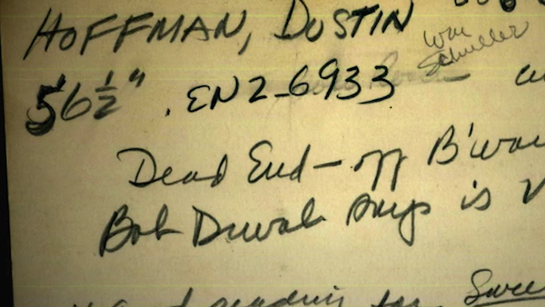 Screen capture from the movie
-
-
www.biorxiv.org www.biorxiv.org
-
Reviewer #3 (Public Review):
The authors demonstrate in mice that the amount of sleep is related to stress resilience, and specifically that increased sleep after stress exposure supports resilient behavior. The aims are achieved through an array of methodologies, which highly strengthens the conclusions of the work. The question of whether sleep is related to stress resilience is highly significant and in the current research, the authors tackle the questions by evaluating differences in sleep homeostasis in stress-resilient compared to stress-susceptible mice. To induce more stress-susceptibility, the authors challenge the mice with sleep restriction, and to induce more stress-resilience, the authors chemogenetically induce increased NREM sleep. Mechanistically the authors demonstrate that cortically mediated NREM sleep is sufficient to promote resilience. Despite the challenging nature of the technological approaches at hand, particularly in mice, the experiments are well-designed and the authors are commended for the execution of these studies.
It is very difficult to separate sleep loss and stress responses, as losing sleep is inevitably a stressful experience. The authors attempt to quell the notion that sleep restriction was also stressful to the animals by measuring fecal corticosterone, however, the measurements were from fecal pellets collected during an entire 24 hr period raises concerns that acute changes in HPA response may not be evident through this measurement. This is a challenging notion to tackle and deserves a bit more consideration.
Chemogenetic experiments induce a beautiful increase in NREM sleep at the expense of REM loss, yet all the animals treated with chemogenetic agents are resilient to social defeat stress. The authors conclude that because sleep restriction also reduced REM, yet opposite effects occur on social interaction, REM sleep is unlikely to be related to resilience. In this context, it would be beneficial to discuss the theories of sleeping to forget and sleeping to remember, and supporting literature that REM sleep is critical to the consolidation of memories, particularly upon stressful experiences.
-
-
www.biorxiv.org www.biorxiv.org
-
Reviewer #3 (Public Review):
This study addresses fundamental aspects of the eco-evolutionary dynamics of highly fecund organisms experiencing huge mortality rates during early life stages. In such species, a mechanism called "sweepstakes reproductive success" (Hedgecock, 1994) has been proposed to understand the dynamics of recruitment, in which individual reproductive success shows high variance and skewed distribution. Sweepstakes reproductive success can be either neutral due to random environmental variation influencing the recruitment of reproducing offspring, or selective because genetic variation at particular loci influences the likelihood of successful recruitment. Unfortunately, empirical tests of sweepstakes reproduction remain scarce due to the difficulty of studying individual reproductive success directly, particularly in highly fecund marine organisms.
By analysing genome-wide genetic diversity data under different coalescent models representing alternative recruitment dynamics, this pioneering study specifically tests whether random or selective sweepstakes reproduction occurs in the highly fecund Atlantic cod. Using the classical Kingman coalescent and two multiple-merger coalescent models approximating random sweepstakes (the Xi-Beta-coalescent model) and selective sweepstakes (the Durrett-Schweinsberg model), the authors show that genetic diversity in the Atlantic cod genome is most likely shaped by pervasive selective sweeps of new beneficial mutations. The best-fit selective sweepstakes model is able to reproduce the main characteristics of the allele frequency spectrum of each Atlantic cod population, while alternative models include either random sweepstakes or other biologically plausible scenarios (i.e. historical demographic changes, cryptic breeding structure, and background selection) show a much poorer fit.
These findings have a broad impact on evolutionary genomics since they provide a new and exciting perspective on the choice of appropriate coalescent models for the study of highly fecund organisms that may experience high rates of selective mortality during early life stages. The low-fecundity low-variance reproductive success model classically used in evolutionary genetics may simply not apply in highly fecund organisms with skewed offspring distribution.
By confronting different alternative models of coalescence with genome-scale genetic diversity data, this work provides a roadmap for exploring fundamental processes at the crossroads between ecology and evolution. It highlights the importance of (i) understanding the potential impact of species-specific biological characteristics when inferring demography and selection from molecular data, and (ii) being aware of the potentially significant effects of unaccounted aspects of the data (e.g. variant misorientation, past admixture) on the interpretation of results.
-
-
www.biorxiv.org www.biorxiv.org
-
Reviewer #3 (Public Review):
The manuscript by Ronzano et al presents a rigorous neuroanatomical study to convincingly demonstrate that there is no difference in the medio-lateral organization of flexor and extensor premotor interneurons. The study uses monosynaptic restricted transsynaptic tracing from ankle flexor and extensor muscles with several (4) strategies for delivery of the G protein complement and delta G Rabies virus, and additional (2) variations that consider titer and cre line. The authors went to great lengths here in attempt to replicate prior studies for which they had initial conflicting findings. Further, the experiments are performed in laboratories in four different locations. The variations on the Rabies and complement delivery, regardless of lab performing the experiment and analysis, all converge on the same conclusion. Aside from the primary conclusion, the paper can be used as a manual for anyone considering transsynaptic tracing as it details the benefits and caveats of each strategy with examples.
The initial conflicting results put the onus on the authors to demonstrate where the divergence occurred. The authors took a highly comprehensive approach, which is a clear strength of the paper. All of the data is fully and transparently presented. Standardizations and differences between experiments run or analyzed in each lab are well laid out. Figure 1 and Table 2 provide a great summary of the techniques and their limitations. These are also well thought out and discussed within each section of results.
The only thing missing is a likely explanation for the differences seen. Although the authors made several attempts to provide such explanation, the question remains - how did two groups who published independent studies using different strategies demonstrate flexor and extensor separation in the dorsal horn, when this study, using several strategies in multiple labs, show that the premotor neurons are in complete overlap? Additional small differences in methodologies could be identified which are not discussed and may provide potential explanations, but only for discrepancies in results of single techniques, not for all of the strategies used. The lack of reason for the discrepancy with prior studies despite the extensive efforts is unsatisfying, but, most importantly, the experiments were rigorously performed and the data support the conclusions presented.
-
-
www.biorxiv.org www.biorxiv.org
-
Reviewer #3 (Public Review):
Zhao and Sharpe identified telocytes in the periodontium. To address their contribution to periodontal diseases, they conducted scRNA-seq analysis and lineage tracing in mice. They demonstrated that telocytes are activated in periodontitis. The activated telocytes send HGF signals to surrounding macrophages, converting M2 to M1/M2 hybrid status. The study implies that targeting telocytes and HGF signal for the potential treatment of periodontitis.
The significance of the study could be improved by authors testing if targeting telocytes or HGF signals could ameliorate periodontitis in the mouse model. The current form of the manuscript lacks the data that demonstrate the actual contribution of telocytes in the homeostasis of periodontium or progression of periodontitis.
Major comments:
1) I see the genetic validation of the role of telocytes or HGF signals are crucial to assure the significance of this manuscript. I recommend either of two experiments. a. testing the role of HGF signals by deleting the Hgf gene in telocytes. Using Wnt11-Cre; Hgf f/f mice, the authors could address the role of HGF signals in periodontitis. CX3CR1-Cre; cMet f/f mice will delete HGF signals in monocyte-derived macrophages. This will be another verification, but not sure if the PDL macrophages are derived from yolk sac or monocytes. b. measuring the contribution of telocytes in the homeostasis or disease progression. The mouse model could be challenging though, the system if achieved will be very informative. The authors could first check the expression of telocyte enriched genes, such as Lgr5 or Foxl1 reported previously in other tissue telocytes. Delete those genes under the Wnt1-Cre driver and check if telocyte lineage is removed. The system would be very useful for next-level study. DTA model could be an alternative, but Wnt1-Cre is vastly expressed in neural crest lienage.
2) This paper points out that the M1/M2 hybrid state of macrophages appears upon periodontitis. The authors could further characterize the hybrid macrophages by the expression of more markers, production of cytokines, and morphology. Need to clarify if this means some macrophages are in M1 state and others are in M2 state, or one macrophage possesses both M1 and M2 phenotype. Please conduct either FACS or immunofluorescence to demonstrate if one macrophage expresses both markers. Please introduce more information about the M1/M2 hybrid state of macrophage based on other present literature.
3) In the introduction part, the author lists several markers that can be used for telocyte identification, such as CD34+CD31-, CD34+c-Kit+, CD34+Vim+, CD34+PDGFRα+. Could authors explain why they chose CD34 CD31, but not other markers?
4) In figure 5g, I don't think the yellow color cell shows the reduction trend in the Tivantinib treatment group compared with a control group. Please validate the observation by gene expression analysis, WB, etc. In addition, please show c-Met+ cells level in the Tivantinib treatment group and control group.
-
-
www.biorxiv.org www.biorxiv.org
-
Reviewer #3 (Public Review):
In this MS by Sefton et al., the authors investigate the role of HGF/MET pathway, as well as the cellular source of these molecules, during diaphragm development. In particular, the authors address the function of this pathway on muscle progenitors and phrenic nerve. They further provide evidence for the expression of HGF in pleuroperitoneal folds and for its requirement for muscle progenitor recruitment and maintenance during diaphragm muscle formation. This study is interesting and in general the results support the conclusions. The work could be improved by (1) providing appropriate controls for the role of HGF in the connective tissue and (2) linking the muscleless diaphragms and HGF to the hernia phenotype.
-
-
www.biorxiv.org www.biorxiv.org
-
Reviewer #3 (Public Review):
This manuscript aimed to reveal the difference and similarity of sharp-wave ripples in awake vs. sleeping mice. To do this, the authors used wide-view voltage and glutamate activity imaging in awake head-fixed mice. The two-photon Ca imaging was applied to examine the spiking activity of the retrosplenial cortex.
They showed that the mean membrane potential and glutamatergic transmission of the neocortex's superficial layers were suppressed and enhanced, respectively, just after the sharp-wave ripples in awake mice, contrary to the same authors' previous findings in urethane-anesthetized and naturally sleeping mice. The retrosplenial cortex was most strongly modulated in membrane potential and glutamatergic transmission by awake sharp-wave ripples. The authors also found two groups of retrosplenial cortical neurons, whose spiking activity, measured by Ca dynamics, was suppressed and enhanced by awake sharp-wave ripples. These findings revealed the critical difference between sharp-wave ripples during waking vs. sleep, which would impact the field of memory research.
This manuscript's strength is that it compares the dynamics of membrane potential and glutamate transmission using wide view imaging. Both experimental and analytical methods were appropriate and supported their main conclusions.
-
-
www.biorxiv.org www.biorxiv.org
-
Reviewer #3 (Public Review):
The authors attempted to, and succeeded at, testing the recent hypothesis that the large theropod dinosaurs Spinosaurus was a fast and capable swimmer and diver.
The strengths of the paper are the extent of the analyses which address and test numerous aspects of the 'aquatic hypothesis' with some depth, though in places this needs a better explanation of the details of the process and organisation. The results do support the conclusions, though these should be more specific and clear and relate to the recent literature on Spinosaurus habits to say what is, and is not, possible/plausible based on their analyses.
Overall this is likely to be a major step forwards in resolving the biology of this animal (and kin) and add considerable depth to the discussion by adding new data and results.
-
-
www.biorxiv.org www.biorxiv.org
-
Reviewer #3 (Public Review):
In the manuscript by Bosada et al, the authors present work identifying and interrogating two cis-regulatory elements at TBX5 associated with atrial fibrillation. Mouse models lacking both copies of either element, but particularly one in the last intron of the Tbx5 gene, referred to as RE(int), results in increased Tbx5 expression, changes to cardiac electrophysiology, and downstream gene expression changes. Of interest, RE(int) induces expression of Prrx1, also associated with atrial fibrillation, and compound mutants partially rescues the RE(int) phenotype. Overall, this paper is of interest and advances our understanding of TBX5 in atrial fibrillation risk in humans. Critically, this study focuses on the impact of risk SNPs, which increase TBX5 expression in patients, while previous studies involving TBX5 in atrial fibrillation have focused on decreased expression.
The authors' work presents the following major claims:<br /> Figure 1. Identification of two, independently segregating risk regions at TBX5, which are conserved in humans and mice with predicted cis-regulatory functions termed RE(int) and RE(down).<br /> Figure 2. Homozygous RE(int) and RE(down) mutants, with a particular focus on RE(int) mutants, resulting in increased expression of Tbx5. The functional SNP appears to be rs7312625 A>G.<br /> Figure 3. Homozygous RE(int) mutants demonstrate several cardiac electrophysiological changes consistent with increased atrial fibrillation risk in humans.<br /> Figure 4. Cellular electrophysiology of RE(int) mutant cardiomyocytes demonstrates additional supportive changes to explain whole organ phenotypes presented in Figure 3.<br /> Figure 5. Transcriptional profiling of RE(int) and RE(down) homozygous mutants demonstrates many significant differences from control samples that are suggestive of specific mechanisms disrupting cardiac electrophysiology, including calcium and potassium regulators and gap junctions.<br /> Figure 6. Homozygous RE(int) and Prrx1(enh) mutant alleles genetically interact and result in partial rescue of phenotypes from RE(int) alone.
-
-
www.biorxiv.org www.biorxiv.org
-
Reviewer #3 (Public Review):
Experimental and computational works have proposed that neurons in the hippocampus represent a predictive map of the environment called the successor representation. This theoretical study examines how plasticity in a model network of recurrently connected neurons can lead to such a representation. The main conclusion is that any plasticity rule that encodes transition probabilities in synaptic weights gives rise to the successor representation at the level of neural activity. This fundamental theoretical insight gives additional credibility to the idea that the hippocampus can implement the successor representation.
Strengths:<br /> - elegantly designed theoretical study<br /> - very clear writing that progressively introduces the main result and argues for its generality<br /> - comparison of the model with data in a random foraging task
Weaknesses:<br /> - certain technical choices need additional motivation
-
-
www.biorxiv.org www.biorxiv.org
-
Reviewer #3 (Public Review):
The present study is a comparison of brain magnetic resonance imaging (MRI) of mice who developed in an enriched environment laboratory environment, in which some mice become habituated while other mice maintain active exploration of the environment over time. Between these groups, differences are shown in the pattern of correlations between brain regions of interindividual variability, which may indicate differences in brain connectivity or other shared maturational processes between regions. Because the mice are genetically inbred and have the same shared environment, these differences are attributed to individual-level differences in environment and behavior, which are extremely difficult to isolate in non-laboratory settings.
My comments are focused on aspects of the paper that overlap with my area of expertise which is human brain MRI methods. The strengths of the paper include the unique environmental paradigm that provides support for important hypotheses about individual-level variation. The imaging methods are rigorous and sound, and there is a nice convergence with human work. The application of structural covariance is interesting. The weaknesseses of the paper are that the writing could be clearer. Alternative explanations for structural covariance and alterations in "down roamers" should be more fully considered. The statistical approaches could be more rigorous in places. The areas of novelty relative to past work should be more explicitly articulated.
-
-
www.biorxiv.org www.biorxiv.org
-
Reviewer #3 (Public Review):
Lillvis et al present a new method for quick targeted analysis of neural circuits through a combination of tissue expansion and (lattice) light sheet microscopy. Three color labeling is available which allows to label neurons of a molecularly specific type, presynaptic and/or post-synaptic sites.
Strengths:<br /> - The experimental technique can provide much higher throughput than EM<br /> - All source code has been made available<br /> - Manual correction of automatic segmentations has been implemented, allowing for an efficient semi-automatic workflow<br /> - Very different kinds of analyses have been demonstrated<br /> - Inclusion of electrical connections is really exciting, what a great complement to the existing EM volumes!
Weaknesses:<br /> - Limitations of the method are not really discussed. While the approach is simpler and cheaper than EM, it's still important to give the readers a clear picture of the use cases where it's not expected to work before they embark on the journey of acquiring tens of terabytes of data. Here are just a few examples of the questions I would have if I wanted to implement the method myself - I am a computational person and can easily imagine my "wet lab" colleagues would have even more to ask about the experimental side:
-- It is not very clear to me if the resolution of the method is sufficient to disentangle individual neurons of the same type. It has been demonstrated for a few examples in the paper, but is it generally the case? Are there examples of brain regions/neuron types where it wouldn't be possible? If another column was added to the table in Figure 1, e.g. "individual neuron connectivity", EM would be "+", LM "-", what would ExLLSM be?<br /> -- Similarly, the procedures for filling gaps in the signal could result in falsely merged neurons. Does it ever happen in practice?<br /> -- How long does semi-manual analysis take in person-hours/days for a new biological question similar in scope to the ones demonstrated in the paper?<br /> -- How robust are the networks for synaptic "blob" detection? The authors have shown they work for different reporters, when are they expected to break? Would you recommend to retrain for every new dataset? How would you recommend to validate the results if no EM data is available?
-
-
www.biorxiv.org www.biorxiv.org
-
Reviewer #3 (Public Review):
Almedia and Macklin sought to characterize oligodendrocyte behavior at the earliest onset of myelination in the central nervous system. By sparsely labeling oligodendrocytes with transgenic fluorescent reporters in zebrafish larvae, they show that oligodendrocytes in the dorsal and ventral spinal cord have characteristic numbers of sheaths and sheath lengths. With impressive and technically laborious time-lapse imaging, they demonstrate that oligodendrocytes repetitively sample axon segments before stabilizing a nascent sheath, with most (~90%) immature sheaths failing to stabilize. They quantify differences in dorsal and ventral oligodendrocyte sampling and convincingly show that dorsal oligodendrocytes form more sheaths due to increased sampling relative to ventral, with similar rates of sheath retraction. Finally, the authors conclude that Rab5 and Rab11 promote myelination locally and cell-autonomously in oligodendrocytes, showing specifically that Rab5 is critical for stabilization of nascent sheaths but is dispensable for sampling. Altogether, the authors provide novel and detailed visualization of early myelin sheath development by oligodendrocytes.
Strengths:<br /> This is one of the first studies to closely examine early oligodendrocyte behavior at high resolution and adds to a body of work showing how oligodendrocytes initiate and maintain myelin sheaths. The authors find that sampling is widespread while most immature sheaths destabilize. This is an intriguing finding, as sampling is likely an energetically intensive process, but its prevalence in wild-type animals suggests that it is an important part of development.
The authors' claims are substantiated by technically challenging mosaic labeling experiments that monitored individual oligodendrocyte development over the course of days. Importantly, the authors have measured the sheath accumulation and loss phase for a single oligodendrocyte over the course of several days, and can pinpoint when individual sheaths are maintained with high resolution.
The imaging acquisition and data analyses are thorough, and the labor-intensive nature of the experiments is commendable. The authors carefully use statistics to validate their conclusions, including power analyses to determine appropriate sample size.
Limitations:<br /> Prior studies suggested that the potential for each oligodendrocyte to produce myelin sheaths is at least partially dependent upon the diameter of axons enwrapped. While axons are labeled to demonstrate ensheathment, axonal diameter is not measured, and it is unclear whether dorsal and ventral oligodendrocytes behavior could be explained by regional or individual differences in axon caliber.
Other recent studies suggest that early myelination is driven by axonal factors as much as oligodendrocyte-intrinsic factors. For instance, neuronal activity stabilizes myelination for a subset of early-born neuronal types. Because of the sparse labeling techniques and focus on oligodendrocyte behavior, it is unknown how or whether axonal subtypes and activity influence early oligodendrocyte sheath sampling and stabilization.
While the authors provide a tantalizing suggestion that early oligodendrocyte sampling primes axonal segments for myelination, it is not tested directly here. Thus, the paper does not address why, or even if, repeated sampling is important in development.
-
-
www.biorxiv.org www.biorxiv.org
-
Reviewer #3 (Public Review):
The authors have constructed a large-scale interaction perturbation network from 2,167 CRC tissues and 308 normal tissues, deciphering six GINS subtypes with particular clinical and molecular peculiarities. In addition, the GINS taxonomy was rigorously validated in 19 external datasets (n =3,420) with distinct conditions. From an interactome perspective, this study identified and diversely validated a high-resolution classification system, which could confidently serve as an ideal tool for optimizing decision-making for CRC patients. The multifariously biological and clinical peculiarities of GINS taxonomy improve the understanding of CRC heterogeneity and facilitate clinical stratification and individuation management. Additionally, candidate specific-subtype agents provide more targeted or combined interventions for six subtypes, which also need to be validated in clinical settings.
-
-
www.biorxiv.org www.biorxiv.org
-
Reviewer #3 (Public Review):
In this report, the authors examined 3 mutations in the BRC-repeat region of BRCA2 in a series of functional assays. They found that two of the mutants showed severe defects in BRCA2 function, whereas the third mutant had no clear phenotype. The two mutants with functional defects are tested most thoroughly. The assays used here a numerous and have been validated and performed with appropriate controls and statistics. There are no concerns about the experiments themselves or the conclusions. So, the strength of the study is the number of assays performed in a rigorous manner.
However, the weakness of the study is that it is unclear why these results are impactful. Several reports over the years, including some recent studies mentioned at the end of the Discussion, have involved parallel functional analysis of hundreds of alleles of BRCA2, with a clear end goal of improving medical decision-making for carriers of these BRCA2 alleles. Certainly, these studies have usually focused on other domains of BRCA2, like the DNA binding domain, but nonetheless, since these studies have typically involved testing hundreds of BRCA2 alleles, it is unclear how this manuscript studying 3 alleles fits into a broader population science effort to categorize BRCA2 variants of unknown significance (VUS). Perhaps the authors would argue that their study involves a comprehensive analysis of the 3 alleles, whereas other studies typically involve one or two functional assays. However, if that is the case, then is the argument that multiple assays are needed for accurate characterization of VUS? If so, what is the evidence for that assertion? Are there particular assays that are more likely to be predictive of pathogenicity based on their analysis?
The mechanistic insight of the study is also unclear. These alleles are in conserved residues of the BRCA2 BRC repeats, which have been established as being important for BRCA2 function. Indeed, in the Discussion, it appears that the findings here are largely confirmatory for other mechanistic studies of the BRC repeats of BRCA2. What new information has been determined about the role of the BRC2 and BRC7 repeats from this study?
-
-
www.biorxiv.org www.biorxiv.org
-
Reviewer #3 (Public Review):
This study was designed to test the hypothesis that output from a subpopulation of neurons (PKCδ neurons) in the central nucleus of the amygdala (CeA) inhibits ZI neurons in a neuropathic pain condition and this ZI inhibition results in pain-related behaviors (Fig. 5).
First, the targets of CeA-PKCδ neurons were identified using cre-dependent viral vector for anterograde labeling with red-shifted channelrhodopsin (CrimsonR-tdTomato) or mCherry, and cholera toxin B (CTB) in PKCδ-tdTomato mice for retrograde labeling. The ZI was identified as one of the targets with approximately 19% of CTB+ CeA neurons identified as PKCδ- tdTomato positive, which is significant and makes this pathway worth exploring.
Next, electrophysiological (patch-clamp) studies showed monosynaptic inhibitory transmission from CeA to both VGAT+ and VGAT- neurons of the ZI and found no significant difference between these projections (from CeA to GABAergic or non-GABAergic ZI neurons).
Finally, chemogenetics are used to activate or silence GABAergic ZI neurons and determine behavioral consequences. Inhibition of GABAergic ZI neurons induced hypersensitivity in naïve mice and activation of these neurons reversed hypersensitivity in a neuropathic pain model. Interestingly, these effects were modality specific.
The combination of tracing techniques, electrophysiology, chemogenetics and behavior is a strength of this study, and so this the impressive amount of high-quality data. The focus on CeA-PKCδ neurons in the modulation of ZI is an important novelty of the present study.
However, slice physiology and behavioral data presented here do not actually link CeA-PKCδ neurons to ZI. Electrophysiological data show inhibitory transmission from CeA to ZI, but not specifically from CeA-PKCδ neurons to ZI. Behavioral studies assess the effects of modulation of ZI neurons but not of CeA-PKCδ to ZI projections. Previous data already showed the effects of activation and inhibition of GABAergic ZI neurons on pain behaviors, including in a neuropathic pain model.
Therefore, although the proposed model of CeA-PKCδ to ZI interactions in pain (Fig. 5) is novel and significant, additional experiments focusing on CeA-PKCδ neurons and their ZI projections would be needed to fully support this concept and enhance impact of the work.
-
-
www.biorxiv.org www.biorxiv.org
-
Reviewer #3 (Public Review):
The authors have used both overall and local genetic correlations to understand how genes associated with two traits relate to those same traits. Their work focuses on understanding why in some cases local genetic correlations may disagree with overall correlation in terms of the direction of effect and exploit known biology to understand why and when this arises.
Overall the work is solid methodologically as it relies on well-established statistical methods and known biology. I don't see particular weaknesses in this work limited to the presented examples. It remains unclear how these observations will generalise to other less well-known biology or traits, but this is a matter of future work.
The work is in my opinion highly impactable as it creates a framework to be used to investigate the pleiotropic effects of genes and could help understand their biological role.
-
-
www.biorxiv.org www.biorxiv.org
-
Reviewer #3 (Public Review):
This study reports on the phenotypes of a CRISPR-engineered zebrafish mutants in kinesin light chain 4 (KLC4). KLC4 is expressed prominently in spinal cord sensory neurons, and mutants have defects in peripheral axon branching/stabilization and branch repulsion, as well as make occasional ectopic axon branches. Imaging also demonstrates that axonal microtubule growth dynamics are altered. These axonal phenotypes are nicely characterized with beautiful light sheet time-lapse microscopy and clever image analyses methods. Additionally, the growth of adult KLC4 mutants is stunted, and they exhibit a variety of behavioral defects.
The strengths of this paper are the creation of a new mutant for studying axonal transport, the impressive imaging methods, and the development of image analysis methods for characterizing axonal trajectories across a population.
The main weaknesses is the lack of a specific mechanistic explanation for how kinesin dysfunction leads to axonal defects-what kinesin cargoes play a role in branch stabilization and branch repulsion? How does kinesin-mediate transport affect microtubule growth?
Another weakness is the lack of a connection between the cellular defects characterized in larval sensory neurons, and the behavioral defects in adults. Since the adult behavioral defects likely do not involve sensory neurons, these two parts of the paper don't fit together. The authors may want to consider moving the behavior to a different paper. Additionally, the cellular basis of the adult behavioral defects is unknown, and likely involves a complex combination of defects in multiple cell types.
-
-
www.biorxiv.org www.biorxiv.org
-
Reviewer #3 (Public Review):
In this manuscript, Zhou et al describe basal cell heterogeneity in the mouse trachea. They describe how dorsally vs ventrally located tracheal basal cells which are supported by different stromal cell populations show differential potential to undergo squamous metaplastic differentiation. Furthermore, they suggest that the differences in these basal cells might be epigenetically programmed as they are maintained after these basal cells have been isolated and cultured in vitro. However, it is not clear whether dorsal vs ventral supporting stromal cell populations made it into the culture medium.
-
-
www.biorxiv.org www.biorxiv.org
-
Reviewer #3 (Public Review):
This work is important for understanding both how immune cells are regulated and how alterations in receptor signaling can affect the balance of health and development of autoimmune diseases. The work uses CRISPR-based genetic manipulation of the autoimmunity associated PTPN22 gene in single donor human cord-derived naïve T cells to analyze T-cell receptor functions. The authors conclude that the autoimmunity associated PTPN22 variant PTPN22(620W) is a loss-of-function mutant as T cells expressing PTPN22(620W) phenocopies PTPN22 deficient T cells. The use of a single donor minimizes potential other effects that would be observed when comparison cellular functions from multiple donors.
-
-
www.biorxiv.org www.biorxiv.org
-
Reviewer #3 (Public Review):
Angueyra et al. tried to establish the method to identify key factors regulating fate decisions in the retinal visual photoreceptor cells by combining transcriptomic and fast genome editing approaches. First, they isolated and pooled five subtypes of photoreceptor cells from the transgenic lines in each of which a specific subtype of photoreceptor cells are labeled by fluorescence protein, and then subjected them to RNAseq analyses. Second, by comparing the transcriptome data, they extracted the list of the transcription factor genes enriched in the pooled samples. Third, they applied CRISPR-based F0 knockout to functionally identify transcription factor genes involved in cell fate decisions of photoreceptor subtypes. To benchmark this approach, they initially targeted foxq2 and nr2e3 genes, which have been previously shown to regulate S-opsin expression and S-cone cell fate (foxq2) and to regulate rhodopsin expression and rod fate (nr2e3). They then targeted other transcription factor genes in the candidate list and found that tbx2a and tbx2b are independently required for UV-cone specification. They also found that tbx2a expressed in the L-cone subtype and tbx2b expressed in L-cones inhibit M-opsin gene expression in the respective cone subtypes. From these data, the authors concluded that the transcription factors Tbx2a and Tbx2b play a central role in controlling the identity of all photoreceptor subtypes within the retina.
Overall, the contents of this manuscript are well organized and technically sound. The authors presented convincing data, and carefully analyzed and interpreted them. It includes an evaluation of the presented data on cell-type specific transcriptome by comparing it with previously published ones. I think the current transcriptomic data will be a valuable platform to identify the genes regulating cell-type specific functions, especially in combination with the fast CRISPR-based in vivo screening methods provided here. I hope that the following points would be helpful for the authors to improve the manuscript appropriately.
1) The manuscript uses the word "FØ" quite often without any proper definition. I wonder how "Ø" should be pronounced - zero or phi? This word is not common and has not been used in previous publications. I feel the phrase "F0 knockout", which was used in the paper cited by the authors (Kroll et al 2021), is more straightforward. If it is to be used in the manuscript, please define "FØ" and "CRISPR-FØ screening" appropriately, especially in the abstract.
2) Figure 1-supplement 1 shows that opn1mw4 has quite high (normalized) FPKM in one of the S-cone samples in contrast to the least (or no) expression in the M-cone samples, in which opn1mw4 is expected to be detected. The authors should address a possible origin of this inconsistent result for opn1mw4 expression as well as a technical limitation of using the Tg(opn1mw2:egfp) line for detection of opn1mw4 expression in the GFP-positive cells.
3) The manuscript lacks a description of the sampling time point. It is well known that many genes are expressed with daily (or circadian) fluctuation (cf. Doherty & Kay, 2010 Annu. Rev. Genet.). For example, the cone-specific gene list in Fig.2C includes a circadian clock gene, per3, whose expression was reported to fluctuate in a circadian manner in many tissues of zebrafish including the retina (Kaneko et al. 2006 PNAS). It appears to be cone-specific at this time point of sample collection as shown in Fig.2, but might be expressed in a different pattern at other time points (eg, rod expression). The authors should add, at least, a clear description of the sampling time points so as to make their data more informative.
-
-
www.biorxiv.org www.biorxiv.org
-
Reviewer #3 (Public Review):
At the heart of this manuscript is a debate concerning the role of the orbitofrontal cortex (OFC) in goal-directed behavior. One commonly sees a paper in which Ostlund and Balleine placed large OFC lesions in behaviorally-experienced rats cited as irrefutable evidence that OFC is not involved in goal-directed behavior because these rats could perform typically in a simple devaluation task. Meanwhile, others have argued that the ventrolateral OFC (VLO) sits at a nexus between the medial PFC structures (which are attuned to reinforcer value, etc.) and the far lateral regions (which appear to be more specialized in Pavlovian associations) and may therefore play a role in goal-directed behavior (e.g., this argument is put forward in Gourley and Taylor, 2016, Nature Neuroscience). The present team published a crucial manuscript a couple of years ago showing that selective VLO lesions do indeed disrupt goal-seeking behaviors, particularly when value and contingency information needs to be integrated and/or updated (Parkes 2018). Because this sophisticated process is not tested in simple devaluation assays, it would have been missed in the older study. The Parkes 2018 paper, meanwhile, supports other investigations that also selectively manipulate the VLO and require animals to integrate new information into existing instrumental response strategies.
Here, the team first depleted NE fibers in the OFC and found that rats were unable to encode new associations in an instrumental reversal. This same deficit was not observed with parallel DA manipulation. They found that LC-OFC and not mPFC projections had the same effect. Throughout, important control experiments were conducted, and the tools being used were largely well-validated. The conclusions are sensible, and the writing is clear.
I would be curious about the authors' thoughts regarding the recent Duan ... Robbins Neuron paper (https://pubmed.ncbi.nlm.nih.gov/34171290/), in which marmosets displayed paradoxical responses to VLO inactivation and stimulation in contingency degradation tasks. Are there ways to reconcile these reports?
-
-
www.biorxiv.org www.biorxiv.org
-
Reviewer #3 (Public Review):
The authors constructed a single-cell transcriptome atlas of bone marrow in normal and R-ISS-staged MM patients. A group of malignant PC populations with high proliferation capability (proliferating PCs) was identified. Some intercellular ligand receptors and potential immunotargets such as SIRPA-CD47 and TIGIT-NECTIN3 were discovered by cell-cell communication. A small set of GZMA+ cytotoxic PCs was reported and validated using public data.
For scRNA-seq data analysis, the authors did QC and filtering and removed low quality cells, including some doublets and followed by batch effect correction. Malignant PC populations were identified using the copy number analysis tool - "inferCNV".
The authors have done lots of analysis. But I think the results can be improved if they can do more analyses. I would recommend to 1) analyze doublets; 2) remove cell cycle effect; 3) GO and pathway analysis for genes with copy number change; 4) do cell-cell communication with more cell type/clusters.
Data analysis of public data was sufficient to prove the small set of GZMA+ cytotoxic PCs. More data analysis or wet experiment proof is required.
-
-
www.biorxiv.org www.biorxiv.org
-
Reviewer #3 (Public Review):
The authors identified a splicing factor that regulates mitochondrial homeostasis by regulating the alternative splicing of the pro-apoptotic protein BAX, which induces basal upregulation of interferon stimulated genes and sensitizes cells to apoptotic cell death. They report that loss of Serine/Arginine Rich Splicing factor 6 (SRSF6) results in accumulation of an alternatively spliced form of BAX known as BAX-, which results in increased release of mitochondrial DNA (mtDNA). The released mtDNA is sensed by cGAS, which leads to upregulation of interferon stimulated genes via IRF3. Importantly, the increase in BAX- sensitizes macrophages to apoptosis and various pathogens decreased the expression of SRSF6 during infection, which served a protective role. Interestingly, Mycobacterium tuberculosis decreases SRSF6 expression, but this resulted in a replication advantage. Overall, these findings add new mechanistic insight into the role of alternative splicing in regulating immunity and cell death. This work can potentially open novel avenues of inquiry into the role of BAX in regulating apoptosis.
Strengths:
The paper is well written, and the major conclusions are rigorously tested by numerous experiments. The data supports the major conclusions, which are that loss of SRSF6 increases ISG and leads to accumulation of alternatively spliced BAX, sensitizing cells to death.
Weaknesses:
The authors make a very interesting discovery that SRSF6 KD sensitizes macrophages to a caspase independent death by up regulating an alternatively spliced variant of BAX, a protein that has a well-established role in mediating caspase dependent death, but they did not rigorously test whether it was truly caspase independent.
-
-
www.medrxiv.org www.medrxiv.org
-
Reviewer #3 (Public Review):
In the manuscript, the authors provided the development of a sensitive and rapid diagnostic tool for detection of pathogenic bacteria in respiratory infections given the limitations of traditional cultures in the clinical settings. Rapid identification and treatment of bacterial infections can impact the prognosis in sepsis. This work highlights how a new rapid diagnostic tool may be beneficial in the treatment of patients with bacterial pneumonia given the time-consuming nature and low sensitivity of traditional culture methods.
Strengths:
The manuscript authors created a diagnostic tool using CRISPR-Cas12 with bacterial species-specific DNA-tags to 10 epidemic bacteria at their local intensive care unit (ICU). The appendix data provided detailed reports of the reaction conditions, sample preparations and reaction incubation time.
A 2-stage validation process was used. The initial validation stage compared the use of the novel diagnostic tool to traditional cultures from bronchoalveolar lavage samples from ICU patients. Once the accuracy of the diagnostic tool was evaluated, the second validation stage was pursued in the form of a randomized controlled trial at the ICU of the study. The second validation stage demonstrated that the proposed novel diagnostic tool had faster results and correlated with improved APACHE II scores and more effective antibiotic coverage rates in the experimental group.
The use of the novel diagnostic test highlighted limitations traditional culture modalities may have in identifying polymicrobial infections which were identified more frequently in the two validation stages
Weaknesses:
Although the study has many strengths, a potential weakness could lie in the unclear use of next-generation sequence (NGS) testing where samples were reported to be sent at random. However, similar to the novel diagnostic tool proposed in this manuscript, NGS testing has been noted to have high sensitivity and specificity and both had similar results in the manuscript.
Additionally, the novel diagnostic testing demonstrated increased detection of polymicrobial infection when compared to traditional cultures; however, clinical evaluation will remain important to help decipher potential "false positive" results or identification of non-pathogenic colonization.
Based on the author's proposed aims to develop a rapid and sensitive diagnostic tool for bacterial pathogens in pneumonia; the authors demonstrated a highly sensitive and specific test when compared to gold-standard testing. Random samples were assessed against NGS testing technology with similar reported results. The development of this rapid, sensitive diagnostic tool can have wide-spread clinical implications to guide management in patient care where earlier time to effective treatment can have important impacts on prognosis.
-
-
www.biorxiv.org www.biorxiv.org
-
Reviewer #3 (Public Review):
In the manuscript by Gao et al, the authors were trying to achieve an understanding of how Kiaa1024L/Minar2 is necessary for hearing in vertebrates. It is known that the Kiaa1024L/Minar2 mutation causes deafness in mice but not much beyond that is known.
Strengths:<br /> - In this manuscript, they were successful in making two zebrafish mutant zebrafish strains in the Kiaa1024L/Minar2 gene using Crispr/Cas9. The mutant(s) has defects in hearing (using the C-start assay and determining thresholds) and reduced hair cell numbers in the ear (phalloidin labeling to determine hair cell density in utricle and saccule) and the lateral line (including using the AM1-43 assay). From these data, they demonstrate that hair cells are defective in these mutants.
- The authors show that Lamp1-GFP labeled lysosomes change in size in the minar2fs139 mutant. In addition, they show that GFP-Minar2 localizes to lysosomal membranes in cultured cells (human and monkey).
- They performed primary amino acid sequence analysis on Minar2 and showed that it contained a putative CSD of caveolin, which is known to interact with cholesterol. They then show that when Minar2 is expressed in cells in culture, there is an increase in cholesterol detection in the region that contained Minar2, supporting the idea that cholesterol interacts with Minar2.
The experiments in figure 5 seem to show that lowering cholesterol levels using pharmacology exacerbates hair cell defects in a minar2 mutant.
Weaknesses:<br /> 1. The authors attempt to show localization (Fig 2 A and B) of Minar2 to the stereocilia and the apical region of hair cells using GFP-MINAR2 fusion protein expression in hair cells of transgenic animals. Although this is a typical way of demonstrating localization, it is usually used to validate location after a similar pattern has been shown using an antibody (usually in mice.) So, special precautions must be taken when interpreting this kind of transgenic data. According to the authors, GFP-MINAR2 localized to the stereocilia and the apical region of hair cells. This needs to be validated by some other means. I can also see the localization of the green signal at the basolateral area of the cells in Fig 2a. Moreover, it's important to note that other mislocalized fusion proteins localize to the apical region of hair cells.
2. Figure 2C and D. The defects in the hair bundles are plausible but not convincing. Electron microscopy should be used to validate. Also, are hair bundle defects seen in the neuromast? EM would be easier to do there.
3. Fig 1A do prim 1- and prim 2-derived neuromasts express minar2? Do anterior neuromasts express minar2?
4. It's my impression that the authors don't take into account that there is much more plasma membrane in the stereocilia than in the basolateral membrane. So, this statement, "These data suggested that there are high levels of accessible cholesterol located to the stereocilia membranes, while the accessible cholesterol levels are marked lower in the basolateral membranes in the hair cells" based on Figure 4 needs to be reconsidered. The authors need to show that the little reporter that is present in the basolateral membrane is not equal to the reporter present in a single sheet of the plasma membrane in a stereocilium. I can see basolateral labeling in the lateral line hair cells.
5. It's not clear if there is a paralog of the Kiaa1024L/Minar2 gene.
-
-
www.biorxiv.org www.biorxiv.org
-
Reviewer #3 (Public Review):
The manuscript by Sureshchandra et al is a very extensive analysis of monocyte function and their molecular landscape in cord bloods from lean and obese mothers. They aimed to analyze the effects of pre-pregnancy BMI on the functioning of the innate immune system in newborns in a very extensive way. The combination of functional and molecular analyses strengthens their observations and shows many different sides of monocyte activation. I think this approach needs to be praised and should be an inspiration to many others who study monocyte function. This allows for a broad view on the matter and also shows where potential targeting will be necessary in the future. Overall, the manuscript and particularly the methods section is very well written and extensive, making it easy to study how robust the data are.
-
-
www.biorxiv.org www.biorxiv.org
-
Reviewer #3 (Public Review):
Through a series of rigorous in vitro studies, the authors determined McdB's domain architecture, its oligomerization domains, the regions required for phase separation, and how to fine-tune its phase separation activity. The SEC-MALS study provides clear evidence that the α-helical domains of McdB form a trimer-of-dimers hexamer. Through analysis of a small library of domain deletions by microscopy and SDS-PAGE gels of soluble and pellet fractions, the authors conclude that the Q-rich domain of McdB drives phase separation while the N-terminal IDR modulates solubility. A nicely executed study in Figure 4 demonstrated that McdB phase separation is highly sensitive to pH and is influenced by basic residues in the N terminal IDR. The study demonstrates that net charge, as opposed to specific residues, is critical for phase separation at 100 micromolar. In addition, the experimental design included analysis of McdB constructs that lack fluorescent proteins or organic dyes that may influence phase separation. Therefore, the observed material properties have full dependence on the McdB sequence.
Studies of proteins often neglect short, disordered segments at the N- or C- terminus due to unclear models for their potential role. This study was interesting because it revealed a short IDR as a critical regulator of phase separation. This includes experiments that remove the IDR (Fig 2 & 3) and mutate the basic residues to show their importance towards McdB phase separation. In a nice set of SDS-PAGE experiments, the authors showed that as the net charge of the IDR decreased the construct became more soluble.
One challenge is in the experimental design when mutating residues is to assess their impact on phase separation. The author's avoided substitutions to alanine, as alanine substitutions have synthetically stimulated phase separation in other systems. The authors, therefore, have a good rationale for selecting potentially milder mutations of lysine/arginine to glutamine. A potential caveat of mutation to glutamine is that stretches of glutamines have been associated with amyloid/prion formation. So, the introductions of glutamines into the IDR may also have unexpected effects on material properties. Despite these caveats, the authors show mutation of six basic residues in the short IDR abolished phase separation at 100 mM.
Computational studies (Fig 7) also suggest that this short N-IDR region may play a role as a MORF upon potential binding to a second protein McdA. The formulation of this hypothesis is strengthened by the fact that for other ParA/MinD-family ATPases, the associated partner proteins have also been shown to interact with their cognate ATPase via positively charged and disordered N-termini. This aspect of understanding McdB's N-IDR as a MORF is at a very early stage. This study lacks experimental evidence for an N-IDR: McdA interaction and experimental data showing conformational change upon McdA binding. However, the computation study sets up the future to consider whether and how the phase separation activity of McdB is related to its structural dynamics and interactions with McdA.
In summary, this study provides a strong foundation for the contribution of domains to McdB's in vitro phase separation. This knowledge will inform and impact future studies on McdB regulating carboxysomes and how the related family of ParA/MinD-family ATPases and their cognate regulatory proteins. For example, it is unknown if and how McdB's phase separation is utilized in vivo for carboxysome regulation. However, the revealed roles of the Q-rich domain and N-IDR will provide valuable knowledge in developing future research. In addition, the systematic domain analysis of McdB can be combined with a similar analysis of a broad range of other biomolecular condensates in bacteria and eukaryotes to understand the design principles of phase separating proteins.
-
-
www.biorxiv.org www.biorxiv.org
-
Reviewer #3 (Public Review):
In this manuscript, the authors aim to identify the regulators of epithelial invasiveness upon Lethal giant larvae (Lgl), a basolateral polarity protein, knockdown in the follicular epithelium of the Drosophila ovaries, which can serve as a model system to investigate cellular plasticity when apical-basal polarity is lost. Knockdown (KD) of Lgl causes a multilayered epithelium and through extensive single cell RNA-seq analyses, the authors demonstrate that Lgl-KD triggers the appearance of groups of cells exhibiting tumor-associated molecular signatures and invasive behaviour. Overall, the manuscript is technically sound and the combination of computational and experimental approaches results in a thorough characterisation of the earliest steps of epithelial de-stabilisation upon the loss of apical-basal polarity. In my view, the aims set by the authors are met and the experimental data provided support the claims. Interpretations are balanced and the display items are presented logically and informatively for even non-experts. Together, this work will set the basis for further investigations using apical-basal destabilisation of the follicle epithelium as a model of epithelial tumorigenesis.
-
-
www.biorxiv.org www.biorxiv.org
-
Reviewer #3 (Public Review):
Barsi-Rhyne reports a novel mode of engagement of beta arrestins as endocytic adaptors and associates this novel mode together with the previously known canonical mode to the regulation of endocytosis and signaling by class A versus class B receptors. The manuscript is very well written, very good to read, almost flawless, extremely interesting, and highly relevant to the GPCR field with very well-crafted figures and fantastic microscopy.
-
-
www.biorxiv.org www.biorxiv.org
-
Reviewer #3 (Public Review):
The authors use a previously developed technology, CRISPR activation screening, in which pooled sgRNAs are used to guide an RNA-associated regulatory complex (MS2-p65-HSF1 transcriptional activators) to promoter regions resulting in increased expression of a specific target gene. The authors screen two different pooled libraries TM1 (single pass) and TM2+ (multiple pass) with 20 different recombinant biotinylated soluble ligands and identified 22 novel interactions. These interactions were further characterized by SPR and cell-based binding experiments; however, several of the interactions are low affinity and were not characterized for any activity or function beyond the relatively weak biochemical binding. Therefore, while the data provide evidence of potential novel interactions, the biological relevance remains unclear.
-
-
www.biorxiv.org www.biorxiv.org
-
Reviewer #3 (Public Review):
Aside from one critical reservation, I thought this paper was excellent. The figures are clear, the manuscript is well-written, the scope of the study is well-defined (i.e. it characterizes traveling beta), and the authors were circumspect in all aspects of the work, with the authors' consideration of wave propagation along different cortical meshes being but one example in a generally deft and careful approach.
However, the inverse problem remains the inverse problem, and I believe there is one thorny issue to treat regarding the 3D geometry of the central sulcus as it pertains to synchronized beta events before I can accept the authors' conclusions. After this subtle issue is treated, I believe the work will be an important step forward and generally impactful on the community interested in human brain rhythms.
The authors were gracious enough to raise the issue of spatially synchronized events themselves in their discussion: Their argument, with which I mainly agree, is that the beamformer method essentially removes synchronous components from consideration, leaving the traveling component for analysis.
However, synchronization across the sulcus introduces a further bias into event detection by means of physical source-cancellation. I will here defer to Ahlfors et al (2010), who state that "Substantial cancellation occur also for locally extended patches of simulated [cortical] activity, when the patches extended to opposite walls of sulci and gyri."
With that in mind, let's look at Figure 1, where the authors seem to show a higher density of beta events relatively deep in the sulcus compared to the sulcal walls. This is certainly an interesting result if true! But even given only the occasional synchronization of mesoscale cortical neighborhoods, it appears that events in the sulcal walls will still be systematically undersampled and those deep in the sulcus oversampled here, by vice or virtue of cortical geometry as it pertains to the magnetic field.
This spatial sampling bias could impact nearly all aspects of the event propagation analysis that follows, and so I believe it must be considered in some detail before I can fully agree with the manuscript's conclusions.
-
-
www.medrxiv.org www.medrxiv.org
-
Reviewer 3 (Public Review):
This is an interesting study that describes a single cell RNAseq analysis of human menisci. The study describes cell profiling of healthy and degenerated menisci divided into two zones, inner and outer meniscus.
-
-
www.biorxiv.org www.biorxiv.org
-
Reviewer #3 (Public Review):
This article by Roberts, Hayden and colleagues expands on an interesting high-throughput experimental approach developed by Kobori and Yokobayashi (2016; Angew Chem) by determining the relative activity for every possible single and double mutant of five known self-cleaving ribozymes. While this approach is not in itself new, the fact that the authors analyze their data by looking at epistasis (non-additive effects between pairs of mutations) provides an additional opportunity for extracting meaningful structural information that is proposed to be similar to chemical or enzymatic probing experiments obtained on these self-cleaving ribozymes. In fact, this type of high throughput mutagenesis analysis might provide data closer to comparative sequence analysis and as such, might provide even more reliable structural information than structural probing experiments, especially when a relative activity can be properly assessed for the studied RNAs.
(1) Overall, the experiments have been carefully performed and the data seem to be highly reliable.<br /> (2) The strength of this article is that it demonstrates the generality of the approach initially developed by Kobori and Yokobayashi (2016; Angew Chem) by validating its usefulness in identifying most (if not all) the structural features of the studied ribozymes. The determination of positive and negative epistasis is very useful as it can facilitate the identification of base pairs covariations that are indicative of RNA structural elements.<br /> (3) At the present time, the authors have not really discussed how their data analysis compares to comparative sequence analysis. This aspect is important.<br /> (4) It is necessary to mention more clearly that this article builds on the method of Kobori and Yokobayashi (2016). Overall, with the exception of a few experimental details, the experimental method described herein is almost identical to the one of Kobori and Yokobayashi (2016) and this should be better emphasized.<br /> (5) Most importantly, this article provides an analysis of self-cleaving ribozymes for which the three-dimensional structures are known. Considering the scope of this article, instead of mostly focusing on the 2D structural aspect, it would be absolutely necessary to provide more 3D structural information.<br /> (6) When a self-modifying enzymatic activity is associated with the studied RNA, a relative activity could potentially be derived from high throughput sequencing. Could the authors expand on the generality and requirement of their high throughput approach for the study of RNA?
-
-
www.biorxiv.org www.biorxiv.org
-
Reviewer #3 (Public Review):
Menjivar et al. present an analysis of the role of immunosuppressive Arginase 1 in myeloid cells in pancreatic cancer. They show that depletion of Arg1 in macrophages leads to attenuation in progression from PanIN to PDAC and use single cell analysis to understand underlying changes in immune activation, including an increase in cytotoxic T cells. Interestingly, the authors observed what seems to be a compensatory upregulation in Arg1 in epithelial cells and used arginase1 inhibitor to assess the therapeutic potential of targeting Arg1 systemically. This study is overall well performed and generates novel mouse models to study immunosuppression in pancreatic cancer. While the notion that Arginase1 is immunosuppressive is not novel, the observation that Arg1 is upregulated in epithelial cells is interesting. There are several instances of overstating conclusions that, if addressed in the main text (not just relegated to the discussion section), could significantly strengthen the manuscript.
-
-
www.biorxiv.org www.biorxiv.org
-
Reviewer #3 (Public Review):
The central conclusion of this beautiful experimental study is that bumblebees prefer flowers on the basis of their remembered ranking in their context, but are insensitive to their absolute properties. Thus, let's say that there 4 flower types, ranked as follows in nectar concentration: A>B>C>D. However, when the bee learns about these flowers, it does in either of two 'contexts', populated as follows: A & B, or C & D. Thus, the bee experiences that B is the worse option in the context in which it is found, and C is the better one in its own context. If, at a later time, the bee has to make a novel choice, this time between B and C, its memory for ranking leads it to prefer C over B, while its (putative) memory for nectar concentration should favour B over C. The authors find, in a variety of different treatments, evidence for the influence for ranking, but they do not find any evidence for sensitivity to absolute properties (i.e., concentration).
One difficulty that permeates the argument is the ubiquitous difficulty in proving the null hypothesis as true: lack of significant evidence for a putative effect in one or a few experiments, does not mean reliable absence of the effect.
Another difficulty is that in my view memory for absolute properties was not given a full chance: bees were always trained in situations where both dimensions (concentration and ranking) were present. In such situations, they preferentially used ranking. However, to learn ranking between flower types in sequential encounters, they must remember the absolute properties, so that in each encounter they contrast the present flower with the memory for others. Say the bee encounters a type B flower. How does it store its ranking if it doesn't remember the properties of A at all? To take this objection into account and still maintain the claim, it is necessary to say that it remembers the properties of A when in the A & B context, but it erases it from memory when in the context B & C.
Neglecting memory for concentration may be an overshadowing effect. Overshadowing is known in learning studies, and it means that, when more than one cue is paired with an outcome, the most salient between them may reduce learning about the predicting power of the other. In this case, bees may remember and use concentration when trained in contexts where there is only flower type, so that there is no chance of using ranking, and then offered choices between pairs of them. In this case, the bees would not have access to ranking, so that there would be a stronger opportunity for absolute memory to manifest itself.
In experiment 4, during training, they could move between two zones representing the 'contexts', each with 2 flower types, and they were then given choices between the 4 types, rather than just binary choices as previously. In this case, the bees did prefer the top-quality flower type (type A), which is consistent with memory for absolute concentration and with ranking, because A offered the highest concentration of the 4-type context. Why this happened is not clear, but it indicates that the context of choice may be crucial. It is known from other studies that the number of options at the time of choice can be very influential. For instance, in one study, it was shown that starlings appeared to be risk prone when offered a binary choice and risk averse when offered a trinary choice, even if the choices were all intermingled in the same sessions. In any case, this experiment raises doubts as to the claimed insensitivity to memory for nectar concentration. Another possibility is that the separation between contexts in this experiment (a partially avoidable wall) was not extreme as in the previous ones, so that the bees could now establish a ranking among the 4 types because they were all encountered intermingled to an extent.
There is one potential mechanism that may also be discussed. It is known from other species, that state at the time of learning influences subjective value of alternatives. To explain this effect I will exemplify the problem with a non-eusocial consumer. Say that food sources B and C are of equal caloric value. Say, further, that B is encountered when the subject is less food deprived than when it encounters C. Then the hedonic (conditioning) power of B will be lower, because it causes a smaller improvement in fitness (this was Daniel Bernoulli's argument regarding the concept of utility). In animal studies this effect is called State-Dependent Valuation Learning (SDVL). Since in the present experiments the context A & B was richer than the context C & D, the bees would have been in a consequently more favourable state (maybe carrying bigger sugar loads), so that each encounter with B would cause a smaller improvement than each encounter with C. This effect is totally different from remembering the ranking of flower types. The two alternative explanations for preference of C over B (ranking and SDVL) can, fortunately, be confronted because it is possible to change the state of the bees by a common 3rd source that could be used to equate or manipulate the average richness of the contexts.
All the reasons mentioned above should make it clear that this reviewer finds the study of very great interest and much merit, but considers that the conclusion for exclusive impact of ranking on preference should be tempered, or at least defended more strongly against these doubts.
-
-
www.biorxiv.org www.biorxiv.org
-
Reviewer #3 (Public Review):
In this ms Li et al. examine the molecular interaction of Rabphilin 3A with the SNARE complex protein SNAP25 and its potential impact in SNARE complex assembly and dense core vesicle fusion.
Overall the literature of rabphilin as a major rab3/27effector on synaptic function has been quite enigmatic. After its cloning and initial biochemical analysis, rather little new has been found about rabphilin, in particular since loss of function analysis has shown rather little synaptic phenotypes (Schluter 1999, Deak 2006), arguing against that rabphilin plays a crucial role in synaptic function.
While the interaction of rabphilin to SNAP25 via its bottom part of the C2 domain has been already described biochemically and structurally in the Deak et al. 2006, and others, the authors make significant efforts to further map the interactions between SNAP25 and rabphilin and indeed identified additional binding motifs in the first 10 amino acids of SNAP25 that appear critical for the rabphilin interaction.
Using KD-rescue experiments for SNAP25, in TIRF based imaging analysis of labeled dense core vesicles showed that the N-terminus of SN25 is absolutely essential for SV membrane proximity and release. Similar, somewhat weaker phenotypes were observed when binding deficient rabphilin mutants were overexpressed in PC12 cells coexpressing WT rabphilin. The loss of function phenotypes in the SN25 and rabphilin interaction mutants made the authors to claim that rabphilin-SN25 interactions are critical for docking and exocytosis. The role of these interaction sites were subsequently tested in SNARE assembly assays, which were largely supportive of rabphilin accelerating SNARE assembly in a SN25 -terminal dependent way.
Regarding the impact of this work, the transition of synaptic vesicles to form fusion competent trans-SNARE complex is very critical in our understanding of regulated vesicle exocytosis, and the authors put forward an attractive model forward in which rabphilin aids in catalyzing the SNARE complex assembly by controlling SNAP25 a-helicalicity of the SNARE motif. This would provide here a similar regulatory mechanism as put forward for the other two SNARE proteins via their interactions with Munc18 and intersection, respectively.
While discovery of the novel interaction site of rabphilin with the N-Terminus of SNAP25 is interesting, I have issues with the functional experiments. The key reliance of the paper is whether it provides convincing data on the functional role of the interactions, given the history of loss of function phenotypes for Rabphilin. First, the authors use PC12 cells and dense core vesicle docking and fusion assays. Primary neurons, where rabphilin function has been tested before, has unfortunately not been utilized, reducing the impact of docking and fusion phenotype.
In particular the loss of function phenotype in figure 3 of the n-terminally deleted SNAP25 in docking and fusion is profound, and at a similar level than the complete loss of the SNARE protein itself. This is of concern as this is in stark contrast to the phenotype of rabphilin loss in mammalian neurons where the phenotype of SNAP25 loss is very severe while rabphilin loss has almost no effect on secretion. This would argue that the N-terminal of SNAPP25 has other critical functions besides interacting with rabphilin. In addition, it could argue that the n-Terminal SNAP25 deletion mutant may be made in the cell (as indicated from the western blot) but may not be properly trafficked to the site of release.
-
-
www.biorxiv.org www.biorxiv.org
-
Reviewer #3 (Public Review):
The manuscript by Le T.D.V. et al used in vitro cell culture and inhibitors for cellular signaling molecules and found that GLP-1 receptor activation stimulated the phosphorylation of Raptor, which was PKA-mediated and Akt-independent. The authors reported the physiological function of this GLP-1R-PKA-Raptor in liraglutide stimulated weight loss. This timely study has high significance in the field of metabolic research for the following reasons.
(1) The authors' findings are significant in the field of obesity research. GLP-1 receptor (GLP-1R) is a successful target for diabetes (and weight loss) therapeutics. However, the mechanisms of action for the weight-loss effect of GLP-1 agonists are not fully understood. Therefore, mechanistic studies to elucidate the signaling pathways of GLP-1 receptors pertaining to weight loss at the cellular level are timely.
(2) G protein-coupled receptors (GPCRs) induces various signaling activities, which could be cellular and tissue specific. As these are an important protein family for drug targeting, understanding the basic biology of these receptors is of interest to a broad readership.
(3) The authors have made important discoveries that Exendin-4 stimulated mTORC1 signaling was essential for the anorectic effect induced by Exendin-4. The study reported in this current manuscript provides more details of brain GLP-1R signaling pathways and is innovative.
Overall, the authors have presented sufficient background in a clear and logically organized structure, clearly stated the key question to be addressed, used the appropriate methodology, produced significant and innovative main findings, took potential caveats into consideration, and made a justified conclusion.
The manuscript can be further strengthened with more clarification on the following points.
1. In Figure 1 panels B and C, please provide the quantification for pCREB/CREB. In Figure 1 panel D, please provide the quantification for pAkt/Akt.
2. The western blots to assess the signaling activities revealed the phosphorylation status of the key signaling molecules at a single time point. Whether the overall signaling dynamics have been affected is unclear.
3. Figure 3 panels A and B demonstrated the remarkable importance of the Ser791 Raptor. However, this PKA-resistant mutant did not completely abolish the weight loss effect of Liraglutide. The authors pointed out the importance of AMPK in mTORC1 signaling. Other pathways that may complement GLP-1R-PKA-Raptor signaling can be further discussed.
4. Food intake was decreased on day 2 in Figure 3D but became comparable between WT and S791A Raptor groups on the following days. Could this be due to some compensatory mechanisms?
-
-
www.biorxiv.org www.biorxiv.org
-
Reviewer #3 (Public Review):
The authors identify tse1, a gene located in the type 6 secretion system (T6SS) locus of the bacterium Pseudomonas chlororaphis, as necessary and sufficient for induction of Bacillus subtilis sporulation. The authors demonstrate that Tse1 is a hydrolase that targets peptidoglycan in the bacterial cell wall, triggering activation of the regulatory sigma factor sigma-w. The sporulation-inducing effects of sigma-w are dependent on the downstream presence of the sensor histidine kinases KinA and KinB. Overall, this is a well-structured paper that uses a combination of methods including bacterial genetics, HPCL, microscopy, and immunohistochemistry to elucidate the mechanism of action of Tse1 against B. subtilis peptidoglycan. There are some concerns regarding a few experimental controls that were not included/discussed and (in a few figures) the visual representation of the data could be improved. The structure of the manuscript and experiments is such that key questions are addressed in a logical flow that demonstrates the mechanisms described by the authors.
To begin, we have concerns regarding the sporulation assays and their results. The data should be presented as "Percent sporulation" or "Sporulation (%)" - not as a "sporulation rate": there is no kinetic element to any of these measurements, so no rate is being measured (be careful of this in the text as well, for instance near lines 204). More importantly, there is no data provided to indicate that changes in percent spores are not instead just the death of non-sporulated cells. For example, imagine that within a population of B. subtilis cells, 85% of the cells are vegetative and 15% are spores. If, upon exposure to tse1, a large proportion of the vegetative cells are killed (say, 80% of them), this could lead to an apparent increase in sporulation: from 15% for the untreated population to ~50% of the treated, but the difference would be entirely due to a change in the vegetative population, not due to a change in sporulation. The authors need to clearly describe how they conducted their sporulation assays (currently there is no information about this in the methods) as well as provide the raw data of the counts of vegetative cells for their assays to eliminate this concern.
A related concern is regarding the analysis of the kinases and the effects of their deletions on the impact of Tse1. Previous literature shows that the basal levels of sporulation in a B. subtilis kinA or a kinB mutant are severely defective relative to a wild-type strain; these mutants sporulate poorly on their own. Therefore, the data presented on Lines 394+ and the associated Supplemental Figure regarding the sporulation defects of these two mutants are not compelling for showing that these kinases are required for this effector to act. It is likely that simply missing these kinases would severely impact the ability of these strains to sporulate at all, irrespective of the presence of Tse1, and no discussion of this confounding concern is discussed.
Another concern is regarding the statistical tests used in Figure 2. For statistical tests in A, B, and D, it should be stated whether a post-test was used to correct for multiple comparisons, and, if so, which post-test was used. For C, we suggest the inclusion of a mock control in addition to the two conditions already included (i.e., an extraction from an E. coli strain expressing the empty vector) to provide a stronger control comparison.
An additional concern regarding controls is that there is an absence of loading controls for the immunoblot assays. In Figure 5D and all immunoblot assays, there is no mention of a loading control, which is a critical control that should be included.
Some of the visualizations could be improved to help the reader understand and appropriately interpret the data presented. For instance, in Figures 3 and 4 the scale bars are different across each of the Figure's imaging panels. These should be scaled consistently for better comparison. Additionally, the red false colorization makes the printed images difficult to see. Black-and-white would be easier to see and would not subtract from the images.
An additional weakness of the paper is that the RNA-seq data is not fully investigated, and there is an absence of methods included regarding the RNA-seq differential abundance analysis (it is mentioned on L379-380 but no information is provided in the methods). As stated by the authors, 58% of differentially regulated genes belonged to the w regulon, but the other 42% of genes are not discussed, and will hopefully be a target of future investigations.
Another methodological concern in this paper is the limited details provided for the calculation of the permeabilization rate (Figure 4, L359, L662-664). It is not clear how, or if, cell density was controlled for in these experiments.
Finally, one weakness of the paper is the broad conclusions that they draw. The authors claim that the mechanism of sporulation activation is conserved across Bacilli when the authors only test one B. subtilis and one B. cereus strain. They further argue (lines 469+) that Tse1 requires a PAAR repeat for its targeting, but do not provide direct evidence for this possibility.
-
-
www.biorxiv.org www.biorxiv.org
-
Reviewer #3 (Public Review):
Leander et al used deep mutational scanning to assess the effect of nearly all possible point mutations on four homologous bacterial allosteric transcription factors (aTFs). In particular, they identified mutations that abrogated the transcription factor response to a small molecule effector. The authors go on to use machine learning to determine which physicochemical properties distinguish mutations with allostery-eliminating effects from those without an effect. They report that mutations that eliminate the allosteric response to small molecules are quite variable across homologs and that global features are more predictive of which mutations will break allostery relative to local properties. Overall, the experimental strategy is well-chosen, and a comprehensive comparison of mutational sensitivity across allosteric homologs is highly important to understand how conserved (or not) the implementation of allostery is across homologs. Moreover, the idea to use machine learning to assess which features are most predictive of "allosteric hotspots" is very nice, and provides some insight into what physical properties distinguish mutations that influence allostery. The authors include some interesting results on transfer learning (evaluating whether models trained on one protein predict allostery in another), and the use of alternate sequence representations (e.g. UniRep) in their machine learning analyses. However - at least in the manuscript's present form - the paper suffers from key conceptual difficulties and a lack of rigor in data analysis that substantially limits one's confidence in the authors' interpretations. More specifically:
1) A key conceptual challenge shaping the interpretation of this work lies in the definition of allostery, and allosteric hotspot. The authors define allosteric mutations as those that abrogate the response of a given aTF to a small molecule effector (inducer). Thus, the results focus on mutations that are "allosterically dead". However, this assay would seem to miss other types of allosteric mutations: for example, mutations that enhance the allosteric response to ligand would not be captured, and neither would mutations that more subtly tune the dynamic range between uninduced ("off) and induced ("on") states (without wholesale breaking the observed allostery). Prior work has even indicated the presence of TetR mutations that reverse the activity of the effector, causing it to act as a co-repressor rather than an inducer (Scholz et al (2004) PMID: 15255892). Because the work focuses only on allosterically dead mutations, it is unclear how the outcome of the experiments would change if a broader (and in our view more complete) definition of allostery were considered.
2) The experimental determination of which mutations impacted allostery is given only a limited description in the methods, but if we understand what was done, the analysis seems to neglect both (1) important caveats due to assay specifics and (2) more general limitations of deep mutational scanning data. In particular:<br /> a. The separation in fluorescence between the uninduced and induced states (the assay dynamic range, or fold induction) varies substantially amongst the four aTF homologs. Most concerningly, the fluorescence distributions for the uninduced and induced populations of the RolR single mutant library overlap almost completely (Figure 1, supplement 1), making it unclear if the authors can truly detect meaningful variation in regulation for this homolog.<br /> b. The methods state that "variants with at least 5 reads in both the presence and absence of ligand in at least two replicates were identified as dead". However, the use of a single threshold (5 reads) to define allosterically dead mutations across all mutations in all four homologs overlooks several important factors:<br /> i. Depending on the starting number of reads for a given mutation in the population (which may differ in orders of magnitude), the observation of 5 reads in the gated non-fluorescent region might be highly significant, or not significant at all. Often this is handled by considering a relative enrichment (say in the induced vs uninduced population) rather than a flat threshold across all variants.<br /> ii. Depending on the noise in the data (as captured in the nucleotide-specific q-scores) and the number of nucleotides changed relative to the WT (anywhere between 1-3 for a given amino acid mutation) one might have more or less chance of observing five reads for a given mutation simply due to sequencing noise.<br /> iii. Depending on the shape and separation of the induced (fluorescent) and uninduced (non-fluorescent) population distributions, one might have more or less chance of observing five reads by chance in the gated non-fluorescent region. The current single threshold does not account for variation in the dynamic range of the assay across homologs.<br /> c. The authors provide a brief written description of the "weighted score" used to define allosteric hotspots (see y-axis for figure 1B), but without an equation, it is not clear what was calculated. Nonetheless, understanding this weighted score seems central to their definition of allosteric hotspots<br /> d. The authors do not provide some of the standard "controls" often used to assess deep mutational scanning data. For example, one might expect that synonymous mutations are not categorized as allosterically dead using their methods (because they should still respond to ligand) and that most nonsense mutations are also not allosterically dead (because they should no longer repress GFP under either condition). In general, it is not clear how the authors validated the assay/confirmed that it is giving the expected results.<br /> 3) In several places, the manuscript lacks important statistical analyses needed to firmly establish the authors' claims<br /> a. The authors performed three replicates of the experiment, but reproducibility across replicates and noise in the assay is not presented/discussed<br /> b. In the analysis of long-range interactions, the authors assert that "hotspot interactions are more likely to be long-range than those of non-hotspots", but this was not accompanied by a statistical test (Figure 2 - figure supplement 1)
4) Data availability and analysis transparency need improvement. The raw fastq reads do not seem to be publicly available, nor did we see access to the code used to perform the analysis. If the code is not provided, the description of the analysis in the methods section needs to be more detailed for reproducibility.
Overall, these concerns with fundamental aspects of the data analysis make it challenging to assess the reproducibility of the results, the fidelity of the assay (in reporting allosterically dead mutations), and the extent to which the data robustly support the authors' claims.
-
-
www.biorxiv.org www.biorxiv.org
-
Reviewer #3 (Public Review):
The authors assess response to ocean acidification with three populations of mussels encompassing two species: Mytilus trossulus from the intertidal and subtidal and M. galloprovincialis from a subtidal aquaculture farm. All three species received an ambient of low pH treatment prior to a freezing treatment. The authors find species differences in freeze tolerance in mussels, with intertidal M. galloprovincialis showing the least freeze tolerance. The authors go a step further and do a comprehensive assessment of the metabolic capacity and molecular components with analyses of amino acids, fatty acids, and osmolytes and anaerobic byproducts.
The authors hypothesized metabolic changes due to OA and cold temperatures, yet they demonstrated a significant amount of stasis with high similarity among species at the molecular level. The fatty acids in the intertidal M trossulus, the most freeze tolerant, did not change. Further, there is little explanation of molecular/metabolic changes that could explain their results. Because of this somewhat unexpected lack of signal of these stressors, I would like to see an enhanced explanation of animal homeostasis. The authors mention previous results relating to heat stress, and I thought it would be beneficial to discuss how the lack of a molecular response to freezing is related to the strong responses seen in heat stress.
The idea that species in fluctuating environments (here, the intertidal) might respond differently to those in constant environments (here, the subtidal) has been explored in multiple systems. These general concepts could be elaborated on more in the paper to increase the connection to other studies.
-
-
www.biorxiv.org www.biorxiv.org
-
Reviewer #3 (Public Review):
This manuscript presents and analyzes a novel calcium-dependent model of synaptic plasticity combining both presynaptic and postsynaptic mechanisms, with the goal of reproducing a very broad set of available experimental studies of the induction of long-term potentiation (LTP) vs. long-term depression (LTD) in a single excitatory mammalian synapse in the hippocampus. The stated objective is to develop a model that is more comprehensive than the often-used simplified phenomenological models, but at the same time to avoid biochemical modeling of the complex molecular pathways involved in LTP and LTD, retaining only its most critical elements. The key part of this approach is the proposed "geometric readout" principle, which allows to predict the induction of LTP vs. LTD by examining the concentration time course of the two enzymes known to be critical for this process, namely (1) the Ca2+/calmodulin-bound calcineurin phosphatase (CaN), and (2) the Ca2+/calmodulin-bound protein kinase (CaMKII). This "geometric readout" approach bypasses the modeling of downstream pathways, implicitly assuming that no further biochemical information is required to determine whether LTP or LTD (or no synaptic change) will arise from a given stimulation protocol. Therefore, it is assumed that the modeling of downstream biochemical targets of CaN and CaMKII can be avoided without sacrificing the predictive power of the model. Finally, the authors propose a simplified phenomenological Markov chain model to show that such "geometric readout" can be implemented mechanistically and dynamically, at least in principle.
Importantly, the presented model has fully stochastic elements, including stochastic gating of all channels, stochastic neurotransmitter release and stochastic implementation of all biochemical reactions, which allows to address the important question of the effect of intrinsic and external noise on the induction of LTP and LTD, which is studied in detail in this manuscript.
Mathematically, this modeling approach resembles a continuous stochastic version of the "liquid computing" / "reservoir computing" approach: in this case the "hidden layer", or the reservoir, consists of the CaMKII and CaM concentration variables. In this approach, the parameters determining the dynamics of these intermediate ("hidden") variables are kept fixed (here, they are constrained by known biophysical studies), while the "readout" parameters are being trained to predict a target set of experimental observations.
Strengths:
1) This modeling effort is very ambitious in trying to match an extremely broad array of experimental studies of LTP/LTD induction, including the effect of several different pre- and post-synaptic spike sequence protocols, the effect of stimulation frequency, the sensitivity to extracellular Ca2+ and Mg2+ concentrations and temperature, the dependence of LTP/LTD induction on developmental state and age, and its noise dependence. The model is shown to match this large set of data quite well, in most cases.
2) The choice for stochastic implementation of all parts of the model allows to fully explore the effects of intrinsic and extrinsic noise on the induction of LTP/LTD. This is very important and commendable, since regular noise-less spike firing induction protocols are not very realistic, and not every relevant physiologically.
3) The modeling of the main players in the biochemical pathways involved in LTP/LTD, namely CaMKII and CaN, aims at sufficient biological realism, and as noted above, is fully stochastic, while other elements in the process are modeled phenomenologically to simplify the model and reveal more clearly the main mechanism underlying the LTP/LTD decision switch.
4) There are several experimentally verifiable predictions that are proposed based on an in-depth analysis of the model behavior.
Weaknesses:
1) The stated explicit goal of this work is the construction of a model with an intermediate level of detail, as compared to simplified "one-dimensional" calcium-based phenomenological models on the one hand, and comprehensive biochemical pathway models on the other hand. However, the presented model comes across as extremely detailed nonetheless. Moreover, some of these details appear to be avoidable and not critical to this work. For instance, the treatment of presynaptic neurotransmitter release is both overly detailed and not sufficiently realistic: namely, the extracellular Ca2+ concentration directly affects vesicle release probability but has no effect on the presynaptic calcium concentration. I believe that the number of parameters and the complexity in the presynaptic model could be reduced without affecting the key features and findings of this work.
2) The main hypotheses and assumptions underlying this work need to be stated more explicitly, to clarify the main conclusions and goals of this modeling work. For instance, following much prior work, the presented model assumes that a compartment-based (not spatially-resolved) model of calcium-triggered processes is sufficient to reproduce all known properties of LTP and LTD induction and that neither spatially-resolved elements nor calcium-independent processes are required to predict the observed synaptic change. This could be stated more explicitly. It could also be clarified that the principal assumption underlying the proposed "geometric readout" mechanisms is that all information determining the induction of LTP vs. LTP is contained in the time-dependent spine-averaged Ca2+/calmodulin-bound CaN and CaMKII concentrations, and that no extra elements are required. Further, since both CaN and CaMKII concentrations are uniquely determined by the time course of postsynaptic Ca2+ concentration, the model implicitly assumes that the LTP/LTD induction depends solely on spine-averaged Ca2+ concentration time course, as in many prior simplified models. This should be stated explicitly to clarify the nature of the presented model.
3) In the Discussion, the authors appear to be very careful in framing their work as a conceptual new approach in modeling STD/STP, rather than a final definitive model: for instance, they explicitly discuss the possibility of extending the "geometric readout" approach to more than two time-dependent variables, and comment on the potential non-uniqueness of key model parameters. However, this makes it hard to judge whether the presented concrete predictions on LTP/LTD induction are simply intended as illustrations of the presented approach, or whether the authors strongly expect these predictions to hold. The level of confidence in the concrete model predictions should be clarified in the Discussion. If this confidence level is low, that would call into question the very goal of such a modeling approach.
4) The authors presented a simplified mechanistic dynamical Markov chain process to prove that the "geometric readout" step is implementable as a dynamical process, at least in principle. However, a more realistic biochemical implementation of the proposed "region indicator" variables may be complex and not guaranteed to be robust to noise. While the authors acknowledge and touch upon some of these issues in their discussion, it is important that the authors will prove in future work that the "geometric readout" is implementable as a biochemical reaction network. Barring such implementation, one must be extra careful when claiming advantages of this approach as compared to modeling work that attempts to reconstruct the entire biochemical pathways of LTP/LTD induction.
-
-
www.biorxiv.org www.biorxiv.org
-
Reviewer #3 (Public Review):
Accumulating evidence supports the expression of anterior pituitary glycoprotein hormone family of receptors, namely FSHR, TSHR, and luteinizing hormone/human chorionic gonadotropin receptor (LHCGR), in various brain regions, and their function in regulating peripheral actions. However, the link between the stimulation of these receptors in the brain and the regulation of peripheral physiological processes remains poorly understood. Using RNAscope, a cutting-edge technology that detects single RNA transcripts, the authors created a comprehensive neuroanatomical atlas of glycoprotein hormone receptors in the mouse brain. Overall, these are a very comprehensive and well-done set of studies that offer new insights into the distributed brain network of anterior pituitary hormone receptors. The atlas provides an important resource for scientists to explore the link between the stimulation or inactivation of these receptors on somatic function.
-
-
www.medrxiv.org www.medrxiv.org
-
Reviewer #3 (Public Review):
The authors combine outcomes data from patients hospitalised with COVID-19 across 30 countries to investigate differences in likelihood of death from the Omicron variant vs pre-Omicron variants. Data are from the ISARC COVID-19 database; variant status is inferred from country-specific GISAID data. The principal finding is a 36% reduced risk of 14-day death in the Omicron period (OR 0.64 (0.59 - 0.69)) compared with the pre-Omicron period, after multiple adjustment.
The strengths of this paper are the large N and large number of participating countries from different regions, and also the careful and thorough analytical approaches. The main findings are stress-tested through a range of sensitivity analyses using different variant-dominance thresholds and statistical approaches and found to be robust. The figures are clear, well-chosen and easily interpretable.
The principal weaknesses, as acknowledged in the discussion, are the imbalance in the data sources (96.6% of the observations came from GBR or SA), and the lack of fidelity of data on vaccination (vaccination status is limited to a binary 'one or more vaccinations received Y/N' variable). This latter means that conclusions about the innate severity of Omicron vs pre-Omicron variants cannot be drawn.
Nonetheless the findings represent a useful contribution to the literature on the severity of COVID-19 variants, and the approach establishes a template for rapid international collaboration, using GISAID data to infer variant status, that will be useful for formulating policy in response to new variants in the future.
-
- Aug 2022
-
www.biorxiv.org www.biorxiv.org
-
Reviewer #3 (Public Review):
In this study, Dai and colleagues used genetic models combined to electrophysiological recordings and behavior as well as immunostaining and immunoblotting to investigate the role of trans-synaptic complexes involving presynaptic neurexins and cerebellins in shaping the function of central synapses. The study extends previous findings from the same authors as well as other groups showing an important role of these complexes in regulating the function of central synapses. Here, the authors sought to achieve two main objectives: (1) investigating whether their previous findings obtained at mature CA1-> subiculum synapses (Aoto et al., 2013; Dai et al., Neuron 2019; Dai et al., Nature 2021) extend to different synapse subtypes in the subiculum as well as to other central synapses including cortical and cerebellar synapses and (2) investigating whether Nrx-Cbln-GluD trans-synaptic complexes play a role in synapse formation as previously proposed by other groups.
Overall, the study provides interesting and solid electrophysiological data showing that different Nrxns and Cblns assemble trans-synaptic complexes that differently regulate AMPAR and NMDA-mediated synaptic transmission across distinct synaptic circuits (most likely through binding to postsynaptic GluD receptors).
However, the study has several important weaknesses:
(1) The novelty of the findings appears limited. Indeed, previous studies from the same authors with similar experimental paradigms and readouts already demonstrated the role of Nrxn-Cbln-GluD complexes in regulating AMPARs versus NMDARs in mature neurons (Aoto et al., Cell 2013; Dai et al., Neuron 2019; Dai et al., Nature 2021). Moreover, the absence of role of Cblns and GluD receptors in synapse formation was already suggested in previous studies from the same authors (Seigneur and Sudhof, J Neurosci 2018; Seigneur et al., PNAS 2018; Dai et al., Nature 2021).
(2) The conclusion made by the authors that the Nrxn-Cbln-GluD trans-synaptic complexes do not play a role in synapse formation/development is not sufficiently supported by their data, while previous studies suggest the opposite. Actually, this conclusion is essentially based on the two following measurements taken as a 'proxy' for synapse density: (1) 'the average vGluT1 intensity calculated from the entire area of subiculum' and (2) the 'synaptic proteins levels' assessed by immunoblotting. None of these measurements (only performed in the subiculum) allow to precisely assess synapse density on the neurons of interest. While the average vGluT1 intensity over large fields of view does not directly reflect the density of synapses and does not take into account the postsynaptic compartment, the immunoblotting data only reflects the overall expression of synaptic proteins without discriminating between intracellular, surface and synaptic pools and between cell types. In the subiculum from Cbln1+2 KO mice, the authors performed mEPSCs recordings and found an increase in frequency. However, this increase may reflect the unsilencing and/or potentiation of AMPAR-EPSCs above the detection threshold, irrespectively of the actual synapse number. Finally, the decrease in NMDAR-EPSCs is not discussed by the authors while it could actually reflect a decrease in synapse number.
(3) The authors do not provide sufficient data in order to interpret the increase in AMPAR-EPSCs and decrease in NMDAR-EPSCs amplitudes. Are the changes in AMPARs and NMDARs occurring at pre-existing synapses or do they result from alterations in the number of physical synapses and/or active synapses (see point#2)? In particular, the increase in AMPAR/NMDAR ratio accompanied by the increase in mEPSCs frequency might be well explained by the unsilencing of some synapses and/or by the fact that the available pool of AMPARs is distributed over a smaller number of synapses, resulting in higher quantal size. These effects could explain the blockade of LTP, i.e., through an occlusion mechanism.
(4) The authors did not demonstrate (or did not cite relevant studies) that the deletion of Cbln1 and/or Cbln2 does not affect the expression of the remaining Cblns isoforms (Cbln2 and/or Cbln4) or Nrxns1/3 and GluD1/2. This verification is important to preclude the emergence of any compensatory effect.
-
-
www.biorxiv.org www.biorxiv.org
-
Reviewer #3 (Public Review):
This manuscript details a methodological approach for the characterisation of ligands based on nuclear receptor conformational ensembles. Using ancestral steroid receptor AncSR2 and atomistic MD simulations, the authors generated ensembles of the WT and mutants of the conserved Methionine residue at position 75. The mutation, as well as the ligands (3-ketosteroid hormones and estradiol), shifted the populations into distinct conformational clusters. These clusters were then well correlated to ligand activation, making use of the cell-based luciferase assay. Next, the binding affinities of the ligands to the WT, M75L, and M75I were probed by fluorescence polarization assay to understand the extraordinary activation of M75L by estradiol (inactive ligand). The decreased binding affinity of M25L for the ligands was further investigated using differential hydrogen-deuterium exchange (HDX). The deprotection pattern observed for the M25L mutant compared to WT and decreased binding affinity of the ligands for this mutant led to the conclusion that this specific mutation shifts the ensemble conformation to a ligand-bound state.
This approach can be useful for the prediction of ligand responses, understanding underlying mechanisms, and their detailed characterisation based on the population shifts of the nuclear receptor conformational ensembles. It is commendable that the results obtained from computational techniques are well supported by a range of biochemical and biophysical techniques. Logical correlation is established between the results and light is shed on the underlying molecular mechanism through in-depth discussion. The control of the mutants based on secondary structure, melting temperature, and purity through SDS-PAGE is appreciable. The techniques are well chosen and appropriate to reach the conclusions.
-
-
www.biorxiv.org www.biorxiv.org
-
Reviewer #3 (Public Review):
The goal of Barendregt et al. is to extend the normative model of decision thresholds to changing environments. The immediate precursors of this work are Drugowitsch et al (2012) and Malhotra et al (2018), both of which derive optimal decision boundaries using dynamic programming. However, both those papers assumed a stationary environment. Barendregt et al. relax this assumption and show that non-stationary environments predict some very strange decision boundaries - decision boundaries can be non-monotonic or infinite, depending on the change in the environment. They consider two types of changes: change in reward and change in signal-to-noise ratio. Decision boundaries for a change in reward are particularly intriguing. To show empirical support for their theory, Barendregt et al. compare decision boundaries derived from their task with the Urgency Gating Model (UGM) and show their model shows a better fit to the data, at least under some conditions.
Here are my thoughts on the paper:
1. The theory of the paper is elegantly developed and clearly presented. While I can't be certain that there are no errors in the theory or simulation, the results presented based on this theory make intuitive sense.
2. The authors have developed the theory diligently and explored different predictions. They not only present some example thresholds for a few selected conditions but explore the space of possible types of thresholds (Figure 2C & 3C). They go further and explore the benefits of adopting this theory over UGM and constant thresholds (Figure 3) and they also show some evidence that participant behaviour is more in line with their model than UGM in a previous study (the "Tokens task").
3a. As much as I appreciate the authors' efforts (and the elegance of the theory) it seems to me that the notion of 'changing environments' explored by authors is quite limited. The decision thresholds are derived from a world in which an observer makes a (large) sequence of decisions and every decision has the exact same form of change. For example, in one of the reward-change tasks, the reward switches from low to high during every trial. In other words, the environment changes repeatedly in every trial (and in the exact same manner). There may be some circumstances in the natural world where such a setup is justified - the authors identify one where change is a function of the time of the day. But in many circumstances, the environment changes at an entirely different timescale - over the course of a sequence of trials. For example, a forging animal may make a sequence of decisions in a scarce environment, followed by another sequence of decisions in a plentiful environment. That is the statistics of the environment change over several trials. As far as I can see, the assumptions made by the authors mean that the results of the model cannot be applied to changes that occur at this timescale.
3b. One particular area where the integrate-to-threshold models have been particularly successful is perceptual decision-making. For example, in motion perception (Shadlen & Newsome, 1996) or brightness perception (Ratcliff, 2003). This is where we have evidence of something like an integration signal in the cortex. However, these decisions are typically really fast, occurring at sub-second intervals. Another area is lexical decision tasks (e.g. Wagenmakers et al, 2008), where mean reaction times are <1s, frequently a lot faster. It is difficult to imagine that the model developed by the authors has much bearing on these types of decisions - firstly because it is unlikely that the reward structure in natural environments fluctuates at these timescales and secondly because participants are unlikely to pick up on such changes over the course of a small sequence of trials.
3c. This does not mean that the model developed by Barendregt et al. is of no value. There will be situations (like the Tokens task) where the model will be the correct normative model. But these limitations are important to clarify for researchers in the field.
4. The weakest part of the paper is its empirical support. The authors apply their model to the Tokens task. First of all, this is by no means the modal task used to study decision-making. The model developed by the authors simply does not apply to most perceptual decision-making tasks (see 3b above). So the ideal case would have been to design a task based on predictions of the model. For example, there is a clear prediction about RTs in Figure 4D, but this has never been tested. (My own view is that this prediction will only bear out under some scenarios - e.g. when decision-making is slow - but not during others). There are also some highly unusual boundaries predicted by the model - e.g. Figure 2i, 2ii, 2iv. I really doubt if participants ever adopt a boundary like this. The authors could have tested this, but haven't. I don't want to ask the authors to design and run these studies at this stage (it seems like a lot of work) but, at the very least, it would be good if the authors discussed whether they predict these highly idiosyncratic boundaries to bear out in empirical data. For example, an "infinite" threshold (Figure 2i, 2ii) means that participants never make a decision in this interval, even if they receive highly informative cues during this interval. Or do the authors believe that participants adopt some heuristic boundaries that approximate these normative boundaries? Currently, the authors seem to be arguing against heuristic models. Or perhaps they have a different heuristic model in mind? It would be good to know.
5. One neat aspect of the paper is showing that there are some participants who show non-monotonic boundaries in the Tokens task. This task was specifically designed to justify the UGM. But the authors show that their model fits some participants better than UGM itself. To the best of my knowledge, this is the first demonstration of the fact that participants can show non-monotonic decision boundaries.
7. Some of the write-ups need to make better contact with existing literature on boundary shapes. Here are some studies that come to mind:<br /> 7a. Some early models to predict dynamic decision boundaries were proposed by Busemeyer & Rapoport (1988) and Rapoport & Burkheimer (1971) in the context of a deferred decision-making task.<br /> 7b. One of the earliest models to use dynamic programming to predict non-constant decision boundaries was Frazier & Yu (2007). Indeed some boundaries predicted by the authors (e.g. Fig 2v) are very similar to boundaries predicted by this model. In fact, the switch from high to low reward used to propose boundaries in Fig 2v can be seen as a "softer" version of the deadline task in Frazier & Yu (2007).<br /> 7c. Another early observation that time-varying boundaries can account for empirical data was made by Ditterich (2006). Seems highly relevant to the authors' predictions, but is not cited.<br /> 7d. The authors seem to imply that their results are the first results showing non-monotonic thresholds. This is not true. See, for example, Malhotra et al. (2018). What is novel here is the specific shape of these non-monotonic boundaries.
8. One of the more realistic scenarios is presented in Fig 2-Figure supplement 3, where reward doesn't switch at a fixed time, but uses a Markov process. But the authors do not provide enough details of the task or the results. Is m_R = R_H / R_L? Is it the dark line that corresponds to m_R=\inf (as indicated by legend) or the dotted line (as indicated by caption)? For what value of drift are these thresholds derived?
9. Figure 4F: It is not clear to me why UGM in 0 noise condition have RTs aligned to the time reward increases from R1 to R2. Surely, this model does not take RR into account to compute the thresholds, does it? In fact, looking at Figure 4B, Supplement 1, the thresholds are always highest at t=0. Perhaps the authors can clarify.
-
-
www.biorxiv.org www.biorxiv.org
-
Reviewer #3 (Public Review):
In this manuscript, Kuwabara and colleagues use genetic ablation to reduce the number of fibroblasts resident to the heart. At baseline, the authors observe that fibroblast numbers stay proportionally low after ablation, but with very minimal effects to the structure or composition of the extracellular matrix. Fibroblast ablation prior to myocardial infarction is shown to be beneficial to cardiac function without affecting relative abundance of scar tissue, whereas in an Ang/PE model of fibrosis collagen deposition is impaired and systolic function is preserved.
-
-
www.biorxiv.org www.biorxiv.org
-
Reviewer #3 (Public Review):
In this paper, Proux-Giraldeaux et al. develop evolutionary simulations to study how size control can emerge. In the first part of the paper, the authors initiate cell cycle simulations with a simple network that does not allow cell size sensing and ask what molecular networks can lead to size control after evolution. Results show that a wide range of network types allows size control, some of which are comparable to experimentally identified networks such as the dilution inhibitor model in budding yeast. In the second part of the paper, the authors use their framework to ask how the structure of the cell cycle, including the duration of G1 vs. S/G2/M and the form of size control in each of these phases (i.e. 'sizer' or 'adder'), affects the overall size control. While this is a very important question and the authors bring comprehensive and interesting answers, it is less clear that framing the findings in the context of evolution is meaningful. Indeed, the solutions for how the combination of strength of size control, noise levels, and respective duration of the phases can be found analytically/with simulations that are not 'evolving' the cell cycle structure. Additionally, the finding that a sizer in G1 can lead to an overall adder if it is followed by a timer in S/G2/M is only true if a significant amount of noise is added during the timer phase. At present, this finding is discussed as a result of 'evolution' which is confusing and the dependency of this conclusion on the level of noise during S/G2 does not appear very clearly.
With more cautiously formulated conclusions and a better discussion of already established theoretical and experimental work, this paper will become more accessible to experimentalists and will be a very valuable contribution to the field of cell size control.
Major suggestions:
1) Fig 4-5. While the use of the evolution simulation seems interesting to identify which underlying network(s) can result in size control, the use of the same framework to compare the result of sizer+timer vs. timer+sizer is less easy to interpret. Previous analytical/simulation approaches have explored how noise & duration of the timer phase can alter the 'sizer' or 'adder' signature (see doi.org/10.1016/j.celrep.2020.107992, doi.org/10.3389/fcell.2017.00092, for example) and what evolutionary simulations add to this question is unclear.<br /> - What is the authors' interpretation of why the optimization of Pareto vs. number of divisions yield different size control results (Fig. 4A)? Is it possible that these different fitness parameters allow for the evolution of different levels of noise/duration of the timer phase?<br /> - In the conclusion: 'G1 control is more conducive to the evolution of adders, while G2 control is more conducive to sizers', do the authors really believe that this is an evolutionary acquired trait, or are their observations instead the natural consequence of having a noise-adding phase (timer + multiplicative noise) after a phase with size control?<br /> - A perfect sizer in G1, followed by a timer (with exponential growth) in S/G2/M would simply give an overall 'noisy sizer' (i.e. the slope of final volume vs. initial volume would still be 0 but with some variability around the slope). Only beyond a certain level of noise added in S/G2/M, would the sizer signature be lost. Would it be possible for the authors to perform simulations with different levels of noise (on the timer in S/G2) to help understand this conclusion better? This conclusion could be one of the most valuable to experimentalists studying different organisms.
2) Some aspects of the mathematical formalism were unclear:<br /> - Working with the hypothesis that growth is exponential and at a constant rate is reasonable. However, the description of the scenario where growth modulation contributes to size homeostasis is incorrect. E.g. the statement 'cells further from the optimum size grow slower' is not accurate. If size control occurs via growth regulation, what is expected is a negative correlation between size and growth rate (big cells grow slow, small cells grow fast).<br /> - 'the quantity I is produced with a rate proportional to volume, degraded at a constant rate, diluted by cell growth': why is I diluted? Concentration should be constant if I increases at the same rate as volume. 'the quantity of I does not initially depend in any way on the volume'. Does the quantity of I not increase with volume (since concentration is constant)?
3) Fig. 2, The rescaling of the variables to tau and Veq was difficult to understand. Fig. 2A: If T_S/G2/M is at ~0.5 of the doubling time tau, how relevant is it to look at the behaviour of T_(Vc) for values of T_(Vc)/tau above 0.5 (and beyond 1)? Fig 2B: for which value of T(Vc) is the prediction made?
4) Discussion:<br /> - Including a discussion of previous theoretical work that explored the consequences of varying the relative duration of the timer and sizer phases would be valuable.<br /> - A reason commonly evoked to explain why cells might show sizer vs. adder behaviour is the role of the growth mode: S. pombe is a sizer but is thought to grow linearly, E. coli behaves like a sizer when it grows slower than usual (see Walden et al. 2015). It would be helpful to mention this when discussing S. pombe and remind the reader that the findings of this paper are limited to exponential growth mode.<br /> - The paper seems to be focusing on the noise of the size control mechanism (i.e. probability of transitioning through G1/S based on levels if I) but does not address the question of other sources of noise (i.e. asymmetry at division). What do the authors think about the role of such sources of noise as selective pressure on size control mechanisms evolution?
-
-
inews.co.uk inews.co.uk
-
Gye, H. (2021, November 29). All adults to be offered booster jabs after 3 months, and over-12s to get second doses. Inews.Co.Uk. https://inews.co.uk/news/politics/covid-booster-jabs-to-be-offered-all-adults-after-3-months-and-over-12s-second-doses-1325151
Tags
- second dose
- United Kingdom
- vaccine
- demand
- Omicron
- NHS
- rollout
- lang:en
- 3 month gap
- is:news
- booster
- health service
- danger
- over 12 year olds
- COVID-19
Annotators
URL
-
-
www.biorxiv.org www.biorxiv.org
-
Reviewer #3 (Public Review):
This paper addresses whether the sequences of neural activity that are believed to underlie song production in songbirds emerge as a result of experience-dependent tutoring or rather preceded tutored song production. The primary approach relies on calcium imaging in HVC in untutored zebra finches. The key results include the detection of neural sequences in untutored birds, and that after late tutoring the sequences associated with the tutored song can be partially attributed to pre-existing sequences. This is a short paper that addresses an important question and seems to provide significant support for the notion that neural sequences in HVC emerge independent of tutored song, and that rather than being created by tutoring, learning exploits the presence of pre-existing sequences for song generation. The results of the paper rely in large part on the extraction of neural sequences in an unsupervised fashion, while the method used does require some assumptions (such as sequence length) the conclusions seem well supported by the data.
-
-
www.biorxiv.org www.biorxiv.org
-
Reviewer #3 (Public Review):
Truman et al. investigated the contribution and remodeling of individual larval neurons that provide input and output to the Drosophila mushroom body through metamorphosis. Hereto, they used a collection of split-GAL4 lines targeting specific larval mushroom body input and output neurons, in combination with a conditional flip-switch and imaging, to follow the fates of these cells.
Interestingly, most of these larval neurons survive metamorphosis and persist in the adult brain and only a small percentage of neurons die. The authors also elegantly show that a substantial number of neurons actually trans-differentiate and exert a different role in the larval brain, compared to their final adult functionality (similar to their role in hemimetabolous insects). This process is relatively understudied in neuroscience and of great interest.
Using the ventral nerve cord as a proxy, the authors claim that the larval state of the neuron would be their derived state, while their adult identity is ancestral. While the authors did not show this directly for the mushroom body neurons under study, it is a very compelling hypothesis. However, writing the manuscript from this perspective and not from the perspective of the neuron (which first goes through a larval state, metamorphosis, and finally adult state), results in confusing language and I would suggest the authors adjust the manuscript to the 'lifeline' of the neuron.
In general, this manuscript does not explain how the larval brain has evolved as the title suggests but instead describes how the larval brain is remodeled during metamorphosis. It thus generates perspectives on the evolution of metamorphosis, rather than the larval state. Additionally, this manuscript would benefit from major rearrangements in both text and figures for the story to be better comprehended.
The introduction is very focused on the temporal patterning of the insect nervous system, while none of the data collected incorporate this temporal code. Temporal patterning comes back in the discussion but is purely speculative.
Furthermore, the second part of the introduction describes one strategy for remodeling and why that strategy is not likely but does not present an alternative hypothesis. The first section of the results might serve as a better introduction to the paper instead, as it places the results of the paper better and concludes with the main findings. The accompanying Figure 1 would also benefit from a schematic overview of the larval and adult mushroom bodies as presented in Fig. 2A (left).
In the second results section, the authors show the post-metamorphic fates of mushroom body input and output neurons and introduce the concept of trans-differentiation. Readers might benefit from a short explanation of this process. I also encourage the authors to revisit this part of the text since it gives the impression that the neurons themselves undergo active migration (instead of axon remodeling).
The discussion starts with a very comprehensive overview of the different strategies that neurons could use during metamorphosis (here too, re-writing the text from the neurons' perspective would increase the reflection of what actually happens to them).
The discussion covers multiple topics concerning trans-differentiation, metamorphosis, memory, and evolution and is often disconnected from the results. It could be significantly shortened to discuss the results of the paper and place them in current literature. Generally, the figures supporting the discussion are hard to comprehend and often do not reflect what the text is saying they are showing.
-
-
www.biorxiv.org www.biorxiv.org
-
Reviewer #3 (Public Review):
The manuscript by Huelsz-Prince et al. studies the fate of intestinal crypt cells in organoids and, to some extent, in vivo, through a combination of live cell tracking (in organoids), static in vivo lineage tracing, and mathematical modelling. They find through live imaging that the vast majority of divisions in the crypt are symmetric with respect to the proliferative potential of daughter cells (something that has previously been shown indirectly). Furthermore, they show that fate outcomes depend on the distance of the mother cell from Paneth cells, but not on the position of daughter cells relative to the latter, and the fluctuations of numbers of proliferating cells are much less than would be expected from a naive cell fate model. They suggest a two-compartment model where one compartment represents the niche with a high propensity for divisions with two proliferating daughter cells and another compartment with a high propensity of divisions with two non-proliferating daughter cells, which is consistent with the data and the observed small fluctuations.
The work is very interesting and solid and establishes its main claims through a variety of measurements supported by mathematical modelling. The methodology is strong, using cutting-edge imaging, statistical and image analysis, and mathematical modelling. The methods firmly establish that cell divisions in the crypt are predominantly symmetric and that the propensity towards proliferating divisions increases with the proximity of the mother cell (but not of the daughter cells) to Paneth cells, a mechanism that maintains homeostatic control. Their theoretical finding that such a mechanism minimises fluctuations in cell numbers is nice but has already been reported in the authors' previous work (Kok et al. bioRxiv 2022). My only concern is that while their two-compartment model is consistent with the data, other models cannot be excluded. Most models with symmetric divisions and contact inhibition, or niche crowding control (negative feedback), where cells are expelled from a crowded niche via a differentiation rate that increases with cell numbers, would lead to similar results. The presented model can rightly be seen as a simplified paradigmatic representative of such model types, and it is a valid approach to use a simplified model to demonstrate qualitative features of this mechanism but to describe the real mechanism one should not take the two-compartment aspect too literally. Instead, the direct measurements presented in this work, showing that the propensity towards divisions with non-proliferating daughters increases with the distance of mother cells from Paneth cells, show that a model where the proliferative potential decreases continuously rather than abruptly is probably better suited to describe that mechanism.
Apart from that, the findings are very solid and certainly of high interest to any developmental biologist working on adult stem cell fate. While here the authors only establish this mechanism for intestinal cells, it can be reasonably suggested that a similar mechanism of homeostatic control is also present in other tissues, as the prevalence of symmetric divisions has been shown for many mammalian tissues.
-
-
www.biorxiv.org www.biorxiv.org
-
Reviewer #3 (Public Review):
This study highlights the functional consequences of combined genomic losses of CIC and ERF which results in the activation of ETV1, in the absence of the canonical fusion event involving TMPRSS2 in a subset of prostate cancer. ETV1 is an oncogenic driver of cell growth and metastatic behaviour in many cancer types including prostate cancer. The experiments performed provided tantalizing evidence on the biological and functional consequences of combined losses of CIC and ERF and appeared to support the findings of the mined publicly available cancer genomic datasets.
The manuscript could be improved by providing evidence of proteomic interactions between CIC and ERF proteins in the form of immune-precipitation and Western protein blots. The authors had provided predominantly genomic, transcriptomic, and functional data. In most parts, the manuscript is logical and thorough and leveraged available genomic data. This is followed by genomic-functional experimentations. Given the postulate of co-operativity between CIC and ERF, it would be logical to investigate their potential proteomic interactions.
-
-
www.biorxiv.org www.biorxiv.org
-
Reviewer #3 (Public Review):
The authors study mammalian heart regeneration and study the connection between Yap and β-adrenergic receptor (β-AR) blockade. Interestingly, metoprolol robustly enhanced cardiomyocyte proliferation and promoted cardiac regeneration post myocardial infarction, resulting in reduced scar formation and improved cardiac function. The conclusion was also supported by genetic deletion of Gnas. CMs had an immature cell state with enhanced activity of Hippo-effector YAP. They also find that increased YAP activity is modulated by RhoA.
Overall, the data are supportive of the conclusions and this may provide new insight into treating heart disease. The final mechanisms connecting Hippo signaling to Rho activity remain incompletely defined.
-
-
www.biorxiv.org www.biorxiv.org
-
Reviewer #3 (Public Review):
This study utilizes 46 species of Drosophila and 4 closely related species to try and determine the relative role of specific hydrocarbons on desiccation resistance. The use of many species of Drosophila that have variations in hydrocarbon profiles and variations in natural desiccation resistances allowed the researchers to draw conclusions about the relative role of specific hydrocarbons contributing to preventing water loss through the cuticle. By using a statistical package they were able to conclude that methyl-branched hydrocarbons are the most important in those species that were more desiccation resistant. This is not surprising since a previous study has shown that 2 methyl-branched hydrocarbons have the highest melting temperatures. In addition, it seems that desiccation resistance also involves other factors since some species that had lower desiccation rates had similar amounts of methyl branched hydrocarbons. It is also difficult to extrapolate to other insects that have a variety of lipids on their cuticular surface. Probably most insects will have hydrocarbons but some have a variety of other lipids on the cuticular surface that will contribute to preventing desiccation. The use of Drosophila species in this study is fortuitous because apparently only hydrocarbons are found on the cuticular surface.
-
-
www.biorxiv.org www.biorxiv.org
-
Reviewer #3 (Public Review):
In the study "Spatiotemporal properties of glutamate input support direction selectivity in the dendrites of retinal starburst amacrine cells", Srivastava, deRosenroll, and colleagues study the role of excitatory inputs in generating direction selectivity in the mouse retina. Computational and anatomical studies have suggested that the "space-time-wiring" model contributes to direction-selective responses in the mammalian retina. This model relies on temporally distinct excitatory inputs that are offset in space, thereby yielding stronger responses for motion in one versus the other direction. Conceptually, this is similar to the Reichardt detector of motion detection proposed many decades ago. So far, however, there is little functional evidence for the implementation of the space-time-wiring model.
Here, Srivastava, deRosenroll and colleagues use local glutamate imaging in the ex-vivo mouse retina combined with biophysical modeling to test whether temporally distinct and spatially offset excitatory inputs might generate direction-selective responses in starburst amacrine cells (SACs). Consistent with the space-time-wiring model, they find that glutamatergic inputs at proximal SAC dendrites are more sustained than inputs at distal dendrites. This finding was consistent across different sizes of stationary, flashed stimuli. They further linked the sustained input component to the genetically identified type 7 bipolar cell and showed that the difference in temporal responses across proximal and distal inputs was independent of inhibition, but rather relied on excitatory interactions. By estimating vesicle release rates and building a simple biophysical model, the authors suggest that next to already established mechanisms like asymmetric inhibition, excitatory inputs with distinct kinetics contribute to direction-selective responses in SACs for slow and relatively large stimuli.
In general, this study is well-written, the data is clearly presented and the conclusion that (i) the temporal kinetics of excitatory inputs varies along SAC dendrites and that (ii) this might then contribute to direction selectivity is supported by the data. The study addresses the important question of how excitation contributes to the generation of direction-selective responses. There have been several other studies published on this topic recently, and I believe that the results will be of great interest to the visual neuroscience community.
However, the authors should address the following concerns:<br /> - They should demonstrate that differences in response kinetics between proximal and distal dendrites are unrelated to differences in signal-to-noise ratio.<br /> - To demonstrate consistency across recordings/mice, the authors should indicate data points from different recordings (e.g. Fig. 2C).<br /> - The authors mention in the introduction that the space-time-wiring model is conceptually similar to other correlation-type motion detectors that have been experimentally verified in different species. It would be great to expand on the similarity and differences of the different mechanisms in the Discussion, especially focusing on Drosophila where experimental evidence at the synaptic level exists.<br /> - The authors use stationary spot stimuli of different sizes to characterize the response kinetics of excitatory inputs to SACs. I suggest the authors add an explanation for choosing only stationary stimuli for studying the role of excitatory inputs in direction selectivity/motion processing. In addition, the authors use simulated moving edges to stimulate the model bipolar cells. They should provide details about the size of the stimulus and the rationale behind using this size, given their previous results.<br /> - Using the biophysical model, the authors show that converting sustained bipolar cell inputs to transient ones reduces direction selectivity in SACs. I suggest the authors also do the opposite manipulation/flip the proximal and distal inputs or provide a rationale why they performed this specific manipulation.<br /> - In each figure, the authors should note whether traces show single trial responses or mean across how many trials. If the mean is presented (e.g. Suppl. Fig. 2a), the authors should include a measure of variability - either show single ROIs in addition and/or add an s.d. shading to the mean traces.
-
-
www.biorxiv.org www.biorxiv.org
-
Reviewer #3 (Public Review):
In invertebrates, learning-dependent plasticity was reported to take place predominantly in presynaptic neurons. In Drosophila appetitive olfactory learning, cholinergic synapses between presynaptic Kenyon cells and postsynaptic MBONs undergo behaviourally relevant associative plasticity, and it was shown to reside largely in Kenyon cell output sites. This study provided several lines of evidence for postsynaptic plasticity in MBONs. The authors nicely showed the requirement of Kenyon cell output during training, strongly suggesting that behaviourally relevant associative plasticity also resides downstream of Kenyon cell output. This is further supported by impaired appetitive memory by downregulating nAChR subunits (a2, a5) and scaffold protein Dlg in specific MBONs. Live imaging experiments demonstrated that the learning-dependent depression in M4-MBON was reduced upon knocking down the a2 nAChR subunit. Using in-vivo FRAP experiments, the authors showed recovery rates of nAChR-a2::GFP were altered by the co-application of olfactory stimulation and DA. All these lines of evidence point to the significance of nAChR subunits in MBONs for postsynaptic plasticity.
On the technical side, this study achieved a very high standard, such as the measurement of low-expressed receptor mobility by in-vivo FRAP. The authors conducted a wide array of experiments for collecting data supporting postsynaptic mechanisms. The downside of this multitude is somewhat compromised coherence. To give an example, the authors duplicated many behaviour and imaging experiments in different MBONs for non-associative learning (Fig. 7 and 8), which is primarily out of the scope of this paper (cf. title).
-
-
www.biorxiv.org www.biorxiv.org
-
Reviewer #3 (Public Review):
The manuscript by Krshnan et al. reports a cellular mechanism akin to the endoplasmic reticulum-associated degradation (ERAD) that degrades SUN2, a nuclear inner membrane protein. The authors previously identified the Asi ubiquitin ligase complex that mediates the degradation of inner nuclear membrane proteins in budding yeast. In this manuscript, they identified the SCF β TrCP, and SCF as another ligase that regulates the ubiquitination and degradation of SUN2 in mammalian cells. The key findings include the identification of a substrate recognition motif that appears to undergo casein kinase (CK) dependent phosphorylation. Mutagenesis studies show that mutants defective in phosphorylation are stabilized while a phosphor-mimetic mutant is more unstable. They further show that the degradation of SUN2 requires the AAA ATPase p97, which allows them to draw the analogy between SUN2 degradation and Vpu-induced degradation of CD4, which occurs on the ER membrane via the ERAD pathway. Lastly, they show that the stability of endogenous SUN2 is regulated by a phosphatase and that over-expression of a non-degradable SUN2 variant disrupts nuclear envelope morphology, cell cycle kinetics, and DNA repair efficiency. Overall, the study dissects another example of inner nuclear envelope protein turnover and the involvement of a pair of kinase and phosphatase in this regulation. The data are of extremely high quality and the manuscript is clearly written. That being said, the following questions should be addressed to improve the robustness of the conclusions and to avoid potential misinterpretation of the data.
1. Since SUN2 is normally incorporated into a SUN2-SYNE2-KASH2 LINC heterohexamer complex, the authors should be cautious with the use of over-expressed SUN2 in this study. Over-expressed SUN2 is expected to stay mostly as unassembled molecules and thus is likely degraded by a protein quality control mechanism that targets unassembled proteins. Consistent with this possibility, CK2 has been implicated in the regulated turnover of aggregation-prone proteins (Watabe, M. et al., JCS 2011). This mechanism would be potentially distinct from the one proposed for endogenous SUN2 degradation.<br /> 2. Certain conclusions appear to be an overstatement. This is particularly the case for the title, which implies that SUN2 is a protein that undergoes regulated turnover (under certain physiological conditions). Given that CK2 is a constitutive kinase and that the authors have not identified the conditions under which the activity of CTDNEP1 is regulated, it is premature to make such a conclusion.<br /> 3. Likewise, the demonstration of the impact of SUN2 accumulation on different cellular pathways mainly relies on the over-expression of a non-degradable SUN2 mutant. Whether similar defects could be seen when the degradation of endogenous SUN2 is blocked remains an open question.
-
-
www.biorxiv.org www.biorxiv.org
-
Reviewer #3 (Public Review):
The goal of this paper is to describe how newly synthesized histones are imported into the nucleus.
Prior biochemical purifications suggest that H3-H4 dimers fold in the cytoplasm, are regulated by the sNASP histone chaperone, and translocate to the nucleus in association with the ASF1 histone chaperone and the importin-4 (Imp4) karyopherin. However, using an imaging-based approach, the authors previously showed that histones H3 and H4 can be imported into the nucleus as monomers.
Here, the authors show that new, cytoplasmic H3.1 and H4 monomers are bound by HSPA8 and importin-5 (Imp5). Imp5 then translocates monomeric histones into the nucleus and transfers H3.1 to sNASP. They further propose that the previously observed cytosolic H3-H4 dimers are not new histones but rather old nucleosomal histones that diffuse into the cytoplasm, which are then re-imported via Imp4. Therefore, folding of H3-H4 dimers exclusively occurs in the nucleus.
The authors certainly provide compelling evidence that monomeric histones are imported into the nucleus via Imp5. Constitutively monomeric histone mutants co-purified with Imp5 and the association was recapitulated in vitro. A wide range of exciting techniques is used to address how monomeric histones are handled in cells (i.e., biochemical, FRAP, imaging of cytoplasmic tethered and released histones, proximity-dependent protein labeling, etc). The aim of finding how monomeric histones are imported into the nucleus is certainly attained. More data could however support some of the conclusions regarding the association of histones to ASF1 and Imp4 and whether they truly exclusively represent evicted nucleosomal histones that diffused out of the nucleus.
Otherwise, the data shown here is certainly important for the field, as it provides an explanation of how monomeric histones are handled in the cytoplasm.
-
-
-
Forcertainlyagreatervarietyofcards,clippings,andsuchlikecan befiledbehind 4x6slipsthan behind3x5's.
A benefit of 4 x 6" cards is that clippings and other items can often be more easily filed along with them as opposed to the smaller 3 x 5" cards.
-
shall I adopt the 3x5 slip or the 4x61
Dow indicates in 1924 that 3 x 5" and 4 x 6" are both commonly had in a range of materials the US as well as boxes or cases to keep them in. He does mention that one can also cut their own paper, indicating that this is a possibility.
-
-
www.biorxiv.org www.biorxiv.org
-
Reviewer #3 (Public Review):
The authors show miR-23a and miR-27a as an important regulator of bone homeostasis. They observed that miR 23a and miR27a regulates osteoclast function and loss of miR 23a and miR27a causes severe osteopenia conditions in mice without affecting osteoblast function. It has been already reported that miR27a regulates osteoclast function and inhibits osteoclast mediated bone resorption and F action formation (Guo L, et al). But the novelty of this manuscript is that single deletion of miR27a causes severe osteoporosis without affecting cortical bone. Reports suggest that p62 is an important regulator of osteoclastogenesis and deficiency of p62 impaired osteoclast differentiation. In paper, authors established a link between miR27a and p62 in osteoclast cells which could be a potential target for treatment of bone related disorders. Importantly, the mechanism of miR27a-p62 is not well explored in osteoclast cells.
-
-
www.medrxiv.org www.medrxiv.org
-
Reviewer #3 (Public Review):
The work is of general interest to audiences of public policy and public health. The data found some evidence that mobile health interventions may be affected by the type of mobile used but failed to substantiate the claim conclusively on how the lack of mobile ownership may hinder their rollout process. The claim about gender or geographic inequality must be elaborated in detail and many countries in developing countries are now connecting more users in rural areas through unconventional methods such as village phones instead of just mobile ownership.
Strengths:
The main strength of this paper is the usage of the cross-sectional data from the R7 Afrobarometer survey which is a newly available dataset and contains comprehensive data from more than 50 African countries. The usage of the Bayesian Logistic Regression (BLR) model produced some useful findings.
Weakness:
1) The authors have generalized a lot of things in a very simple manner. For example, they have assumed if participants have access to the internet means they own a smartphone and if they don't then they are basic phone users. It is possible a lot of smartphone owners do not have subscriptions to the internet due to the high cost of internet in African countries.
2) They have consistently talked about inequalities in gender, and rural-urban geographic regions based on the odds ratio derived from the BLR. A regression decomposition technique can quantify these differences more elaborately in detail.
3) They failed to explain why a lot of poor people own smartphones. This could be due to the usage of village phones (first implemented by Grameen Phone in Bangladesh). This has expanded in African countries as well where multiple users communicate through a community phone connecting more users in rural areas.
4) Basic phones may also be effective for mobile health interventions through voice-enabled systems and disseminating important messages to communities. (For e.g. there is extensive literature on how community-level messages, such as instructions on personal hygiene and usage of masks, were transmitted through basic phones during the beginning of covid19 in developing parts of Asia).
5) Further clarification of why lack of ownership of a mobile phone may propagate inequalities in health is needed beyond just simple associations. A latent factor may also cause these differences.
-
-
www.biorxiv.org www.biorxiv.org
-
Reviewer #3 (Public Review):
Llobet-Rosell et al. use Drosophila to decipher the relationships between factors in the Wallerian degeneration pathway and the metabolite NMN, an activator of the central pathway enzyme dSarm. NMN had previously been proposed to be a crucial regulator of Sarm, but there was a shortage of good in vivo evidence, especially in the crucial Drosophila system. The authors addressed this here by generating optimized fly lines, including a strongly expressing transgenic line for the NMN-consuming enzyme NMN-deamidase (NMNd). This variant conferred extremely strong protection against degeneration both in morphological and functional studies, thus confirming the key role of NMN as an activator of the degeneration pathway. They also confirm that NMNd alters NMN/NAD metabolism using mass spec of Drosophila heads, and then use Drosophila genetics to show that dSarm is the crucial NMN target. In a reverse experiment, the authors also use overexpression of murine NAMPT, an NMN-producing enzyme, to speed up degeneration. As in mammals, NMNd delays degeneration induced by loss of Nmnat.
A clear strength of the fly system is the degree of rescue conferred by the optimized NMN-D reagent which essentially establishes NMN as a crucial regulator in the pathway. The rigor of experimentation is also very high. Essentially all reagents are optimized, and most conclusions are backed by complementary analyses. The manuscript also nicely describes a metabolomic analysis of NAD biosynthetic pathways from fly heads.
-
-
www.biorxiv.org www.biorxiv.org
-
Reviewer #3 (Public Review):
In this manuscript, the authors investigate the behavioral and brain transcriptional alterations associated with short- and long-term partner separation in the monogamous male prairie vole. Male prairie voles continue to show affiliative behavior after short- (2 days) and long-term (4-weeks) partner separation, with similar effects for same and opposite-sex pairs. However, the transcriptional signature in the nucleus accumbens exhibits marked alterations after long-term separation.
Strengths:<br /> 1) A key strength of this manuscript is its use of the monogamous prairie vole to investigate transcriptional alterations associated with pair bonding and subsequent pair separation. This sort of behavior cannot be investigated in typical rodent model systems (e.g., mice, rats), and the choice of using prairie voles allows for dissection of potential mechanisms of social bonding with relevance to partner loss in humans.<br /> 2) Investigation of behavioral measures and transcriptional alterations at both short- and long-term time points after pairing and separation is a strength of the manuscript. These time points were selected based on previous studies in laboratory and wild prairie voles related to the time it takes to form a pair bond and for the male prairie vole to leave the nest after the loss of the female pair. The datasets generated will be of great use to the scientific community.<br /> 3) The authors investigate the behavior and transcriptional profiles after same-sex as well as opposite-sex pairing. This is considered a thoughtful decision on the authors' part which allows them to tease apart the effects of same vs. opposite sex.<br /> 4) The use of numerous behavioral measures to assess both affiliative and aggressive behaviors is a strength of the approach.<br /> 5) The authors use many biostatistical approaches (e.g., RRHO, WGCNA, Enrichr) to probe the transcriptomics data. These approaches allow the authors to move beyond simply assessing transcriptional profiles separately, but to look for patterns that are similar or different across datasets.<br /> 6) The authors use rigorous statistical methods to assess behavioral measures.<br /> 7) The TRAP approach in prairie voles is novel and will provide a great resource to the research community.
Weaknesses:<br /> 1) The methods state that prairie voles were treated differently in the behavioral and transcriptomics studies. Specifically, for the separation in the behavioral studies, prairie voles were separated by sight, but not necessarily by the smell from partners (i.e., partners were kept ~1 foot apart). However, prairie voles in the transcriptomics studies were separated by both sight and smell (i.e., partners were sacrificed after separation). Thus, it is possible that the lack of degradation of pair bond-related behavior after long-term separation might be due to these prairie voles being able to smell their partners after separation. This is considered a moderate flaw in the design of the studies which limits the integration of results between behavior and transcriptomics. This might be why the authors do not see a strong behavioral degradation of pair bond-related behavior after long-term separation but do see a strong transcriptional signature.<br /> 2) While RRHO is helpful to assess overall patterns of transcriptional signatures across datasets, its utility for determining the exact transcripts is limited. This is because of how RRHO determines the overlapping transcripts for its Venn diagram feature (by taking the point where the p-value is most significant and taking the list to the outside corner of that quadrant).<br /> 3) TRAP expression was verified in only one animal. Thus, the approach has not been appropriately confirmed.
-
-
www.biorxiv.org www.biorxiv.org
-
Reviewer #3 (Public Review):
In this manuscript, Dodd et al. measure the internalization of exogenous fluorescently-labelled tau by cultured HEK cells and iPSC-derived neurons, as well as the aggregation of fluorescent fusion proteins of the repeat domain of tau with the P301S mutation (tau RD) expressed in these cells. They find that inhibition or reduction of V-ATPases and Rab5A reduces tau internalization and increases tau RD aggregation, as does culturing the cells at cold temperatures. The authors also find that exogenous fluorescently-labelled tau is internalized by HEK cell-derived GPMVs. All conditions are dependent on HSPGs, which presumably act as cell-surface attachment factors, similar to their role in the attachment of viruses to the cell surface. Based on the involvement of V-ATPases and Rab5A in endocytosis, the authors conclude that endocytosis of tau does not contribute to the aggregation of expressed tau. In addition, based on the lack of endocytosis in GPMVs, the authors conclude that tau can translocate across membranes and that this contributes to the aggregation of expressed tau.
The observation that conditions that decrease the overall internalization of exogenous tau can increase the aggregation of expressed tau suggests that multiple internalization routes exist, some of which are non-productive for the aggregation of expressed tau. This has important consequences for therapeutic strategies aiming to limit the internalization of tau. However, the conclusions that tau can translocate across membranes and that this contributes to the aggregation of expressed tau, whereas endocytosis of tau is non-productive for the aggregation of expressed tau, are not fully supported by the data.
Major comments:<br /> 1. There appear to be several alternative interpretations other than a reduction of endocytosis for the effects of perturbing V-ATPase and Rab5A function and culturing cells at cold temperatures. First, internalized tau was measured 4 h after the addition of exogenous tau to the cells. This seems like a long time for the study of endocytosis, which occurs in minutes. By 4 h, degradation of tau may have an effect on the amount of measurable internalized tau. This is important because, in addition to their roles in endocytosis, V-ATPases and Rab5A also have roles in protein degradation via the endolysosomal system. Similarly, culturing cells at cold temperatures for 4 h is expected to have many effects beyond the inhibition of endocytosis. In addition, the authors do not control for humidity and CO2 concentration, which could also affect their measurements. Perturbation of V-ATPases and Rab5A could also be exerting their effects by reducing the translocation of tau across endolysosomal membranes, instead of endocytosis. The authors found that the expression of dominant-negative dynamin increased the amount of internalized tau. Is this unexpected, given that dynamin is required for most forms of endocytosis and has been previously reported to be required for tau endocytosis (Wu et al. 2013. J. Biol. Chem. 288, 1856-1870; Falcon et al. 2018. J. Biol. Chem. 293, 2438-2451; Evans et al. 2018. Cell Rep. 22, 3612¬-3624)?
2. It is difficult to draw parallels between the experiments using cells and those using GPMVs. The authors use 25 nM tau for cell experiments, but 500 uM tau for GPMV experiments. This is a huge difference in concentration. The authors should carry out the GPMV experiments using the same concentration of tau as in the cell experiments. 500 uM is also a very high concentration and raises the question of if the GMPVs are completely sealed. GMPVs have recently been reported to be permeable to hydrophilic macromolecules (Skinkle et al. 2020. Biophys. J. 118, 1292-1300). Tau and the TAT peptide are more hydrophilic than the two negative controls used, transferrin and albumin.
3. It is not clear which molecular species of tau (monomers, oligomers, or fibrils) are being studied. The authors refer to tau fibrils, but the species of recombinant tau they are using are never characterised. Incubation of tau with heparin can be expected to result in a mixture of fibrils, oligomers, and monomers. Sonication may also change the distribution of tau species by liberating oligomers and monomers from fibrils. Similarly, key details about the immunoprecipitation are lacking, including neuropathological characterization of the human cases, the brain region, the amount of brain tissue, the lysis buffer, the epitope of the Tau B antibody, the amount of Dynabeads, and analysis of the immunoprecipitated sample to show what species of tau are present.
-
-
www.biorxiv.org www.biorxiv.org
-
Reviewer #3 (Public Review):
Vignogna et al. used yeast genetics, experimental evolution and biochemistry to tackle human congenital disorders of glycosylation (CDG), a disease mostly caused by mutations in PMM2. They took advantage of the observation that the budding yeast gene SEC53 is almost identical to human PMM2, and used experimental evolution to find interactors of SEC53/PMM2. They found an overrepresentation of mutations in genes corresponding to other human CDG genes, including PGM1. Genetic and biochemical characterizations of the pgm1 mutations were carried out. This work is solid, although authors did not reveal why reduction of pgm1 activity could compensate for defects of a particular mutant allele of sec53.
Out of curiosity, if the authors were to simply focus on the preexisting mutations, would they have gotten the materials for most of the experiments in this article? In other words, how important is the experimental evolution?<br /> A strain table with full genotypes is needed.
-
-
www.biorxiv.org www.biorxiv.org
-
Reviewer #3 (Public Review):
The authors report the structure of CPSF30 bound to 2 molecules of FIP1, as well as the structure of FIP1 bound to CSTF77. Their data supports a model in which two molecules of FIP1, are present in the mPSF subcomplex of CPSF, although only one PAP may be bound to this complex. TheCstF77 binding to Fip1, which likely inhibits polyadenylation since it interferes with PAP binding, would occur as the complete complex assembles on the substrate, and result in the active cleavage complex not containing PAP or active in polyadenylation, only becoming active in polyadenylation after cleavage and loss of CstF from the complex.
-
-
www.biorxiv.org www.biorxiv.org
-
Reviewer #3 (Public Review):
The authors explored the net patterns of selection in cancers as measured from tumor:normal exome and whole genome sequencing data. They found that by stratifying tumors on total mutation load, tumors with a low mutation burden exhibited net diversifying selection on previously identified oncogenic driver genes and net purifying selection on non-driver genes. Somewhat counter-intuitively both of these patterns decayed with increasing total mutation burden to the point where for tumors with the highest mutation burden, no net selection signals were identifiable. These findings were replicated using two dN/dS based approaches (with distinct means of defining the null expectation) and also using structural rearrangements as an orthogonal approach. The findings seem well demonstrated.
The proposed explanation for these observations is that of Hill-Roberson interference, where the (almost) perfect linkage disequilibrium of the whole genome in a clonally expanding population of cells provides little opportunity to separate mutations of opposing fitness effects leading to the accumulation of deleterious mutations without opportunity for their removal by selection. An important implication of this conclusion is that tumors, particularly those with a high mutation load, carry a high burden of deleterious mutations.
The modelling of clonal evolution demonstrates that Hill-Robertson like processes can in principal explain the decay of selection signals wither a high mutation burden, though this modelling by the authors own admission has lax parameter constraints and are gross simplifications of reality. As a proof of principal this modelling seems sufficient, and the estimated fitness effects appropriately qualified as "highly provisional".
The authors present the up-regulation of heat-shock/chaperone/protein-degradation pathways as a plausible mechanism through which cancers could manage the accumulation of many deleterious mutations and provide correlative evidence for increased expression of such genes in tumors with higher mutation burdens (Fig 2G). By considering only one such scenario the authors are perhaps placing too much emphasis on that one mechanistic hypothesis for (amino acid changing) mutational tolerance. Other plausible mechanisms include suppression of epitope presentation (adaptive immune evasion), replication stress etc.
Understanding that tumors carry substantial deleterious mutation loads and some prelimiary quantitative estimates of that will be of broad interest to the cancer genomics and also wider fields. The preprint is already being cited and found to be useful. The work also raises an important question - what are the main mechanisms employed to tolerate that deleterious mutation load, if there are predominant mechanisms such as the proposed protein-misfolding response, they become interesting targets for therapeutic suppression in a broad spectrum of cancers.
Tags
Annotators
URL
-
-
www.biorxiv.org www.biorxiv.org
-
Reviewer #3 (Public Review):
Tarasov et al have undertaken a very extensive series of studies in a transgenic mouse model (cardiomyocyte-specific overexpression of adenylyl cyclase type 8) that apparently resists the chronic stress of excessive cAMP signaling for around a year or so without overt heart failure. Based on the extensive analyses, including RNAseq and proteomic screening, the authors have hunted for potential "adaptive" or "protective" pathways. There is a wealth of information in this study and the experiments appear to have been carefully performed from a technical viewpoint. Many interesting pathways are identified and there is plenty of information where additional experiments could be designed.
General comments<br /> 1. Ultimately, this is a descriptive and hypothesis-generating study rather than providing directly proven mechanistic insights. T<br /> -Given several prior studies reporting a detrimental effect of chronically increased cAMP signaling, what is it that is different in this model? Is it something specific about AC8? Is it to do with when (in life) the stress commences?<br /> - Is the information herein relevant to stress adaptation in general or is it just something interesting in this specific mouse model?
2. None of the pathways that are apparently activated were directly perturbed so their mechanistic role requires further study.
Specific<br /> 1. The strain of the mice and their sex needs to be stated as well as the exact age at which the various assays were performed.<br /> 2. The hearts of the Tg mice have more cardiomyocytes but which are smaller. Since there is no observed increase in proliferation of cardiomyocytes, how (or when) did this increase in cell number occur?<br /> 3. While the mice do not show an increased mortality up to 12 months of age, HR/CO/EF are poor indices of contractile function. Data on end-systolic elastance or perhaps echo-based LV strain indices which will be relatively load-independent would be useful.<br /> 4. Quite a lot of conclusions are made relating to metabolism. However, this is entirely based on gene expression or protein levels. Given the substantial role of allosteric regulation in metabolic control, as well as the interconnectedness of metabolic pathways, ultimately any robust conclusions need to be based on an assessment of activity of key pathways.
-
-
www.biorxiv.org www.biorxiv.org
-
Reviewer #3 (Public Review):
The manuscript adds useful information about how structural properties of the brain are related to individual differences in autobiographical memory. A novel metric is used to assess features of white matter in tracts that are important for information exchange between the hippocampus and other brain regions. In one of these, the parahippocampal bundle, a relationship between the MR g-ratio and autobiographical memory recall is identified. This represents new and interesting information. The authors interpret the results in line with the theory that speed of signal transmission is important for cognitive function.
-
-
www.biorxiv.org www.biorxiv.org
-
Reviewer #3 (Public Review):
Krug et al. used emerging model species in biomedical research, Nothobranchius furzeri, to construct a triple mutant line that lacks all three major pigments found in fish (melanophores, iridophores, xanthophores). It demonstrates clearly that multiple genes can be inactivated simultaneously in this species, and that a new line can be a source of additional genetic manipulations. This is because their condition, vigour, and fecundity are standard compared to the wild type, which is convincingly demonstrated.
The introduction is appropriate and results generally correctly report what has been achieved, which is then adequately addressed in the discussion. Methods, as far as I can estimate, are sufficient to replicate the work.
The only substantial point I raise relates to the sexual selection (mate choice) part of the work. While it has no major effect on the overall conclusion, I think their interpretation needs to be reconsidered.
When reporting the results of mate choice experiment (L219ff), the authors state that males of wild and Klara type preferred wild-type females, because 75% of laid eggs belonged to wild-type females. However, another possibility is that Klara females had reduced fecundity, and the lower share of eggs had nothing to do with mate choice. In the same way, "90% of eggs were fertilized by wild-type males" (L223) is used to conclude that they were preferred by females (active mate choice). However, male success in N. furzeri is largely driven by male dominance (and not female mate choice) and it is more likely (and more precise to state) that wild-type males were more successful in male-male competition for access to females (and fertilize their eggs). This is especially so because wild-type males were larger (L. 322) and body size plays a major role in establishing dominance between N. furzeri males. This is then also pertaining to interpretation in discussion (L 318).
While I think this needs to be corrected to avoid misinterpretation, it has a minor impact on the overall high standard of the work or on general interpretation.
-
-
www.biorxiv.org www.biorxiv.org
-
Reviewer #3 (Public Review):
This manuscript uses MEG data acquired from human participants to examine whether representations of competing memories are associated with different phases of the theta rhythm in the human hippocampus. In brief, the authors use a proactive interference task in which subjects are asked to associate a word with two competing images and then subsequently recall the most recent image. Using pattern classifiers on the MEG data, the authors are able to decode reactivated content of the target and competitor memories and find that these patterns appear locked to different phases of the hippocampal rhythm. They also show that those subjects with worse memory performance had fewer differences in the phases to which target and competitor memories are locked. Together, the data provide support for a computational model of competing memories which suggests that oscillatory inhibition can be leveraged to strengthen or weaken inhibition of target and competitor memories (oscillating interference resolution model). One of the main strengths of the manuscript is that this is a pre-registered study, and so the specific hypotheses tested here have previously been reported. The current manuscript does not deviate too significantly from the pre-registered hypotheses and plan and reports the results of those proposed analyses. As such, this manuscript, therefore, presents a valuable addition to the literature, since it reports the results of a clearly established set of hypotheses testing a very specific question regarding memory interference.
-
-
www.biorxiv.org www.biorxiv.org
-
Reviewer #3 (Public Review):
In this manuscript, Jorgensen and colleagues elegantly used cutting-edge technologies to understand how different Ca entries lead to two different types of presynaptic release. They demonstrated that at the worm neuromuscular junctions two different classes of voltage-gated calcium channels, CaV2 and CaV1, mediate the release of distinct pools of synaptic vesicles. CaV2 channels are concentrated in densely packed clusters near the molecularly and EM-defined active zone structures. This type of release is dependent on synaptic vesicle priming protein UNC-13L. By contrast, they found that CaV1 channels are dispersed in synaptic varicosity and are coupled to internal calcium stores via the ryanodine receptor. CaV1 and ryanodine receptors mediate the fusion of vesicles docked broadly in synaptic varicosity and are colocalized with the vesicle priming protein UNC-13S.
The authors were able to direct their hypotheses because they have established powerful experimental methods such as rapid freezing EM coupled with neuronal stimulation. They used genetic null mutants for most of their experiments. They created endogenously labeled proteins to test the localization of proteins in live preparations. They used a combination and electrophysiological and behavioral assays. Since they worked with a system that has a small number of synaptic connections, they can reliably study the same set of synapses. The rigor of these experiments is extremely high.
The comprehensive approaches and the clear-cut results made this manuscript easily the top two or three papers I have read in the last couple of years of any journals.
-
-
www.biorxiv.org www.biorxiv.org
-
Reviewer #3 (Public Review):
In this work, the authors employ a deep convolutional neural network approach to map protein sequence to function. The rationales are that (i) once trained, the neural network would offer fast predictions for new sequences, facilitating exploration and discovery without the need for extensive computational resources, (ii) that the embedding of protein sequences in a fixed-dimensional space would allow potential analyses and interpretation of sequence-function relationships across proteins, and (iii) predicting protein function in a way that is different from alignment-based approaches could lead to new insights or superior performance, at least in certain regimes, thereby complementing existing approaches. I believe the authors demonstrate i and iii convincingly, whereas ii was left open-ended.
A strength of the work is showing that the trained CNNs perform generally on par with existing alignment based-methods such as BLASTp, with a precision-recall tradeoff that differs from BLASTp. Because the method is more precise at lower recall values, whereas BLASTp has higher recall at lower precision values, it is indeed a good complement to BLASTp, as demonstrated by the top performance of the ensemble approach containing both methods.
Another strength of the work is its emphasis on usability and interpretability, as demonstrated in the graphical interface, use of class activation mapping for sub-sequence attribution, and the analysis of hierarchical functional clustering when projecting the high-dimensional embedding into UMAP projections.
However, a main weakness is the premise that this approach is new. For example, the authors claim that existing deep learning "models cannot infer functional annotation for full-length protein sequences." However, as the proposed method is a straightforward deep neural network implementation, there have been other very similar approaches published for protein function prediction. For example, Cai, Wang, and Deng, Frontiers in Bioengineering and Biotechnology (2020),<br /> the latter also being a CNN approach. As such, it is difficult to assess how this approach differs from or builds on previous work.
A second weakness is that it was not clear what new insights the UMAP projections of the sequence embedding could offer. For example, the authors mention that "a generalized mapping between sequence space and the space of protein functions...is useful for tasks other than those for which the models were trained." However, such tasks were not explicitly explained. The hierarchical clustering of enzymatic proteins shown in Fig. 5 and the clustering of non-enzymatic proteins in Fig. 6 are consistent with the expectation of separability in the high-dimensional embedding space that would be necessary for good CNN performance (although the sub-groups are sometimes not well-separated. For example, only the second level and leaf level are well-separated in the enzyme classification UMAP hierarchy). Therefore, the value-added of the UMAP representation should be something like using these plots to gain insight into a family or sub-family of enzymes.
The clear presentation, ease of use, and computationally accessible downstream analytics of this work make it of broad utility to the field.
-
-
www.biorxiv.org www.biorxiv.org
-
Reviewer #3 (Public Review):
In this manuscript, Ibáñez-Solé et al aim to clarify the answer to a very basic and important question that has gained a lot of attention in the past ~5 years due to fast-increasing pace of research in the aging field and development/optimization of single-cell gene expression quantification techniques: how does noise in gene expression change during the course of cellular/tissue aging? As the authors clearly describe, there have been multiple datasets available in the literature but one could not say the same for the number of available analysis pipelines, especially a pipeline that quantifies membership of single cells to their assigned cell type cluster. To address these needs, Ibáñez-Solé et al developed: 1. a toolkit (named Decibel) to implement the common methods for the quantification of age-related noise in scRNAseq data; and 2. a method (named Scallop) for obtaining membership information for single-cells regarding their assigned cell-type cluster. Their analyses showed that previously-published aging datasets had large variability between tissues and datasets, and importantly the author's results show that noise-increase in aging could not be claimed as a universal phenotype (as previously suggested by various studies).
Comments:
1. In two relevant papers (doi.org/10.1038/s41467-017-00752-9 and doi.org/10.1016/j.isci.2018.08.011), previous work had already shown what haploid/diploid genetic backgrounds could show in terms of intercellular/intracellular noise. Due to the direct nature of age/noise quantification in these papers, one cannot blame any computational pipeline-related issues for the "unconventional" results. The authors should cite and sufficiently discuss the noise-related results of these papers in their Discussion section. These two papers collectively show how the specific gene, its protein half-life and ploidy can lead to similar/different noise outcomes.
2. While the authors correctly put a lot of emphasis on studying the same cell type or tissue for a faithful interpretation of noise-related results, they ignore another important factor: tracking the same cell over time instead of calculating noise from single-cell populations at supposedly-different age points. Obviously, scRNAseq cannot analyze the same cell twice, but inability to assess noise-in-aging in the same cell over time is still an important concern. Noise could/does affect the generation durations and therefore neighboring cells in the same cluster may not have experienced the same amount of mitotic aging, for example. Also, perhaps a cell has already entered senescence at early age in the same tissue. This caveat should be properly discussed.
3. Another weakness of this study is that the authors did not show the source/cause of decreasing/stable/increasing noise during aging. Understanding the source of loss of cell type identity is also important but this manuscript was about noise in aging, so it would have been nice if there could be some attempts to explain why noise is having this/that trend in differentially aged cell types in specific tissues.
4. In the discussion section, the authors say that "Most importantly, Scallop measures transcriptional noise by membership to cell type-specific clusters which is a re-definition of the original formulation of noise by Raser and O'Shea." It is not clear what the authors refer to by "the original formulation of noise by Raser and O'Shea". Intrinsic/extrinsic noise formulations?? Please be more specific.
-
-
www.biorxiv.org www.biorxiv.org
-
Reviewer #3 (Public Review):
The paper emphasizes the importance of testing males and females in parallel when designing mice experiments as well as being consistent with age. In agreement with this, significant differences were observed between mice of different sexes and of varying ages. It also offers many insights into how DIA-PASEF workflows can improve performance in proteomics.
I would suggest to the authors they explain how experiments could be designed in a small scale in case there are time and financial constraints so that both female and male mice can be used simultaneously. It would also be beneficial to read over any challenges associated with the DIA-PASEF analysis. Enrich the discussion with performance comparison between DIA-PASEF and DDA-PASEF for mice proteomics data male versus female.<br /> Were there any unique proteins only found by DIA-PASEF?
-
-
www.biorxiv.org www.biorxiv.org
-
Reviewer #3 (Public Review):
This timely manuscript describes the sex dimorphisms in neonatal development as it applies to muscle injury and denervation. More and more studies are identifying sex differences in skeletal muscle function and dysfunction. This is one more study to point out differences. A missing piece to the field and this study are the mechanistic links between skeletal muscle function/dysfunction and sex differences. This paper starts to point to a mechanism highlighting the non-canonical AKT pathway. This is a very well-written manuscript with a clear experimental plan and workflow. I have no major concerns.
My biggest question is the molecular mechanism linking sex differences and skeletal muscle function and dysfunction. However, this is perhaps a follow-up study to the already complete study the authors present.
-
-
www.biorxiv.org www.biorxiv.org
-
Reviewer #3 (Public Review):
The paper uses a mixture of game-theoretical models and individual-based simulations to study the coevolution of manipulation and resistance to manipulation in social interactions. This is a very impressive piece of theoretical research that will likely open new directions for both theoretical and empirical work.
-
-
www.biorxiv.org www.biorxiv.org
-
Reviewer #3 (Public Review):
The authors investigate range adaptation in the orbitofrontal cortex by taking advantage of an existing data set on willingness to gamble where two different groups experienced a wider or a narrower range of gains but the same range of losses. They find that sensitivity not only to gains but also to losses changes as a function of the gain range, such that for the part that was common to the two groups, people in the wide range group were less willing to gamble than people in the narrow range group. Moreover, a two-layer artificial network with Hebbian plasticity explains the behavioral effects of ranges and multivariate neural representations of value in the orbitofrontal cortex. The authors conclude that range adaptation occurs at the level of the integration layer rather than at the level of the attribute-specific input layer (where gains and losses are separate). The paper provides a welcome addition to the literature on how range adaptations may come about but would benefit from a couple of clarifications.
Major:<br /> 1) It appears like the Gaussian assumption may explain as much or even more of the variance as the plasticity assumption. However, the results do not really address this point. It would be good to provide some information about it for the behavioral findings, check whether the impression also holds for OFC and vmPFC activity, and discuss what the Gaussian assumption implies for the representation of value as such. After all, the monotonicity assumption pervades most previous research on value representation and seems to have been supported reasonably well so far (sometimes with the refinement that positive and negative coding monotonic signals/neurons may be intermixed). Relatedly, one may assume that the Gaussian assumption primarily holds for chosen value cells. But Figure 6 suggests that offer value units are more common in the model. Please explain.
2) The paper dismisses simplistic efficient coding scenarios that operate on neurons that transmit gain/loss information based on either finding common coding of gain and loss information but no difference between range groups or a difference between range groups but no common coding of gain and loss information. Did the authors also consider common coding of a) expected value, b) gains only, and differences between range groups in (a) and (b) signals, instead of looking at both gains and losses? Because the range manipulation primarily concerned gains rather than both gains and losses, there may be more power in looking at gains only. It may also be worth mentioning that at least for simple reward prediction error signals, a within-subject design, and regions other than the OFC, the simplistic analysis approach can find both effects (Kirschner et al., 2018, Brain). Of course, some of the mentioned or other differences may explain the difference in findings.
-
-
www.biorxiv.org www.biorxiv.org
-
Reviewer #3 (Public Review):
The enhancer chromatin-modifying enzyme MLL3 functions as a tumor suppressor in multiple human cancers, however, the mechanisms underlying its tumor suppressive function remain unclear. The manuscript of Soto-Feliciano et al. focused on Myc-driven liver cancer and aimed to address and fill the gap. The authors used an elegant genetic design and approach to manipulate the overexpression of the Myc oncogene and knockout of the Mll3 tumor suppressor gene in mouse liver cancer models. Their genetic mouse models showed that loss of Mll3 constrains Myc-driven liver tumorigenesis, with tumors having a slightly later onset compared to mice with Myc overexpression in conjunction with p53 inactivation. Because MLL3 is a major histone-modifying enzyme for enhancer-associated H3K4 monomethylation and is responsible for enhancer activation and the following target gene transcription, they performed ChIP-seq analysis to study the roles of Mll3 in Myc-driven mouse liver cancer. Interestingly, their ChIP-seq studies revealed that loss of Mll3 preferentially limits Mll3 enrichments at promoters and thereby attenuates promoter-associated H3K4 trimethylation and target gene transcription, whereas the unchanged Mll3 genomic binding between the two genotypes (Myc;sgp53 and Myc;sgMll3) is largely located within enhancer (intergenic) regions. They further demonstrated that the cdkn2a locus is a genomic and transcriptional target of Mll3 in Myc-driven mouse liver cancer. Supporting their findings, genomic inactivations of MLL3 and CDKN2A displays mutual exclusivity in human liver cancer and many other cancer types. Furthermore, they described a possible mechanism for MLL3's role in MYC-driven liver cancer that MLL3 mediates MYC-induced apoptosis in a CDKN2A-dependent manner by manipulating Myc overexpression, Mll3 function, and Cdkn2a regulation in their genetic mice models. This manuscript describes a potential function of MLL3 in the control of tumor suppressor gene expression via modulating their promoter chromatin landscapes. More importantly, loss of normal function of MLL3 or the downstream effector CDKN2A may impair MYC-induced apoptosis, and in turn, lead to MYC-induced tumorigenesis.
Overall, the manuscript is well written, organized, and focused on an interesting topic, and with data presented supports the authors' claims.
-
-
www.biorxiv.org www.biorxiv.org
-
Reviewer #3 (Public Review):
The manuscript by Inada et al. examines the role of hypothalamic oxytocin (OT) signaling in feeding behavior. They demonstrate that conditional knockout (KO) of OT in the adult paraventricular hypothalamic nucleus (PVH) increases body weight through increases in food intake, and that conditional knockout of the OT receptor in the posterior hypothalamus has a similar effect. The authors therefore conclude that OT signaling in the posterior hypothalamus, presumably through oxytocin produced in the PVH, contributes to energy balance control.
Strengths:<br /> There has been conflicting literature on the role of OT in feeding behavior. Although pharmacological and genetic approaches have suggested an anorexic effect of OT, knockout of OT or OT receptor has minimal effect on feeding. To address this apparent discrepancy, the authors use conditional knockout models to manipulate OT signaling. This allows not only temporal control of OT and OT receptor, but also allows investigation of signaling in different brain regions (versus, for example, whole body or organ). That the conditional knockout mice display hyperphagia and obesity begins to settle this conflict in the literature.
Weaknesses:
1) There is not much conceptual advance in the study. The data largely confirm what pharmacological and RNAi knockdown studies have previously demonstrated.
2) The finding that IP injection of OT partially rescues the phenotype of the KO mouse lacks rigor and proper controls. It is important to show that the dose of OT used does not influence body weight in wildtype mice in order to make the conclusion that it "rescues" the phenotype of the KP mouse.
3) There is little anatomical precision in the manipulation of OT receptors in the "posterior hypothalamus." Understanding which of these brain regions (e.g. ARH, VMH, LHA, DMH, others?) is involved in mediating these effects would be very informative.
-
-
www.biorxiv.org www.biorxiv.org
-
Reviewer #3 (Public Review):
To investigate their role in B cell development and function, the authors conditionally delete of the structure-specific endonucleases GEN1 and MUS81 at early and late stages of B cell development. Using MB1-Cre, the authors find GEN1 and MUS81 play redundant and essential roles in B cell development, leading to an almost complete depletion of B cells in the pro-B and later stages that was rigorously shown. Conditional deletion of Mus81 in transitional B cells by CD23-cre circumvented this developmental delay, but led to a severe defect in germinal center formation in lymph nodes, Peyer's patches and the spleen specifically in double-deficient cells though total B cell numbers were similar to WT. Further characterization by in vitro stimulated cells revealed that loss of both Gen1 and Mus81 dramatically reduces cell proliferation, induces G2/M checkpoint activation, apoptosis and genome instability. The authors conclude that these defects are caused by MUS81/GEN1's shared role in processing recombination intermediates created by replication stress but do not show the cells experience replication stress. Further, there is no characterization of class switch recombination or IgH damage in the cells, which feels noticeably absent. Finally, the DNA damage analyses presented would benefit from being clarified and extended.
Overall this is an elegant and straightforward dissection of the role of GEN1 and MUS81 in B cell development, but in its current form the manuscript does not directly connect the observed phenotypes to the molecular role of GEN1/MUS81 in DSB repair.
-
-
www.biorxiv.org www.biorxiv.org
-
Reviewer #3 (Public Review):
Lucas et al. expand upon their prior work using 2D high-resolution template-matching (2DTM) to localize macromolecules directly in cells. This clearly presented work contains multiple key highlights using the Saccharomyces cerevisiae 60S maturation process as an example. It demonstrates that focused ion beam (FIB)-milling preserves sufficient high-resolution (better than 4 Å) information for the 2DTM to effectively locate macromolecules in the dense cellular environment. In addition, it demonstrates that the classification of the detected macromolecules can be effectively determined by comparison of the signal-to-noise ratios obtained with 2DTM against templates with relatively minor differences. Furthermore, the authors detail a maximum likelihood approach to specify the confidence of the class assignment for a macromolecule within a mixed population. The authors take advantage of extensive prior knowledge of the 60S biogenesis process to thoroughly evaluate and demonstrate the utility of the 2DTM methodology and accompanying classification strategy.
2DTM has great potential to motivate a broader adoption of cryo-EM for those more interested in robust localization of macromolecules of known structure rather than de novo high-resolution structure determination through conventional averaging approaches. Conventional averaging approaches for cryo-EM data notably suffer at the level of classification for which the results can vary greatly based on choice of a multitude of parameters. The classification strategy presented here for 2DTM should be reproducible and the parameter choice (i.e., priors) more straightforward.
-
-
www.biorxiv.org www.biorxiv.org
-
Reviewer #3 (Public Review):
Numerous studies have demonstrated that the neural dynamics on different brain areas encode elapsed time, yet it has proven challenging to examine how these population clocks emerge over the course of learning because most temporal tasks require many training sessions. In this manuscript the authors use a simple timing task that can be learned in a single day, and accompany the changes in neural dynamics in the mPFC and STR of the first and second day on the task. The most interesting finding is a switch in which the mPFC provides a better code than the STR for elapsed time on the first day, but the STR provides a better code than the mPFC on the second day. Consistent with the increased encoding of time in the mPFC early in training, muscimol inactivation of the mPFC impaired learning of the task, but not performance in trained animals. Overall this study provides a number of novel contributions to our understanding of temporal processing, and show the first example of learning-dependent switch from the dynamics of the mPFC to that of the STR encode time.
-
-
www.biorxiv.org www.biorxiv.org
-
Reviewer #3 (Public Review):
In this work, Chen et al. measured the DNA binding dynamics of HIF transcription factors using single-particle tracking. In particular, they examined the impact of heterodimerization between the alpha and beta subunits, the integrity of the DNA binding domain and the nature of the transactivation domain in DNA binding. As expected, they found that the stoichiometry between the heterodimerization partners directly impacts the bound fraction of the beta subunit which is devoid of a DNA binding domain. More interestingly, using domain swaps between HIF-1alpha and HIF2-alpha they found that the transactivation domain of the alpha subunit plays a major role in determining the bound fraction of the beta subunit (and thus the heterodimer). This work is important because it increases our understanding of how TF search the genome, beyond the traditional conception of the "addressing tag" provided solely by the DNA binding domain. This work is thus of interest to the broad audience of scientists studying gene regulation.
-
-
www.medrxiv.org www.medrxiv.org
-
Reviewer #3 (Public Review):
Fang et al. created an atlas for associations between the genetic liability of common risk factors or complex disorders and the abundance of small molecules as well as the characteristics of major apolipoproteins in blood. The whole study is well executed, and the statistical framework is sound. A clear strength of the study is the large array of common risk factors and disease analyzed by means of polygenic risk scores (PRS). Further, the development of an open access platform with appealing graphical display of study results is another strength of the work. Such a reference catalog can help to identify novel biomarkers for diseases and possible causative mechanisms. The authors further show, how such a systematic investigation can also help to distinguish cause from causation. For example, an inflammatory molecule readily measured by the NMR platform and strongly associated in observational studies, is likely to be a consequence rather than a cause for common complex diseases.
However, in its current form, the study suffers from some weakness that would need to be addressed to improve the applicability of the 'atlas'. This includes a distinction of locus-specific versus real polygenic effects, that is, to what extend are findings for a PRS driven by strong single genetic variants that have been shown to have dramatic impact on small molecule concentrations in blood. Further, it is unclear how much NMR spectroscopy adds over and above established clinical biomarkers, such as LDL-cholesterol or total triglycerides. This is in particular important, since the authors do not adequately distinguish between small molecules, such as amino acids, and characteristics of lipoprotein particles, e.g., the cholesterol content of VLDL, LDL or HDL particles, the latter presenting the vast majority of measures provided by the NMR platform. Finally, the study would benefit from more intriguing or novel examples, how such an atlas could help to identify novel biomarkers or potential causal metabolites, or lipoprotein measures other than the long-established markers named in the manuscript, such as creatinine or lipoproteins.
-
-
www.biorxiv.org www.biorxiv.org
-
Reviewer #3 (Public Review):
In this study the authors investigated whether mountain gorillas of varying ages behave differently toward their siblings compared to non-siblings, and, how this bias varied based on whether individuals were full or maternal half-siblings vs. paternal siblings, opposite or same-sex siblings, and close or far apart in age (with age as a continuous variable).
This study has two major strengths:<br /> One is its long-term dataset. Authors document social interactions for 157 individuals over 14 years on wild mountain gorillas. This is amazing!
A second major strength is the opportunity this dataset and study system provides to test predictions about proposed mechanisms for kin recognition in primates. The authors do a good job of making these details about their study system and their predictions clear:
Kin selection is a proposed mechanism for the evolution of cooperative behavior. For it to operate, animals must have some mechanism by which to recognize their genetic kin and affiliate and cooperate differentially with these kin than with non-kin. However primatological studies have revealed that routes to kin recognition that are immediately clear. First, there are many examples of cooperation with non-kin. Second, in certain species, individuals bias affiliation and cooperation toward maternal but not paternal kin. Because these maternal-kin-biased species are ones with low male reproductive skew (many females mate with many males and many males father infants) and where mothers are sole caregivers of offspring, both the mating system and the familiarity of growing up together under the care of the same mother (especially if close in age) are proposed to drive affiliative and cooperative biases. Mountain gorillas provide a strong model to test these predictions because there is low male reproductive skew and individuals may live in cohesive groups with both maternal siblings and paternal siblings of all ages throughout their lives.
However, this study has two major weaknesses.
First, it lacks clarity in the actual measures of kin bias: that is - how dyadic social interactions and relationships manifest in mountain gorillas and how these change throughout life as relevant to the measures used.<br /> For example, the authors provide little information on the ages of the siblings involved in the study (only that the median was 9.7 years). How do these ages match to different developmental stages and dimensions of mountain gorilla social interaction? For instance, the frequency of play, one of the three social affiliative social measures employed, might vary considerably based on age. In many other species, it occurs more often between immature individuals or between a mature and immature individual rather than between mature ones.
Relatedly, siblings who affiliate frequently do not necessarily need to have reduced aggression. Studies of dyadic affiliative bonds in baboons and chimpanzees both indicate that in certain contexts individuals who affiliate more may also have increased conflict. What might distinguish certain more cooperative bonds from others, for example, is what happens after this conflict. This is not something the authors need to measure in this study but it would be helpful to have such nuances of relationships discussed, or at least to provide the reader with more context for interpreting the behavioral results of affiliation and aggression as assays for kin-bias and potential fitness benefits associated with this bias.
Second, relatedly - there was no basis provided for the evolutionary function of sibling affiliation - that is, how might affiliation as measured by proximity, grooming, and play, contribute to cooperation and/or improved fitness in mountain gorillas? The existence of some form of dyadic social bond benefit (such as alliances, or improved survival) is necessary for kin selection to be in play. What might the functions of sibling relationships be in mountain gorillas? What are modes of dyadic cooperation like alliances described in other species (e.g. alliances between cercopithecine monkey mothers and sisters)? Providing some theoretical justification/context for the existence of benefits that might be enabled by kin selection in mountain gorillas would strengthen the study considerably.
One example of where such a nuanced explanation of both social measures and relationship function was provided well is when the authors interpreted their finding that opposite-sex non-siblings showed heightened rates of aggression compared to opposite-sex siblings and same-sex siblings and same-sex non-siblings. Here, they discussed how an opposite-sex non-sibling relationship is one that has functional importance relevant to reproduction and that increased aggression might represent sexual coercion.
-
-
www.medrxiv.org www.medrxiv.org
-
Reviewer #3 (Public Review):
The MANTICO trial was a 319-patient randomized comparative effectiveness trial of three monoclonal antibodies for COVID-19, during a period of time when the Delta variant was starting to become replaced by the Omicron variant. Due to this unique time period and patient-level variant typing, the trial was able to compare the three antibodies, stratified by variant. Overall, their clinical findings were consistent with in vitro data regarding these antibodies versus variants; this result is of interest as authorization and treatment decisions are being made based on in vitro data, which do not always prove consistent with clinical outcomes.
The major strength is the randomized design, which allows strong causal inference. The major weakness is the limited sample size, due to 2 of the antibodies becoming unavailable, thus forcing the authors to stop the trial early. In addition, as fortunately almost all patients did well, the primary outcome of hospitalization, need for oxygen, or death was non-informative, as were most secondary outcomes, and the authors hinged their conclusions on 1 of multiple secondary outcomes (thus raising the possibility of false discovery due to multiple comparisons).
Nonetheless, the authors largely achieved their aims, and their results generally support their conclusions.
The likely impact of the work is that it reassures the public that authorization and treatment decisions being made on in vitro data (test tube experiments) are likely reliable, as this study found clinical outcomes consistent with in vitro data. Thus, although the current variants are different from the variants treated in this trial, their overall results are compelling.
-
-
www.biorxiv.org www.biorxiv.org
-
Reviewer #3 (Public Review):
Drs. Volante, Alonso, and Mizzuchi presented a milestone experimental finding on how the distinct architecture of centromere (ParS) on bacterial plasmid drives the ParABS-mediated genome partition process. Rather than driven by cytoskeletal filament pushing or pulling as its eukaryotic counterpart, the genome partition in prokaryotes is demonstrated to operate as a burnt-bridge Brownian Ratchet, first put forward by the Mizuuchi group. To drive directed and persistent movement without linear motor proteins, this Brownian Ratchet requires two factors: 1) enough bonds (10s' to 100s') bridging the PC-bound ParB to the nucleoid-bound ParA to largely quench the diffusive motion of the PC, and 2) the PC-bound ParB 'kicks" off the nucleoid-bound ParA that can replenish the nucleoid only after a sufficient time delay, which rectifies the initial symmetry-breaking into a directed and persistent movement. Although the time delay in ParA replenishment is established as a common feature across different bacteria, the binding properties of PC-bound ParB vary greatly, which begs the question of how Brownian Ratcheting adapts to different cellular milieu to fulfill the functional fidelity.
The finding in this work presented a new but important twist in the Brownian Ratchet paradigm. The authors showed that in the pSM19035 plasmid partition system, only four contiguous ParB-binding repeats in ParS are required for the ParA-ParB interactions that drive the PC partition. In other words, only four chemical bonds are needed for the PC partition. Crucially, the authors further demonstrated that distinct orientation of the ParB-binding repeats is required for this fidelity by their state-of-art biochemistry and reconstitution experiments. The authors then elaborated on a possible mechanism of how the smaller number of PC-bound ParB can drive directed and persistent PC movement by interacting with nucleoid ParA. If I understand correctly, in their proposed scheme, due to their specific orientations, when two of the ParS-bound ParB molecules bind to the two nucleoid-bound ParA molecules there arises a torsional/distortional stress. Consequently, the thermal fluctuations preload the forming bonds, triggering the dissociation of the two ParB molecules from the PC. And the remaining PC-bound ParBs may kick off the ParAs that have a time delay in DNA-rebinding, while ParB molecules will replenish the ParS to initiate the next round. In this proposal, the key conceptual leap is that not only the substrate but the cargo remodels to underlie the Brownian Ratcheting.
-
-
www.biorxiv.org www.biorxiv.org
-
Reviewer #3 (Public Review):
The manuscript by Dawani et al. extends previous work by the same group and others to dissect brain circuits that implement decision-making in the presence of conflicting motivation using approach-avoidance behavioral tasks. The current investigation introduces multiple behavioral paradigms in which different types of signals or cues are associated with rewarding or anxiety-inducing conditions. The authors then place these cues in conflict in an attempt to identify the involvement of key brain areas in different aspects of motivational conflict. In particular, they compare processing when objects are used as cues vs when "contextual" features of the overall environment (wall color and texture) convey the motivational conflict. They then use optogenetic inactivation of brain areas that have been implicated in this type of behavior to identify their involvement in each of the different task variations. Using these approaches, they find evidence suggesting that the perirhinal cortex is important for processing conflicting motivational signals under certain conditions. While the idea that the perirhinal cortex plays such a role had been proposed in previous models it had not been tested directly making this a novel finding. In addition, the authors are able to contrast the involvement of this circuit with that of the hippocampus, which had previously been considered the major region responsible for this type of conflict processing. Consistent with previous work, their findings suggest that the Hippocampus is involved when cues are contextual but that the Perirhinal cortex, rather than the Hippocampus, plays an analogous role when conflicting signals are communicated by combinations of objects.
In general, the behavioral experiments as well as controls are well designed, and analysis of the resulting data is also consistent with current practices. Despite this overall quality as well as the strength of some of the optogenetic effects, however, the known involvement of the perirhinal cortex in encoding and recognition of object memory, particularly for complex or combined stimuli (e.g. Haskins et al. Neuron 2008, Ohnuki et al. Comm. Bio. 2020) creates a confound that the authors do not completely overcome. Specifically, they do not exclude the possibility that this area may be involved in recognizing objects with different motivational associations when they are presented together. While some of the evidence presented argues against this possibility, additional analysis and experiments are needed to more conclusively establish that the perirhinal cortex is involved in motivational conflict itself, and that the suppression effects they observe are not simply due to its memory-related functions. In particular, it would be beneficial to suppress the region at distinct time periods within the task to isolate different contributions. Beyond this major issue, there are also several minor changes to the figures and text needed for overall clarity.
-
-
www.biorxiv.org www.biorxiv.org
-
Reviewer #3 (Public Review):
This study uses RNA-seq data sets from pre-cancerous Barrett's Oesophagus (BO) and Oesophageal adenocarcinoma (OAC) patients to identify enhancer-associated (e)RNAs that are specifically associated with the transformed OAC state. Integrative genomics and functional analyses using patient data and data from an OAC-derived cell line provide evidence that eRNA-producing regions are bone fide enhancers driving the expression of genes relevant for AOC tumour biology. These analyses defined a 6-gene signature that shows a strong association with the overall survival of AOC patients but did not compare the clinical value of this signature with signatures based on genes differentially expressed in BO and OAC.
The strength of this study lies in using patient RNA-seq data to identify eRNAs and enhancers unbiased pertinent to AOC tumour biology. General application of this approach to other tumours should be possible but may be limited by the availability of high-quality RNA-seq data sets and tumour purity. Nevertheless, this novel approach provided novel insights into AOC biology.
-
-
www.biorxiv.org www.biorxiv.org
-
Reviewer #3 (Public Review):
Understanding how neural representations throughout the brain, including the hippocampus, interact with neuromodulators such as acetylcholine to support flexible and lasting episodic memories is a fundamental question of interest to a broad neuroscientific community. Here, Blair et al. build on existing literature to concurrently characterize the relationships among these elements. Using large-population widefield miniscope recordings combined with systemic scopolamine administration in rats, the authors first demonstrate that localized aversive experiences result in lasting avoidance behavior as well as changes to (a.k.a. 'partial remapping of') the hippocampal neural code, with lasting changes occurring predominantly near the aversive experience, all replicating prior work with high precision. Next, the authors show that systemic administration of the acetylcholine antagonist scopolamine during the aversive experience gives rise to a different but reliable hippocampal code during that experience. Moreover, rats on scopolamine did not exhibit lasting avoidance behavior or changes to their hippocampal codes from before or after the experience, suggesting that the instantiation of a different hippocampal code during the aversive experience shielded the existing representation and its associated behavior from experience-induced changes. Together, these results demonstrate novel, provocative links between episodic memory, the plasticity of hippocampal neural codes, and the neuromodulator acetylcholine, with a number of important implications for how this memory system functions.
In my eyes, this work has a number of strengths. One major strength is the power and precision afforded by the use of the large-field miniscope recordings. While this may leave questions of fine temporal structure unaddressable, many of the questions of interest here are best addressed with large populations of simultaneously-recorded neurons that can be confidently tracked across at least a week, all of which are strengths of this technique. Another strength of this work is the replicate and extend approach to addressing the relationships among this work's components. The links to prior work in all of these cases are well noted, the replications of prior results are often with significantly more statistical power than the original result had, and these replications raise confidence in the quality of the data and the novel results reported here.
As with all work, this too has its limitations. One fundamental limitation is the inability to speak to functional localization. That is, although this work points to provocative correlational links among acetylcholine, the plasticity of hippocampal codes, and behavioral memory expression (all of which are well-motivated by existing literature) because the administration of scopolamine is systemic and only one region can be monitored it is impossible to draw causal conclusions from this work. While it is tempting to infer that manipulating acetylcholine modulation of hippocampal plasticity is necessary and sufficient to produce these results, it is also possible that the behavioral impact of the acetylcholine manipulation is driven by regions outside of the hippocampus and that changes to the hippocampal plasticity are not behaviorally relevant, or that these changes are necessary but not sufficient to drive memory expression. A specific version of this limitation is referenced by the authors in the discussion when considering the possible impact of the manipulation on amygdala responses.
Despite its limitations, this work meaningfully complements and extends existing literature probing the links between episodic memory, the plasticity and stability of hippocampal codes, and neuromodulators such as acetylcholine.
-
-
www.biorxiv.org www.biorxiv.org
-
Reviewer #3 (Public Review):
This work provides mechanistic insights into two recently described dominant variants of Arl3, a small GTPase, namely mutations D67V and Y90C. The authors identified a phenotype of these dominant variants during the development of rod photoreceptors by in vivo experiments in mice. They specifically observed a defect in rod nuclear migration to their final outer nuclear layer. This phenotype has been previously observed in another constitutively active variant of Arl3, Q71L. The authors performed a series of extensive and thorough biochemical assays to clarify the mode of action of these variants, mostly the Y90C variant, comparing the behavior of these variants to previously described mutants and combining multiple variants by mutagenesis. They also developed a new in vivo crosslinking strategy to be able to identify transient states of protein-protein interactions. They finally performed phenotypic rescue experiments by co-expression of various relevant proteins interacting/involved with Arl3. They finally propose a model based on differential subcellular compartmentalization of Arl3 activation which when disrupted leads to rod nuclei misplacement. These data add to the current understanding of contribution of different Arl3 variants causing human retinal degeneration, which has strong potential translational implications.
Strengths:<br /> Relevance of Arl3 dominant variants to human retinal degeneration.<br /> Identification of Y90C variant as a "fast cycling" GTPase, and not as a predicted destabilizer of the protein structure.<br /> New method of crosslinking to enable snapshots of endogenous protein-protein interactions.
Weaknesses:<br /> - The relevance of this study is justified by the fact that newly identified dominant variants of Arl3 have been associated to retinal degeneration. However, the authors never assess a degeneration phenotype.<br /> - The authors show new dominant variants of Arl3, namely Y90C and D67V, cause rod nuclear mislocalization. This phenotype is interesting but this was previously observed with other constitutively active mutation of Arl3, Q71L, and therefore is not novel.<br /> - The main claim of this paper is that subcellular compartmentalization of Alr3 activation to the cilium (the so called gradient by the authors) is required for proper rod nuclear migration to their final outer nuclear layer destination. The authors provide multiple experiments to support this model, but this is never directly demonstrated.
-
-
www.biorxiv.org www.biorxiv.org
-
Reviewer #3 (Public Review):
The idea of individual ageing trajectories of single cells is important and the authors provide sufficient evidence that there is some stochasticity that directs individual cells towards certain routes of ageing - at least in budding yeast. Additionally, understanding the connection and dependence of various different processes that occur during ageing is critical and timely. However, despite the fact that the hypothesis laid out in the manuscript is tempting and the approaches taken might be the right way to tackle it, the results presented still fall short of connecting chromatin instability and protein aggregation. I have provided more detailed comments below, but in essence, I miss a clear experiment linking rRNA instability and the role of RBPs with protein aggregation and loss of proteostasis. All experiments that try to achieve this are either too unspecific (e.g. NAM as an inhibitor for Sir2, while it inhibits a wide variety of deacetylases) or do not show protein aggregation (e.g. Nop15-mNeon, which might simply stain a fragmented nucleolus).
-
-
www.biorxiv.org www.biorxiv.org
-
Reviewer #3 (Public Review):
During evolution, eukaryotes lost the biosynthetic pathways that are responsible for the production of 9 amino acids. In this study, Wang et al successfully reintroduce the fully functional biosynthesis of these 9 amino acids back into mammalian cells. To accomplish this task, Wang et al had to introduce, into mammalian cells, >40 genes and reconstruct pathways that are naturally functional only in fungi plants, and bacteria. The entire pathway was synthesized de novo by commercial gene synthesis in 3 kilobase fragments and assembled in yeast. The work is a major bioengineering accomplishment that will serve for fundamental research into evolution and metabolism.
-
-
www.biorxiv.org www.biorxiv.org
-
Reviewer #3 (Public Review):
The report is a major leap in understanding the Ca2+-central pathways underlying egress and invasion of Apicomplexa, using T. gondii as a model organism. Temporal phosphoproteomics is novel, yet even more innovative is to apply temperature stability profiling using various Ca2+ concentrations and temperatures. This provides a really unprecedented depth in the Ca2+ protein network, revealing several dynamic trends in the responses, reveals many new proteins with stability shifts in absence of apparent Ca2+-binding, and ties together many previous observations on putative channels and transporters and signaling pathways. The dynamics of PP1 are intriguing, first accumulating apical of the nucleus (secretory pathway compartment?) and then transitioning apically and to the cortex. Although this is characterized as 'pleiotrophic' I am not sure that is a correct term if this is a PKG-dependent trajectory (but can be bypassed by Ca ionophore) - all of which are somewhat artificial stimulations and therefore could present pleiomorphic under these conditions: some more caution in the results/discussion would be warranted.
-
-
www.medrxiv.org www.medrxiv.org
-
Reviewer #3 (Public Review):
In this manuscript, the authors attempt to identify risk factors for PUV, a rare disease with unclear pathophysiology. The study design is a well-designed GWAS, although performed on sequence data rather than SNP array data with imputation; the sequence data also allows for study of structural variants. Strengths of the study include an exemplary design and analytical approach, as well as the novelty of applying a GWAS to a rare disease. Weaknesses include a somewhat thin exposition as to what is known and unknown about the genetic architecture of PUV, some omitted analyses that could further elucidate the genetic basis of PUV, and some results in the latter half of the manuscript that have unclear impact.
I believe that the primary objective of the study was achieved -- the reported genes have reasonable evidence as candidate genes and the association signals nearby them seem to be robust. I am not familiar with PUV but if these are some of the first genes identified for the disease, they may have a significant impact on the PUV research field. They do face the same limitations of any gene identified from a GWAS, however, in that the evidence implicating them in PUV is still circumstantial, and there is a long way to go to demonstrate the mechanism linking them to disease or whether they or other genes in the same pathway could be targeted by therapeutics.
More generally, while the GWAS methodology applied is not particularly novel, the scenario of applying it to a rare disease is innovative and of value -- as we become increasingly aware that the dividing line between rare and common diseases may be blurry, GWAS for rare disease (and, conversely, sequencing studies for common disease) are important data points for advancing the field. Rare diseases are traditionally studied through very different approaches than are common diseases, so bringing rigorous statistics and analytical approaches to a rare disease is of value to the field.
-
-
www.biorxiv.org www.biorxiv.org
-
Reviewer #3 (Public Review):
In this manuscript, the authors present a method for simultaneous assessment of pharyngeal pumping (feeding) and locomotion in many C. elegans simultaneously. In this technique, imaging of the fluorescent labeled pharynx provides a measure of velocity and pumping rate, through analysis of the spatial variations in fluorescence.
The technique is clearly described, well-validated, and yields some novel results. It has the advantage that it can be performed using microscopes found in many C. elegans laboratories.
Some limitations of the method include its reliance on fluorescence imaging, which is a hindrance to genetic analysis, computational intensiveness, and phototoxic effects of fluorescence excitation that are not fully explored in the manuscript.
The authors show the utility of their method by assessing pharyngeal pumping and motor behavior (1) during development, (2) in the presence or absence of food, and (3) in the presence of two mutations affecting feeding.<br /> Although I understand these are proof-of-principle demonstrations, I still came away feeling underwhelmed by these examples. I did not see any results here that could not have been obtained fairly easily with conventional techniques.
Given these limitations, I feel the method's eventual impact in the field will be relatively small.
-
-
www.biorxiv.org www.biorxiv.org
-
Reviewer #3 (Public Review):
This study aims to determine whether the chromosome defects induced by a bacterial endosymbiont in insects in developing embryos are a direct result of paternal chromosome defects from early embryogenesis or due to a second, independent set of defects that arise later: "we addressed whether defects observed in late CI embryos such as chromosome segregation errors and nuclear fallout are the result of first division errors or a second, distinct CI-induced defect."
Using crosses, genetics, and fluorescent microscopy, the study claims that the defects at different embryonic stages are due to independent processes, and this work thus has mechanistic relevance to how bacteria inflict developmental harm to insect embryogenesis. The claim is not well supported by the weight of the evidence in this paper and the literature.
The work is technically sound and proficiently completed to an expert level with appropriate statistics, but it does not provide straight-line evidence to substantiate the primary claim of the paper that later-stage embryos die for different reasons than early-stage embryos. That is no fault of the experimental rigor but rather to the difficulty of directly answering this question. It appears the field has insufficient information on the reductionist, bacterial mechanism that induces embryonic death, namely what acutely is modified by the bacteria to cause embryonic death? As such, the authors hedge that by studying different developmental stages of the embryonic defects, the answer can be surmised. However, a simple explanation for how late and early-stage embryos could die to similar mechanisms is that host cellular conditions are more or less susceptible to the same bacterial-induced change of the insect chromosomes (e.g., new chemical marks on the DNA). It's just not possible to rule this out until the acute mechanism of killing is known. For instance, some embryos may vary in their transcriptomes, proteomes, physiology, etc within a single family of fly offspring, and as such these varying embryos may be more or less susceptible to the same proximal cause of the bacteria-mediated defects. The difference is just when do they take place in development. Without knowing the bacterial mechanism of death (e.g. changes in chemical marks of the fly DNA), the study here can characterize broad strokes of chromatin biology while speculating on the weight of the evidence for whether or not different mechanisms are at play.
To evaluate the primary question of whether or not there are completely separate defects across development, the study shows several pieces of data that offer a finer resolution of the broad defects of embryos that were previously characterized by the literature. The new follow-up details are robustly supported and include percentages of embryos experiencing a defect, nuclear fallout, determination of haploidy/diploid, sequencing depths, Y chromosome tracking, and developmental-staged characterizations of the chromatin defects. However, according to the text, there is effectively a single type of data that speaks to the main question of the paper - whether or not viable embryos that escaped the first mitosis had increased mitotic errors during later developmental stages.
"Therefore, the significant increase in mitotic errors observed in diploid CI-derived embryos relative to wild-type derived embryos demonstrates the existence of a second, CI-induced defect, completely separate from the first division defect." This was already known; later-stage, chromatin defects do occur in a variety of insect species cited in the paper. In effect, the question answers itself because, in order to traverse an early lethal state that does not occur, there must be defects that ensue later in development, several of which have already been characterized, though to a lesser resolution than this study.
Moreover, the study does not link the staged chromatin errors to the CI genes using transgenic tools that are now customary in this field. That work is quite relevant to the conclusion of the paper because the authors speculate in the discussion that additional CI genes may be necessary to explain the later defects in embryogenesis versus the initial defects. This work has been completed to a degree by the papers reporting the initial discovery of the CI genes. CI transgene expression in males causes both 1st mitosis and later chromatin defects, suggesting additional genes are not necessary to explain lethality after the first mitosis. This to me is perhaps the most significant counterpoint of the narrative of the paper's claim because the acute genetic cause of CI can lead to differently timed chromatin errors.
This is solid work and a strong effort to refine the stages and types of embryonic lethality induced by bacteria, however, the claim that there are different acute mechanisms of death during embryogenesis is not well supported.
-
-
www.biorxiv.org www.biorxiv.org
-
Reviewer #3 (Public Review):
Fuchsberger et al. demonstrate that an otherwise LTD-inducing STDP protocol can produce LTP if followed by burst reactivation of post-synaptic neurons in the presence of dopamine. Using computational modeling and single-photon imaging in the CA1 in mice, they propose these findings are relevant to spatial over-representation at a reward location.
This is a follow-up of the two previous studies from the same group (Brzosko et al., 2015 and Andrade-Talavera et al., 2016) where they showed a post-before-pre STDP protocol, which by default induces a (pre-synaptic) LTD, will induce synaptic potentiation in the presence of dopamine and continuous synaptic activity. The main conceptual difference between this manuscript and these previous studies is that continuous synaptic activity can be replaced by post-synaptic burst. This means that reactivation of post-synaptic neurons without any further pre-synaptic instruction is sufficient for successful LTP induction.
Mechanistically, the two protocols (continuous vs burst activation) appear to be similar (but not identical). For example, both require the activation of post-synaptic NMDAr during STDP pairing, and both depend on the AC/PKA pathways. Additionally, there are two new observations here: The activity of voltage-gated calcium channels during bursting is required for potentiation; the burst-induced potentiation also requires protein synthesis.
The evidence provided at this stage is strong.
Major point:
It is not clear to me how the STDP studied here relates to the next part of the study, the reward-based navigation task. My interpretation is that the authors consider the activity before reaching the reward location (approaching time) as resembling the STDP priming protocol, the activity at the reward location as equivalent to the bursting protocol, and consumption of the reward as similar to dopamine application. If so, what is the circumvential evidence that the activity during the approach induces any form of plasticity? The link between the two is not obvious and I see the manuscript as two interesting but not naturally linked stories.
-
-
-
Reviewer #3 (Public Review):
This manuscript identifies specific dominant-negative mutations in the CRMP1 gene encoding Collapsing response mediator protein 1 involved in cytoskeletal remodeling. The authors identified 3 independent probands, each with a de novo CRMP1 mutation-based upon unbiased exome or genome sequencing. Family 1 showed p.P589L/p.P475L, family 2 showed p.T427M/p.T313M and family 3 showed p.A351S/p.A237S. CRIMP1 is known to homo-oligomerize, and the paper attempts to show defects in this ability with the incorporation of patient mutations. Finally, forced expression of patient mutations into neuronal cells show defects in the length of the longest neurite.
Major weakness:
The major weakness is Figure 2, as it is not performed up to high standards like the rest of the paper. Panel A does not show any loading control and does not confirm. Panel B at 720 kDa band is not convincing. Results should be repeated with size exclusion chromatography and/or another method to determine molecular weight and should be quantified from triplicate experiments. Panel C is also not convincing and should be repeated to more carefully show results, and quantified.
-
-
www.biorxiv.org www.biorxiv.org
-
Reviewer #3 (Public Review):
Gupta and colleagues investigate the function of the PgfA (MSMEG_0317) protein in Mycobacterium smegmatis (Msmeg). This protein was of interest due to previous work showing that it interacts with the LamA protein involved in the asymmetric polar elongation of mycobacteria. Evidence is presented that PgfA is essential for the growth of Msmeg and that it localizes primarily to the old cell pole. This asymmetric localization as well as the asymmetric localization of the trehalose monomycolate (TMM) flippase MmpL3 was shown to be dependent on LamA. Co-immunoprecipitation was used to show the MmpL3 and PgfA interact. Moreover, cells depleted of PgfA and MmpL3 were shown to have similar terminal phenotypes - the depleted cells lost cell envelope material from their surface and lysed. PgfA depleted cells were also shown to have defective outer membrane by cryo-electron tomography. Crosslinking studies were also used to show that PgfA interacts directly with TMM. Together, these data make a strong case for the involvement of PgfA in the process of mycolic acid transport to the mycomembrane, which is a significant advance in the field of mycobacterial envelope assembly.
Less convincing were results showing the depletion of PgfA affects the levels of TMM and its derivative TDM (trehalose dimycolate) in cells and that overexpression of PgfA can restore asymmetric polar growth to cells lacking LamA. I was also not convinced by the argument that PgfA and its homolog from related corynebacteria (NCgl2760) have different functions. There are many explanations for the failure of NCgl2760 to complement PgfA inactivation in Msmeg that do not require invoking different functions for the two proteins. Specific protein-protein interactions required for PgfA function could have diverged in the two organisms such that NCgl2760 is unable to interact with its required mycobacterial counterparts. Additionally, the lengths of mycolic acids differ between corynebacteria and mycobacteria, which may make the transporters incompatible across organisms.
Tags
Annotators
URL
-
-
www.biorxiv.org www.biorxiv.org
-
Reviewer #3 (Public Review):
The authors perform a wide range of molecular, cellular tissue, and animal model studies that demonstrate clearly that GCN2 activity impacts amino acid transporter activity and essential amino acid uptake, which is needed for PCa tumor growth in a variety of model systems. As a whole the data are convincing, and the authors have achieved their aims. One potentially translatable finding is that a small molecule inhibitor of GCN2 may be a useful candidate therapeutic tool for certain PCa patients.
-
-
www.biorxiv.org www.biorxiv.org
-
Reviewer #3 (Public Review):
In this manuscript, the authors demonstrate mice can use monocular cues to estimate distance in a new task they developed. They developed an ethologically relevant task in freely moving mice where the animals must estimate the distance of a platform to complete a jump to be rewarded. The task can be coupled to eye tracking and optogenetics. The authors provide evidence that the eye movement compensates the head movement in maintaining gaze and the initiation of the jump depends on V1. The task is in freely moving mice and offers the possibility of genetics and/or electrophysiological interrogation of the brain circuitry in the future.<br /> Strengths:
The authors achieved their aims of demonstrating mice can use monocular cues to estimate distance, and the results are simple and convincing. Regarding the specific claims in the accuracy of mice estimating the distance and whether the monocular condition caused more head movement I have a few specific comments below.
Most of mice behavior is systems neuroscience has been in head-fixed behavior. The electrophysiology and/or imaging equipment do not move with the animals. There has been recent advances in electrophysiological and imaging techniques that allows them to be tethered to the animals. This calls for ethologically relevant behavior in rodents. The authors demonstrated that they can combine eye tracking and optogenetic with the task. As freely moving electrophysiological recording techniques improve in the future. Researchers will be able to combine this with their task to further elucidate the circuitry underlying behavior.
Weaknesses:
Although the paper has a simple message, most of systems neuroscience is interested in how sensory evidence, in this case, monocular cues, are encoded in the brain, and the process in which it is transformed into action. Falling short of the goal to address the circuitry underlying the behavior, we can only judge the merit of how likely the task will be adapted by the community to elucidate insights into the neural circuitry. The behavior in its current form is impossible to speculate which monocular cue the mice used to solve the task, e.g. relative size, occlusion, motion parallax etc., therefore it will be difficult to pinpoint the relevant area of interest to start the interrogation. If the interest is in motor control, the jump has many degrees of freedom and muscles involved than the classical eye movement or arm reaching tasks. It is unclear the advantages this task has. Furthermore the timing of choice and reward is poorly controlled in the trial structure of the task, so it is unclear the additional insights it can offer regarding decision making and motivation.
An important use of mice in system neuroscience is for invasive monitoring of brain activity with electrophysiology and/or imaging. The equipment for electrophysiology and imaging often require the animals to be head fixed. This study does not attempt to expand on the behavior observed, and this will be a limitation for adaptation of the task that the authors presented.
The authors also provide an insufficient amount of details on the task. For example, how were the platform and distance manually changed by the experimenter for each trial? This is an important manual step that limits the number trials and potentially the animals' engagement in the task. In its current form, the task will unlikely be adapted by the community. Head-free behavior and the low trial number might limit the utility of the task to systems neuroscience.
-
-
www.biorxiv.org www.biorxiv.org
-
Reviewer #3 (Public Review):
In this manuscript it has been found that there is a deeply diverged ribonucleotide reductase class that can potentially be the ancestor of both class I and class II ribonucleotide reductases. Furthermore, the structure of a representative member of the new class was characterized with cryo-EM and SAXS. I found the manuscript very interesting and of high relevance. A weakness though was that I did not see anything written about enzyme activity and if the small subunit contains any free radical in the manuscript, which means that we cannot be sure that it really is a ribonucleotide reductase although the homologies and the ability of dTTP to induce dimerization is a strong indicator of that.
Another conclusion in the manuscript was that the last common ancestor of the ribonucleotride reductase classes had the ATP cone-mediated allosteric regulation that we see in approximately half to the ribonucleotide reductase today. However, although the analysis presented is interesting, I think that it is still an open question whether the last common ancestor had an ATP cone or not. Many species contain more than one class of ribonucleotide reductase and because it is a mobile element, it can easily jump from one class to another.
-
-
www.biorxiv.org www.biorxiv.org
-
Reviewer #3 (Public Review):
In this manuscript, Mapps et al. report on the very interesting finding that satellite glia deletion significantly impacts sympathetic neuron function and survival. Specifically, loss of the glia results in reduced mTOR signaling, norepinephrine production, and a loss of neurons. Surprisingly, there was an increase in neuronal activity, leading to increased physiological effects such as increased heart rate and pupil dilation. The authors also demonstrate that many of these effects can be mimicked by glial K+ channel, Kir4.1, deletion, indicating that loss of the glia disrupts K+ buffering around the neurons. This is a very novel finding that reveals an important role for satellite glia in sympathetic physiology. It is comprehensive and well controlled. There are just a few issues that the authors should consider.
In Fig. 1C-D, how many dpi was the TUNEL assay performed? It would be helpful to know how quickly the neurons die after glial depletion and if the cell death continues or plateaus. The authors should also co-label using neuronal and glial markers to evaluate whether the apoptotic cells are primarily neurons or glia. They report a loss of neurons, but how much of that is reflected in the TUNEL labeling is not clear.
In Figs. 1C and 5C TUNEK analysis, there are quite a few TUNEL+ puncta outside of the ganglia, suggesting that there may be apoptosis in other adjacent tissues when the glia removed or Kir4.1 is deleted. The authors should comment on that if it were something consistently observed.
The loss of neurons upon glial cell loss or Kir4.1 deletion is interesting. The authors discuss how neuron death could occur, but did they observe TUNEL+ cells in regions where the glia had been deleted? Given that the diphtheria toxin did not ablate all glia, were the neurons left with little or no surrounding glia more likely to die? This may be difficult to tell, but from the images in 1E, it looks like some neurons lack nearby glia. This would be a potential explanation for why only a fraction of the neurons died; those neurons with associated glia may be more protected.
It would be helpful to clarify a bit more what the control mice used for comparison were. From the text, it seems as if they were the same mice but not treated with tamoxifen. Were they given diphtheria toxin? In addition, did the authors check for any effects of tamoxifen alone? Given that estrogen can affect many physiological parameters, including cardiac function, tamoxifen alone could have some effect, e.g., Kuo et al., PMID: 20392827.
Interestingly, TH levels in BLBP:iDTA mutant axons appeared to be similar to that in controls, despite the marked reduction in TH mRNA and protein levels in neuronal cell bodies (Figure S2A). The Kaplan lab (PMC7164330) showed that TH mRNA trafficking and local synthesis play an important role in synthesizing catecholamines in the axon and presynaptic terminal. Although a bit beyond the scope of this study, it would be interesting to determine whether TH mRNA transport is altered by deletion of the glia. The authors might check to see if TH transcripts are reduced in axons by something like RNAscope.
-
-
www.biorxiv.org www.biorxiv.org
-
Reviewer #3 (Public Review):
This study aims to address the important question of whether working memory can hold multiple conjunctive task representations. The authors combined a retro-cue working memory paradigm with their previous task design that cleverly constructed multiple conjunctive tasks with the same set of stimuli, rules, and responses. They used advanced EEG analytical skills to provide the temporal dynamics of concurrent working memory representation of multiple task representations and task features (e.g., stimulus and responses) and how their representation strength changes as a function of priority and task relevance. The results generally support the authors' conclusion that multiple task representations can be simultaneously manipulated in working memory.
My only concern is that in Figure 4, the strongest priority by task-relevance interaction occurred earlier in the response than the conjunction representation, which seems to be opposite to the assumption that the conjunction representation produces the response and thus requires more discussion on why this is the case. This study expands the working memory research by showing that working memory can simultaneously hold and manipulate multiple task representations. It also provides solid foundation for future work to investigate the control mechanisms on working memory representations of task conjunctions.
-
-
www.biorxiv.org www.biorxiv.org
-
Reviewer #3 (Public Review):
The manuscript examines the neural bases of the exploration/exploitation tradeoff - a crucial component of decision-making, that determines whether we choose the best option or explore less beneficial, but perhaps more informative alternatives. The authors specifically focus on the role of a substructure of the basal ganglia (the globus pallidus internus, or GPi) in modulating the amount of exploration in a simple learning task. This is a straightforward, well-designed study - albeit with a small patient sample, as is often the case in clinical data involving deep brain stimulation - and the computational modelling is rigorous. The presented work convincingly argues for the role of the GPi in suppressing exploration and enhancing exploitative choices.
Strengths of the present work<br /> 1) Testing DBS patients is a somewhat rare opportunity to directly observe the impact of stimulating or inactivating specific neural areas on human behavior. The present task's pallidal-DPS cohort and the ON/OFF stimulation manipulation make for a strong argument that the observed differences in behavior and model parameters are indeed due to the GPi, and the author's proposed neural framework for how the GPi modulates exploration is well-supported and convincing.
2) The computational modelling is rigorous; the authors have shown how their selected model complements the data and model-free analyses, as well as conducted posterior predictive checks to test the extent to which recovered model parameters are actually informative.
3) This line of investigation is always relevant and timely, as most daily decisions from small-scale human decisions to large-scale AI machines involve calibrating exploration and exploitation in some form. Further insight into the neural mechanisms of this tradeoff, therefore, holds significance and countless potential applications.
Other Comments<br /> 1) While historically, 'exploration' was simply defined - as in the present work - as simply choosing the non-greedy/non-maximizing option, in the past decade or so more recent work has crucially distinguished between types of exploration that are explicitly aimed at seeking new information (i.e. directed exploration - specifically choosing the options that are less well-known, in order to build a more accurate world representation) and those that are independent of the informativeness or other properties of the other choice options (i.e. decision noise). Existing literature provides evidence for separate neural substrates for the two, and any model that will enrich our understanding of how the brain calibrates the explore/exploit tradeoff should at least touch on how these separate types of exploration fit into the proposed framework. It would therefore help contextualize and strengthen the presented work to include more discussion on precisely which type of exploration the GPi is modulating.
2) While the proposed model is well-presented and checked, some further clarification for readers who are not familiar with RLDDM might improve clarity. Furthermore, the model-free performance analyses as well as the brain connectivity analyses, while they clearly show a link between GPi stimulation and the overall amount of exploration, do not delve too deeply into the specific patterns of the exploratory behavior (e.g. by showing within-task fluctuations through a moving window of average exploration, or by describing further the differences in decision time between explore and exploit trials, etc.). The basic performance analyses are consistent with the authors' hypotheses and support the conclusions, but a more in-depth check of specific exploration patterns might help clarify the mechanism better.
-
-
www.biorxiv.org www.biorxiv.org
-
Reviewer #3 (Public Review):
In this study, the authors aimed to provide evidence of a novel developmental mechanism regulating brachial arch formation in the little skate. More specifically, the authors leveraged previous studies establishing the role of Hedgehog signaling in early little skate brachial arch development and built upon these studies by discovering the embryonic identity of Shh-expressing cells and the role of canonical Wnt signaling in regulating proper anterior brachial arch formation. The authors nicely combined the use of the spatiotemporal expression of various Hedgehog and Fgf signaling members with transcriptomic analysis and pharmacologic experiments to assess genetic relationships. In general, this manuscript is of high quality and will appeal to a diverse array of scientific disciplines. Moreover, the relationship between Shh-Fgf8 and the importance of Wnt signaling in the context of brachial arch formation in the little skate may be more broadly applied to other cartilaginous fishes or other aquatic vertebrate species in general. As the little skate is largely an unexplored model organism, this study exemplifies the utility of the little skate and emphasizes the wide array of methods that can be implored to further identify this species' development on a molecular basis. Future studies should consider the generation of genetically modified skate species, as current functional interrogation is limited to pharmacological approaches. Although this study has been eloquently conducted, there is some extraneous information that takes away from the major conclusions of the story in addition to some gaps in experimental data that are required to clarify their findings.
-
-
www.biorxiv.org www.biorxiv.org
-
Reviewer #3 (Public Review):
"Obesogenic diet induces circuit-specific memory deficits in mice" by Bakoyiannis et al., investigates the role of specific ventral hippocampal circuits (specifically to nucleus accumbens and mPFC) in high-fat diet-induced memory deficits. The authors had previously shown that increases in activity in the ventral hippocampus accompany high-fat diet-induced memory deficits, and that inhibition of activity thereby normalizes those memory deficits. In this manuscript, the authors extend these findings to specific projections, showing that they normalize different types of memories by inhibiting the two different pathways.
The strengths of the paper include the pathway-specific manipulations that reveal a difference between the two types of memory. The results are a modest step forward for the field of feeding and learning and memory and would be of interest to that subgroup of neuroscientists. However, the paper also has a number of weaknesses which I detail below.
1. First, the authors show an effect of cfos from both pathways in Figure 2 on object learning. However, the inactivation studies show a pathway-specific effect on object recognition and object location, with no experiments to delineate how this divergence occurs. The authors do not specify whether they compared cfos in the control group between NAcc and mPFC projections (presumably they did some controls with each injection), which might reveal differences.
2. Related to this, it is unclear how the pathways end up diverging for memory if they do not show any differences in cfos during training. Perhaps there are pathway-specific differences in cfos following the ORM and OLM tests? It is difficult to support the claim that there are pathway differences in memory following inactivation if we do not see any pathway-specific change in activity.
3. Figure 2 and Figure 3 are also hard to interpret because of the usage of a 1-way ANOVA which is not the appropriate statistical test when there are two independent variables (HFD and DREADD manipulation). Indeed, noticing the statistical test also reveals that a critical control missing: HFD -, hM4di+CNO +. It is possible that inactivation simply brings down cfos levels regardless of diet. While this might benefit memory in the case of HFD, it is critical to know whether the manipulation is specific to the overactivation caused by HFD or just provides a general decrease in activity.
-
-
www.medrxiv.org www.medrxiv.org
-
Reviewer #3 (Public Review):
In this study, Dr Tamai et al. investigated the association between bile acid level and skeletal muscle mass using a rat model and patients with HCCs. The authors found that LCA level was closely associated with skeletal muscle mass in both CLD rats and human patients with HCCs.
-
-
www.biorxiv.org www.biorxiv.org
-
Reviewer #3 (Public Review):
Galdos, et al., have developed a novel lineage tracing technique using genetically encoded fluorophores in human-induced pluripotent stem cells to identify first heart field cells and ventricular cardiomyocytes during differentiation. To label the FHF lineage, the authors use a CRISPR/Cas9 strategy to express a floxed TurboGfp and add a P2A-Cre recombinase sequence at the stop codon of Tbx5 in two well-characterized hiPSC lines. In these same lines, they then added a P2A-tdTomato construct at the stop codon of the ventricular cardiomyocyte-specific sarcomeric protein Myl2. They expected this strategy to allow them to identify cells as they commit to the first heart field lineage and ultimately FHF cells that differentiate into ventricular CMs, which should therefore represent LV CMs by virtue of their lineage. RT-qPCR confirms that over the course of the differentiation protocol cells begin to express well-studied markers of the FHF lineage and eventually markers of ventricular CMs. This matches the flow analysis of their lineage-tracing technique which is suggestive though not conclusive that their technique is identifying the cells it claims to identify.
The authors found, however, that their flow data showed that the differentiation protocol they used gave rise to >90 % FHF lineage cells, most of which were also Tnnt2+ or tdTomato+ by day 30 of differentiation. None of the cells were positive for markers of the second heart field lineage. To confirm this, the authors used scRNAseq data from multiple differentiation time points to identify the paths cells follow through their Wnt-signaling-based small molecule 2D differentiation protocol. What they find suggests there are two distinct path bifurcations using this protocol. The first is between a mesodermal lineage and an endodermal lineage, and the second is, within the mesodermal cells, a bifurcation between myocardial and epicardial lineages. They compare these results to previously published datasets from murine heart field development and see that the mesodermal pathway matches murine FHF lineage development and that there is no good match for SHF lineages. They hypothesize that a 3D differentiation protocol might lead to a subset of cells developing SHF hallmarks and test this by combining the CMs from their own scRNAseq results with those from a group that developed a novel 3D differentiation protocol to form heart organoids. They identify a cluster in the 3D differentiated cells that does not appear in their own dataset and which is enriched for cells expressing SHF markers and markers of outflow tract CMs.
Strengths:<br /> 1. The use of a Cre/lox system to permanently label putative FHF lineage cells with TurboGFP even after reduction of Tbx5 expression will make it possible to both follow the same cells over time to better understand early human heart development and to evaluate novel differentiation protocols for which cell lineages are likely to predominate. This can then be paired with fluorophores tagged to markers of later progenitors or terminally differentiated cell types (as the authors do here with Myl2) allowing isolation of distinct cell types with known lineages at distinct stages of models of human heart development. This is a potentially quite powerful tool given the limited availability of human fetal tissue and the ethical concerns inherent to using it to study development.<br /> 2. The authors have identified a clear weakness of using 2D differentiation protocols based on Wnt-signaling as models of human heart development. They show convincingly for two separate hiPSC lines that while the cells progress through the primitive streak and the emergence of the first heart field cells, the second heart field does not arise in this protocol. This homogeneity of the terminally differentiated cells may be beneficial in regenerative medicine contexts, but it is clear that for studying development and for pushing cells to OFT or RV CM fates, new techniques are required. They then demonstrate the promise of 3D organoid differentiation techniques in overcoming this hurdle.<br /> 3. This manuscript also sets up a powerful workflow for evaluating cell fate decisions over pseudotime in early heart development. The authors used well-published packages to set up their datasets to meaningfully compare scRNAseq results from their own 2D differentiation experiments with those from previously published scRNAseq results of murine heart development and 3D differentiation. For the latter, they were able to combine the datasets to identify a new cluster of cells from the 3D protocol. This workflow will prove extremely beneficial in comparing cell fate outcomes arising from disparate cardiac differentiation protocols.
Weaknesses:<br /> 1. While demonstrating that 2D differentiation of hiPSCs is an imperfect model of development is a valuable outcome of this work, this also makes it an imperfect model in which to test the robustness of their lineage tracing technique. Nearly all of the cells are shown to progress through the FHF lineages using their fluorescent techniques. This is confirmed using scRNAseq, but this means that they are unable to give a proof of principle that their method will distinguish FHF cells from SHF cells since none of the latter arises.<br /> 2. The authors validate their lineage tracing technique with bulk gene expression by RT-qPCR at different time points during differentiation. However, they never directly confirm that isolated TurboGFP+ cells show higher expression or protein levels of their target FHF markers nor that the TurboGFP+tdTomato+ cells are enriched for LV CMs. While their validation as it stands is highly suggestive that their lineage tracing technique works as advertised, the evidence is still only circumstantial.<br /> 3. The section of the paper devoted to the development and validation of their lineage tracing technique is connected to the section analyzing their scRNAseq results only loosely. Having shown by their new technique and its validation that no populations positive for SHF markers are arising during their differentiation, they turn to scRNAseq to confirm this observation. The issue here is that it requires a bit of circular reasoning. Having established that better techniques are required to study human heart development to move away from relying so heavily on our understanding of murine heart development, the authors then draw their conclusion that no SHF lineages arise during the differentiation of their hiPSC lines in part by comparing them to murine heart development. This is in no way a fatal flaw to the work but it limits the ability to use the authors' techniques to draw novel distinctions between human and murine heart development.
-
-
www.biorxiv.org www.biorxiv.org
-
Reviewer #3 (Public Review):
The study by Grone and colleagues proposes to understand how APOE4 contributes to Alzheimer's disease risk by understanding how different cell types within the brain are affected at the level of the transcriptome across the lifespan. There are several strengths of the study, including the concept of profiling different cell types across the lifespan using advanced sequencing methods and the use of a model incorporating neuron-specific deletion of APOE to understand how distinct pools of APOE affect the networks identified according to the form of APOE allele being expressed. There are a number of pathways identified that may inform the field in terms of the elusive role of neuronal APOE in shaping brain function. There are a number of issues in this work that limit many of the conclusions made. For example, the ages chosen to study how APOE alleles affect gene expression in different cell types are limiting and do not unfortunately include earlier ages representing developmental or young adult ages or very advanced age, two ends of lifespan where many functional changes occur in the brain that may be regulated by APOE. Additionally, sex is not studied as a biological variable in the study, leaving the results in question as to whether the findings are limited to one sex. There are a number of other methodological issues, including a lack of clarity on how variance from different sequencing datasets generated at different times for ages within the same comparisons has been handled. In terms of the impact of the study, there is a missing functional validation of key networks that have been identified. We do not know if any of the gene expression differences identified here translate to changes in brain function, limiting our ability to know whether neuronal APOE regulates the brain and may play a role in AD as claimed. Finally, constitutive deletion of APOE within neurons may result in changes in gene expression that are shaped by developmental changes mediated by APOE. Overall, this is an interesting resource that may be useful for scientists seeking to understand the non-canonical roles of APOE in shaping gene expression in the hippocampus.
-
-
www.biorxiv.org www.biorxiv.org
-
Reviewer #3 (Public Review):
The manuscript by Toraason et al investigates the role of BRC-1/BRCA1 and the SMC-5/6 complex in repair pathway choice during C. elegans meiosis. The authors use a recently developed system to detect crossover and non-crossover repair events that use the sister chromatid or the same chromosome for repair of a site-specific induced DSB, a related system to look at repair outcomes using the homolog as a repair template, and a cytological approach to detect inter-sister exchanges. The authors show that BRC-1 and SMC-5 both function during meiosis to limit the formation of inter-sister crossovers but are not essential for interhomolog recombination. BRC-1 also suppresses error-prone DNA repair processes during mid-pachytene and promotes the formation of long non-crossover conversion tracts, functions that may not be reliant on SMC-5/6. Finally, the authors show genetic interactions consistent with a role of BRC-1 regulating theta-mediated end joining in smc-5 mutants; however, BRC-1 and SMC-5 do not appear to regulate one-another's localization.
The manuscript is focused on examining the consequences of brc-1 and smc-5 mutations on repair pathway choice in C. elegans meiosis. It achieves that goal. The experiments are generally well done, and the results will be of interest to investigators studying DNA repair and meiotic recombination in C. elegans.
-
-
www.biorxiv.org www.biorxiv.org
-
Reviewer #3 (Public Review):
This excellent paper is of interest to developmental brain scientists in general and especially those interested in the development of the vital brainstem circuitry that is necessary for postnatal life. The manuscript provides substantial new insight into the crucial role of microglial in the formation of functional neural circuits. Overall, the data are properly controlled, analysed, and presented although other potential functional deficits in the microglia deficient mice (Pu.1-/-) could be discussed.
Microglia, brain-resident macrophages, play key roles during prenatal development in defining neural circuitry function, ensuring proper synaptic wiring, and maintaining homeostasis.
Strengths;<br /> The thorough and well-designed experiments, analysis, and presentation of the results from wild-type and microglia-deficient embryonic and early postnatal mice are convincing. The authors clearly show how microglia deficient mice exhibit lower respiratory activity fewer embryonic active respiration-related neurons as well as less connectivity. Thus their claim that microglia are crucial for vital respiration-related neural networks to function properly is convincing.
Impact:<br /> Further understanding of the role of microglia in brain and brainstem development is important, since environmental pathogens that affect microglia function, may contribute to susceptibility to developmental disorders associated with altered synapse numbers and dysfunctional neural networks.
Weakness:<br /> The paper does not describe any other malformations, that might contribute to the immediate or close to immediate postnatal death of newborn pups.<br /> Please add some more references/discussion or data to underline that the deficits that you show are a major contributor to immediate postnatal death.<br /> Are there any signs of Peripheral deficits; eg upper airway, heart, or lung anatomical /functional abnormalities that might contribute to the immediate postnatal death?
-
-
www.biorxiv.org www.biorxiv.org
-
Reviewer #3 (Public Review):
Blake et al. describe a comprehensive analysis of alternative splicing changes that accompany the activation of primary human T cells with anti-CD3 and anti-CD3/CD28 antibodies. They then focused their attention on 3 genes involved in the regulation of apoptosis that exhibited anti-CD28 enhanced alternative splicing, culminating in functional studies suggesting that the 3 splicing changes make important contributions to T-cell apoptosis/cell survival. They further document a role for JNK signaling in activating the splicing changes. These results should be of considerable interest to both the alternative splicing and T-cell activation fields.
Despite the substantial merits of both the initial comprehensive analysis and the subsequent targeted analysis of genes involved in the regulation of T cell apoptosis and survival, the manuscript has one major limitation (#4 below) and a few lesser limitations. The major limitation makes it difficult to accurately assess the CRISPR-based functional experiments included in the manuscript.
1. The initial analysis in Figure 1D could have been strengthened by the inclusion of additional quantitative information about the distribution of alternative splicing changes. For example, the authors set a threshold of >10% dPSI to be considered a significant event. To appreciate the findings, it would have been helpful to know how many of these start at 0-10 PSI prior to stimulation, how many start at 10-20 PSI, 20-30 PSI, etc. In addition, the distribution of dPSI magnitudes would have been of interest (the scatter plots in Figures 2A and 2B are difficult to evaluate quantitatively).
2. Similar to the above, an evaluation of the data in Figures 2E and 2F would have benefited from a closer look. For example, only a subset of the "significant alternative splicing" events will have the potential to be enhanced 2-fold by CD28 stimulation because the dPSI value with CD3 alone may be in the range of 40 or 50 or more at some genes. It therefore would have been of interest to know the extent to which the distributions shown in Figures 2E and 2F are influenced by the CD3 dPSI. (One thought would be to examine dPSI ratio distributions after separating the splicing events into a few different bins based on CD3 dPSI.)
3. An evaluation of the data in Figure 3 would have benefited from the inclusion of the PSI value from unstimulated cells for each gene.
4. My most significant concern about the results is that, from the data in Figures 5A, 5D, and S5, it isn't clear that the remaining wild-type allele in the CASP9 and BIM heterozygous clones is generating full-length transcripts and protein (unless I'm misunderstanding the experiment). In the images shown, the full-length mRNAs and proteins appear to be entirely absent, despite the genetic evidence that an undeleted allele remains. One possibility is that a CRISPR guide RNA damaged the second wild-type clone without resulting in a large deletion. The strategy employed to create heterozygous clones to examine the impact of moderate changes in protein ratio is admirable, but the results appear to show dramatic changes (rather than moderate changes) in protein ratio due to the absence of transcripts and protein from the undeleted alleles.
-
-
www.biorxiv.org www.biorxiv.org
-
Reviewer #3 (Public Review):
The authors combine comparative genomics and functional approaches to show that wtf are old genes that may drive other Schizosaccharomyces species. Their varied approaches convincingly demonstrate that wtfs exist in S. octosporus, S. osmophilus, and S. cryophilis. While the wtfs are highly diverged in sequence, some of their structural features are conserved across species. One interesting finding is that while in S. pombe wtfs are associated with LTRs, in the other species they associate with a different repetitive DNA locus, the 5S rRDNA. This is interesting, as it suggests that wtfs may have spread through non-allelic gene conversion events within lineages. They have evidence that some of the wtfs in S. octosporus are poison-antidote systems with several parallels to the wtfs in S. pombe.
Overall, this paper makes an exciting contribution to the poison-antidote killers in yeasts and the drive field more generally. The discovery that wtfs are old and are likely to be spore killers in other species, and likely their common ancestor, is interesting as most drive systems are short-lived. Their proposed mechanism for the spread of wtf-like genes through non-allelic recombination shows parallels to repetitive sequences in other taxa, including some other independent drive systems. The tests for a drive phenotype in S. octoporus are especially interesting.
The author's investigation is thorough and the results are sound, with the combination of approaches being the main strength of the study. The functional assays in S. cerevisiae complement the comparative genomic work and suggest that at least a subset of the non-pombe wtfs are poisons/antidotes. It is not clear that examining patterns of protein localization helps the authors understand if there is functional conservation between wtfs in S. pombe and non-pombe species, however. The interpretation of these analyses is unclear in the current manuscript. The paper is generally well organized and reasoned; however, simplifying the discussion to just communicate the main points would strengthen the paper.
-
-
www.biorxiv.org www.biorxiv.org
-
Reviewer #3 (Public Review):
The manuscript by Woods et al. describes a highly interesting study on signalling between the alpha-crystallin domain (ACD) and the disordered N-terminal domain (NTD) in the small heat shock protein HSPB5 (alphaB-crystallin). The authors show that distinct regions in the NTD interact with specific grooves in the ACD. The data are supported with aggregation assays, SEC, HDX, NMR, and X-linking MS experiments. This is a very timely and valuable contribution that will be well received by the community.
-
-
www.biorxiv.org www.biorxiv.org
-
Reviewer #3 (Public Review):
The manuscript by Jera and coworkers describes an internal long-range interaction within the dynein intermediate chain, which can be relieved by light chain binding to provide access for additional protein ligands, and partly by binding of specific protein ligands. The work uses a suite of biophysical methods including AUC, SEC-MALS, and NMR spectroscopy, and a palette of protein constructs and complexes to assess complex sizes and stoichiometries, pinpointing by NMR the molecular details. The molecular auto-inhibition is supported by the data and is likely to be of general interest. The strength of the manuscript is the use of full-length proteins/longer regions and thus the investigation of higher-order complexes within context, which have been crucial to elucidate an important and likely biologically relevant autoinhibitory state in dynein as well as its modulation.
The manuscript by Jera et al is in general very well written, the experiments have been thoroughly conducted and analyzed, and the conclusions are generally well supported by data. The work delivers important new insight into a case where disordered linkers may enable molecular functions. However, the significance of the finding for the biological function of dynein is not clear. How is it anticipated that the observed differential autoinhibition of dynein will affect the biological outcomes?
Below are some recommendations that I find may improve the manuscript.
As a non-dynein expert, I found the introduction into the protein system to be too superficial and the model shown in Fig. 1B, did not help much (e.g. the light chains were hard to acknowledge as they appear to be rather small compared to the IC chain and was at first overlooked at just the binding sites; where is the heavy chain of dynein, why is there no coiled coil of p150, etc?). The biological role of dynein is not explained particularly well in the introduction and the biological relevance of the findings is too briefly addressed. I suggest a much more detailed description of the system at the beginning of the introduction including the biological relevance of the different ligands, which should include an upgrade of figure 1b, with more detail on domains, etc. Also, the abstract would benefit from a more precise description of the biological question and why this study is relevant, and the title is also very broad. Finally, how autoinhibition plays a role in the biological function of dynein should be more clearly discussed in the discussion, e.g., what is the relevance of the differential binding of the two ligands and their differential effects on the autoinhibited state. Which biological outcomes are to be expected?
One of the conclusions is that the internal contacts occur between the C-terminal of the IC and SAH/H2, which is seen from the intensity changes in the HSQC spectra upon addition of the 160-240 construct to the 1-88 construct. However, adding the linker part from 100-160 produces a much more pronounced effect (Fig. 5C, bottom), suggesting that residues in this region, which includes the Tctex and LC8 binding motifs play additional roles. Is the binding of the light chains to IC of higher affinity to the 100-160 protein than to the 1-260? In that case, this could suggest that also inhibitory access to these two sites occurs in the autoinhibited state. The additional effect of the 100-160 residues should be addressed.
Can H3 be excluded as a player in the internal interactions, just because you see binding to the LC7 site when studied in isolation? Once the LC7 regions is bound, H3 may also participate, as also clearly indicated from the data shown in Fig. 4A. Using an H3 peptide would be relevant.
Fig6B and associated text: there is a clear although weak loss in intensity/peak volume in the H2 region for the interaction with NudE. Why assume that there is no interaction? The affinity for NudE is lower, so the concentration of the complex will also be lower at similar conditions compared to that of p150, and this would give rise to the smaller effects in the spectra. In the lower panel, there is a clear indication of binding to H2 as well, and SAH and H2 binding may very well be cooperative as they are sequentially close. What are the relative concentrations of NudE and p150 in the cell? Would they be competitive despite the difference in affinities? Can a mechanism for p150 ability to relieve autoinhibition be proposed - from Fig6B, could it be able to bind first to the H2 region even though SAH is involved in the autoinhibitory interaction?
-
-
www.biorxiv.org www.biorxiv.org
-
Reviewer #3 (Public Review):
The authors have used GWAS summary results for WHR adj. BMI and T2D-risk adj. BMI to identify genome-wide significant loci that show a discordant pattern of association with the traits: higher WHRadjBMI and lower risk of T2Dadj.BMI. They identify 6 discordant loci, for which they perform a series of follow up analyses to connect the genetic variants to their causal genes and their target tissues. They find evidence that THADA-AS and GIN1/PAM may be causal genes in two of these discordant loci.
The strength of the study is the extensive work done by the authors to ensure that the discordant associations between WHRadjBMI and T2DadjBMI are colocalized, to fine-map the genetic loci, and to link the genetic variants to their target genes and tissues. The main weakness is the lack of clear biological and clinical rationale for the analyses that have been performed. Furthermore, there are some remaining concerns about the possibility of allele mismatching, as well as specific gaps in the analysis pipeline and unclear statements in the text, which will require clarification. The paper could be of interest to human geneticists and molecular biologists interested in understanding the function of genetic risk variants of cardiometabolic disease.
-
-
-
How do readers and writers determine and develop relevant, accurate, and complete topics/content?
Here is how I do it:
- Listen and sense the kinesthetic tickle of being on the trail of something that answers an important question. Knowing has a 'feel' to it.
- Adopt the attitude of many futurists: strong opinions, loosely held. Practically speaking, this means that our potential answers to these big questions are filtered through these strong opinions, but that we can change the filters if we remain open to it.
- Share with networks from the inners (personal and face-to-face) to the outers (often many degrees of separation beyond).
Tags
Annotators
URL
-
-
www.biorxiv.org www.biorxiv.org
-
Reviewer #3 (Public Review):
The authors revealed the novel role of the DLL-4-Notch1-NICD signaling axis played in platelet activation, aggregation, and thrombus formation. They firstly confirmed the expression of Notch1 and DLL-4 in human platelets and demonstrated both Notch1 and DLL-4 were upregulated in response to thrombin stimulation. Further, they confirmed the exposure of human platelets with DLL-4 would lead to γ-secretase mediated NICD (a calpain substrate) release. Stimulating platelets with DLL-4 alone triggered platelet activation measured by integrin αIIbβ3 activation, P-selectin translocation, granule release, enhanced platelet-neutrophil and platelet-monocyte interactions, intracellular calcium mobilization, PEVs release, phosphorylation of cytosolic proteins, and PI3K and PKC activation. In addition, Susheel N. Chaurasia et al. showed that when platelets were stimulated with DLL-4 and low-dose thrombin, the Notch1 signaling can operate in a juxtacrine manner to potentiate low dose thrombin mediate platelet activation. When the DLL-4-Notch1-NICD signaling axis was blocked by γ-secretase inhibitors, the platelets responding to stimulation were attenuated, and the arterial thrombosis in mice was impaired.
This study by Susheel N. Chaurasia et al. was carefully designed and used multiple approaches to test their hypothesis. Their research raised the potential of targeting the DLL-4-Notch1-NICD signaling axis for anti-platelet and anti-thrombotic therapies. Interestingly, compared to thrombin, a potent physiological platelet agonist, the signaling cascade triggered by DLL-4 was relatively weak. Given that the upregulation of DLL-4 and Notch1 happened in response to thrombin stimulation, how much DLL-4 mediated signaling could contribute to in vivo platelet activation in the presence of thrombin is questionable. This could potentially limit the application of targeting Notch1 as an anti-thrombotic therapy. Further, the authors showed that Notch1 signaling could operate in a juxtacrine manner to potentiate low dose thrombin mediate platelet activation, which means the DLL-4 mediated platelet signaling can act as an accelerator of early-stage hemostasis. Again, inhibition of Notch1 may slow down the hemostasis process. But given the fact that there are other platelet agonists (ADP, collagen...) existing simultaneously, blocking Notch1 signaling may not have a strong anti-platelet effect.
-
-
www.biorxiv.org www.biorxiv.org
-
Reviewer #3 (Public Review):
Single-molecule tracking is a powerful technique to uncover the dynamic properties of biomolecules at the single-molecule level. However, interpretation of the data is challenged by technical limitations of the fluorophores and image acquisition, such as photobleaching and limited depth of view. Several approaches have been proposed to overcome these challenges and to improve quantitative analysis of single-molecule data. Heckert et al. present in this manuscript novel methods that make use of Bayesian inference to uncover present diffusive states more accurately than common methods such as mean-square-displacement analysis. The advantage of their method compared to existing developed methods such as Spot-On and vbSPT is that it is possible to obtain an estimated diffusion coefficient per tracked molecule. This allows for spatial analysis of diffusion patterns within the cell and to correlate the mobility of molecules directly with underlying cellular organization.
The major strength of this work lies in their presentation of the current technical challenges (limited focus depth, photo bleaching, localization error) in single-molecule tracking and propose useful solutions to these limitations of single-molecule tracking. As an experimental biologist it is difficult for me to assess the analytical approaches entirely, but I do think that they extensively describe the methodology in the main text and in the additional computational methods. Their presentation of several simulations with relevant variables to validate their methods help to appreciate the validity of their approach.
Although I think their methods could be very useful to more accurately describing biological processes, the novel biological insights presented in this paper are limited. While in their simulations it is clear that their methods are more accurate I would suggest the authors to compare the results from their biological experiments with existing methods, such as MSD analysis. I think this could help to convince possible users of this analysis methods to apply these methods in their experiments.
-
-
www.biorxiv.org www.biorxiv.org
-
Reviewer #3 (Public Review):
Yang et al have undertaken a single cell transcriptomic analysis of circulating immune cells from the shrimp, Penes vannamei. They set out to characterize transcriptional differences between circulating immune cell subsets following immune stimulation. Their investigation reveals that shrimp immune cells can be classified into a number of specific subsets defined by unique transcriptional profiles. Using specific marker genes for each cell subset, the authors provide evidence suggesting that shrimp immune cells share transcription factors that define myeloid cell development in mammalian (human) systems.
This study follows an investigative path that is shared by numerous single-cell transcriptomic studies. The authors do an admirable job of synthesizing a complex single-cell transcriptomic analysis into a focused report that highlights important transcripts that define the hemocyte subsets of the shrimp. While I disagree with some of the claims being made related to the evolutionary connection between shrimp hemocytes and mammalian myeloid cells, this dataset will undoubtedly contribute to our understanding of invertebrate immune cell complexity and the relationships these cells have to other invertebrate hemocytes and immune cell evolution.
-
-
www.biorxiv.org www.biorxiv.org
-
Reviewer #3 (Public Review):
This manuscript is using an inducible and skeletal muscle specific Bmal1 knockout mouse model (iMSBmal1-/-) that was published previously by the same group. In this study, they utilized the same mouse model and further investigated the effect of a core molecular clock gene Bmal1 on isoform switching of a giant sarcomeric protein titin and sarcomere length change resulted from titin isoform switching. Lance A. Riley et al found that iMSBmal1-/- mouse TA muscle expressed more longer titin due to additional exon inclusion of Ttn mRNA compared to iMSBmal+/+ mice. They observed that sarcomere length did not significantly change but more variable in iMSBmal1-/- muscle compared to iMSBmal+/+ muscle. In addition, they identified significant exon inclusion in the proximal Ig region, so they measured the proximal Ig length domain and confirmed that proximal Ig domain was significantly longer in iMSBmal1-/- muscle. Subsequently, they experimentally generated a shorter titin in C2C12 myotubes and observed that the shorter titin led to the shorter sarcomere length. Since RBM20 is a major regulator of Ttn splicing, they determined RBM20 expression level, and found that RBM20 expression was significantly lower in iMSBmal1-/- muscle. The reduced RBM20 expression was regulated by the molecular clock controlled transcriptional factor MyoD1. By performing a rescue experiment in vivo, the authors found that rescue of RBM20 in iMSBmal1-/- TA muscle restored titin isoform expression, however, they did not measure whether sarcomere length was restored. These data provide new information that the molecular cascades in the circadian clock mechanism regulate RBM20 expression and downstream titin isoform switching and sarcomere length change. Although the conclusion of this manuscript is mostly supported by the data, some aspects of experimental design and data analysis need be clarified and extended.
Strengths:
This paper links the circadian rhythms to skeletal muscle structure and function through a new molecular cascade: the core clock component Bmal1-transcription factor MyoD1-RBM20 expression-titin isoform switching-sarcomere length change.
Utilization of muscle specific bmal1 knockout mice could rule out the confounding factors from the molecular clock in other cell types
The authors performed the RNA sequencing and label free LC-MS analyses to determine the exon inclusion and exclusion through a side-by-side comparison which is a new approach to identify individual alternative spliced exons via both mRNA level and protein level.
Weaknesses:
Both RBM20 expression and titin isoform expression varies in different skeletal muscles. The authors only detected their expression in TA muscle. It is not clear why the authors only chose TA muscle.
The sarcomere length data are self-contradictory. The authors stated that sarcomere length was not significantly changed in muscle specific KO mice in Line 149, however, in Line 163, the measurements showed significantly longer in muscle specific KO muscle. The significance is also indicated in Figures 2C and 3B.
Manipulating titin size using U7 snRNPs linking to the changes in sarcomere length and overexpressing RBM20 to switch titin size are the concepts that have been proved. These data do not directly support the impact of muscle specific Bmal1 KO on ttn splicing and RBM20 expression
There is no evidence to show if interrupted circadian rhythms in mice change RBM20 expression and ttn splicing, which is critical to validate the concept that circadian rhythms are linked to Ttn splicing through RBM20.
-
-
www.biorxiv.org www.biorxiv.org
-
Reviewer #3 (Public Review):
The authors here describe that the PMO domain of CWR-1 is active on chitin, which is demonstrated with beautiful and solid biochemistry data. Furthermore, they show that the catalytic activity of the PMO domain is dispensable for allorecognition in N. crassa. More specifically, they showed that the side loops of the PMO domain of CWR-1 are important for allorecognition and cell fusion. The chitin catalytic activity of the PMO domain of CWR-1 is not surprising, as other LPMOs from the same family (AA11) had already been characterized. This paper highlights the discovery that LPMOs are involved in cell wall remodeling of filamentous fungi and cell fusion. These findings certainly strengthen the emerging biological roles that LPMOs play in microorganisms, which are still limited.
The strengths of the paper are the interdisciplinary approach, whereby microscopy is combined with genetics and biochemistry.
There are no major weaknesses in the paper.
-
-
www.biorxiv.org www.biorxiv.org
-
Reviewer #3 (Public Review):
Liu et al. investigated the role of Epac2, the "other" less studied cAMP effector (compared to the classical PKA) in dopamine release and cocaine reinforcement using slice electrochemistry, behavior, and in vivo imaging in dopamine neuron-specific Epac2 conditional knockout mice (confirmed by elegant single-cell RT-PCR). Epac2 genetic deletion (Epac2 cKO) or pharmacological inhibition (using the Epac2 antagonist ESI-05, i.p.) reduced cocaine (under both fixed and progressive ratio schedules) but not sucrose, self-administration, supporting an essential role for Epac2 in cocaine reinforcement but not natural reward. Cyclic voltammetry on striatal slices demonstrated that evoked DA release was reduced in Epac2 cKO mice and enhanced by the Epac2 activator S-220 or the PKA activator 6-Bnz independently. Using in vivo chemogenetics and fiber photometry (with the DA fluorescent sensor GRABDA2M), authors showed that DCZ activation of VTA DA neurons expressing rM3D(Gs) increased NAc DA release and cocaine SA in Epac2 cKO mice (rescuing), whereas inhibition of VTA DA neurons expressing hM4D(Gi) decreased DA release and cocaine SA in WT mice (mimicking). Based on these experiments, the authors concluded that Epac2 in midbrain DA neurons contributes to cocaine reinforcement via enhancement of DA release.
The experiments are generally rigorous and the conclusions are mostly well supported by data, but some aspects of behavioral experiments and data analysis need to be clarified or extended.
1. The chemogenetic rescue experiments in Fig. 7 suggested that enhancing DA release in Epac2 cKO mice rescued cocaine SA in mutant mice, but did not necessarily demonstrate that Epac2 mediates this process, thus a causal mechanistic link is missing. This is an important point to clarify because the central theme of the work is that Epac2 regulates cocaine SA via DA release. In addition, it's unclear if chemogenetic activation of DA neurons also enhances sucrose reward. A potentially positive result would not affect the conclusion that enhancing DA release can rescue cocaine SA in mutant mice, but will affect the interpretation and specificity of the rescue data.<br /> 2. Relatedly, chemogenetic inhibition experiments in Fig 8 showed that inhibiting DA neurons reduced DA release and cocaine SA in WT mice, which suggested that the strength of DA transmission was a regulator of cocaine SA. This is expected given the essential role of DA transmission in reward in general, but it did not provide strong insights regarding the specific roles of Epac2 in the process.<br /> 3. Fig 7B. DCZ-induced DA releases enhancement in the fiber photometry recording seems to only last for ~30 min, well short of the duration of a cocaine SA session (3 hrs). It's unclear how this transient DA release enhancement could cause the prolonged cocaine SA behavior.<br /> 4. Fig. 9. working hypothesis: hM4D(Gi) and hM3D(Gs) are shown to inhibit and enhance synaptic vesicle docking, which is not accurate. These DREADDS presumably regulate neuronal excitability, which in turn affects SV release.
-
-
www.medrxiv.org www.medrxiv.org
-
Reviewer #3 (Public Review):
Employing primary myometrial cells, this study investigates molecular actions and cellular pathways regulated by interleukin-33 (IL33) a ubiquitous immune modulator that shapes type 1, type 2 and regulatory immune responses. The rational for this study is the notion that Inflammation is one of the major causes of premature delivery, a hypothesis that is not universally accepted as many investigators suggest that inflammation is a consequence of labour.
The manuscript contains mostly appropriate methodologies, although there are some areas that at present are weak and require additional or more refined approaches.<br /> For example, studies on IL33 expression in human tissues have employed a small number of biopsies of limited potential. I would expect the author to use a substantive number of biopsies and calculate H-scores alongside other parameters of inflammatory pathways and develop various regression models.<br /> Without this crucial evidence once is left to wonder what is the rational for all follow-up studies described.<br /> Also, the authors need to be aware that modern approaches to quantitative PCR require multiple 'housekeeping genes and calculation of geometric means.
-
-
www.biorxiv.org www.biorxiv.org
-
Reviewer #3 (Public Review):
The present study aims to elucidate posterior cingulate cortex (PCC) function with both single-unit and population-level depth electrodes. The results clearly show that the dorsal PCC (dPCC) is involved in executive functions (search and add), but that it also contains neurons that are selective for episodic memory (past and future) and rest conditions. With this impressive study design, the authors are able to reconcile discrepancies between human and primate studies. Furthermore, the derived conclusion that PCC function is more diverse than merely its participation in the DMN is of great importance for the field. Thus, I believe that this work will have a great impact on how we think about the PCC, by (1) emphasizing its participation in executive processes and (2) providing evidence of distinct single-unit response profiles that do not manifest on a population level.
The main strength of this work is the combination of population-level measurements that clearly show the participation of dPCC in executive processes with microelectrode single-unit measurements and an unsupervised hierarchical clustering approach that allows for the identification of 4 distinct SU response profiles within the dPCC. In addition, the population-level electrodes mostly engaged in executive function cluster around an fMRI meta-analysis peak related to executive processing derived from neurosynth, providing a bridge to human fMRI research.
Nevertheless, there is one concern regarding the data collected within the ventral PCC (vPCC) in this study and the way the authors integrated it into their conclusions.
Specifically, the conclusion that "Together, they [the findings] inform a view of PCC as a heterogeneous region composed of dorsal and ventral subregions specializing in executive and episodic processing respectively" may not be completely supported by the data. The dPCC macroelectrode data does clearly show a functional specialization in executive processing, but does the data from vPCC presented in this manuscript also support the claim? While taking a closer look at the vPCC data, several inconsistencies stood out: First, the total number of vPCC electrodes was much smaller (6 vs 29 microelectrodes and one microwire probe that was not analyzed). Second, it is not clear which of the presented electrodes in figure 3 were considered to be ventral. From comparing figure 3 with the dorsal/ventral split displayed in figure 1B, it seems as if only one electrode was unambiguously placed in vPCC. Third, BBG statistics of these 6 electrodes are not presented, thus the claim that they show vPCC functional specialization is not statistically supported.
-