- Dec 2024
-
www.biorxiv.org www.biorxiv.org
-
Reviewer #2 (Public review):
This study uses all-atom MD simulation to explore the mechanics of channel opening for the NOMPC mechanosensitive channel. Previously the authors used MD to show that external forces directed along the long axis of the protein (normal to the membrane) result in AR domain compression and channel opening. This force causes two changes to the key TRP domains adjacent to the channel gate: 1) a compressive force pushes the TRP domain along the membrane normal, while 2) a twisting torque induces a clock-wise rotation on the TRP domain helix when viewing the bottom of the channel from the cytoplasm. Here, the authors wanted to understand which of those two changes is responsible for increasing the inner pore radius, and they show that it is the torque. The simulations in Figure 2 probe this question with different forces, and we can see the pore open with parallel forces in the membrane, but not with the membrane-normal forces. I believe this result as it is reproducible, the timescales are reaching 1 microsecond, and the gate is clearly increasing diameter to about 4 Å. This seems to be the most important finding in the paper, but the impact is limited since the authors already show how forces lead to channel opening, and this is further teasing apart the forces and motions that are actually the ones that cause the opening.
-
Reviewer #3 (Public review):
Summary:
This manuscript by Duan and Song interrogates the gating mechanisms and specifically force transmission in mechanosensitive NOMPC channels using steered molecular dynamics simulations. They propose that the ankyrin spring can transmit force to the gate through torsional forces adding molecular detail to the force transduction pathways in this channel.
3. Constant velocity or constant force<br /> For the SMD the authors write "and a constant velocity or constant force". It's unclear from this reviewer's perspective what is used to generate the simulation data.
Strengths:
Detailed, rigorous simulations coupled with a novel model for force transduction.
Weaknesses:
Experimental validation of reduced mechanosensitivity through mutagenesis of proposed ankyrin/TRP domain coupling interactions would greatly enhance the manuscript. I have some additional questions documented below:
(1) The membrane-parallel torsion force can open NOMPC<br /> How does the TRP domain interact with the S4-S5 linker? In the original structural studies, the coordination of lipids in this region seems important for gating. In this manner does the TRP domain and S4-S5 linker combined act like an amphipathic helix as suggested first for MscL (Bavi et al., 2016 Nature Communications) and later identified in many MS channels (Kefauver et al., 2020 Nature).
(2) Torsional forces on shorter ankyrin repeats of mammalian TRP channels<br /> Is it possible torsional forces applied to the shorter ankyrin repeats of mammalian TRPs may also convey force in a similar manner?
(3) Constant velocity or constant force<br /> For the SMD the authors write "and a constant velocity or constant force". It's unclear from this reviewer's perspective which is used to generate the simulation data.
-
-
www.biorxiv.org www.biorxiv.org
-
eLife Assessment
Maloney et al. offer an important contribution to understanding the potential ecological mechanisms behind individual behavioral variation. By providing compelling theoretical data and convincing experimental data, the study bridges the gap between individual, apparently stochastic behavior with its evolutionary purpose and consequences. The work further provides a testable and generalizable model framework to explore behavioral drift in other behaviors.
-
Reviewer #1 (Public review):
Summary:
In "Drift in Individual Behavioral Phenotype as a Strategy for Unpredictable Worlds," Maloney et al. (2024) investigate changes in individual responses over time, referred to as behavioral drift within the lifespan of an animal. Drift, as defined in the paper, complements stable behavioral variation (animal individuality/personality within a lifetime) over shorter timeframes, which the authors associate with an underlying bet-hedging strategy. The third timeframe of behavioral variability that the authors discuss occurs within seasons (across several generations of some insects), termed "adaptive tracking." This division of "adaptive" behavioral variability over different timeframes is intuitively logical and adds valuable depth to the theoretical framework concerning the ecological role of individual behavioral differences in animals.
Strengths:
While the theoretical foundations of the study are strong, the connection between the experimental data (Figure 1) and the modeling work (Figure 2-4) is less convincing.
Weaknesses:
In the experimental data (Figure 1), the authors describe the changes in behavioral preferences over time. While generally plausible, I identify three significant issues with the experiments:
(1) All of the subsequent theoretical/simulation data is based on changing environments, yet all the experiments are conducted in unchanging environments. While this may suffice to demonstrate the phenomenon of behavioral instability (drift) over time, it does not properly link to the theory-driven work in changing environments. An experiment conducted in a changing environment and its effects on behavioral drift would improve the manuscript's internal consistency and clarify some points related to (3) below.
(2) The temporal aspect of behavioral instability. While the analysis demonstrates behavioral instability, the temporal dynamics remain unclear. It would be helpful for the authors to clarify (based on graphs and text) whether the behavioral changes occur randomly over time or follow a pattern (e.g., initially more right turns, then more left turns). A proper temporal analysis and clearer explanations are currently missing from the manuscript.
(3) The temporal dimension leads directly into the third issue: distinguishing between drift and learning (e.g., line 56). In the neutral stimuli used in the experimental data, changes should either occur randomly (drift) or purposefully, as in a neutral environment, previous strategies do not yield a favorable outcome. For instance, the animal might initially employ strategy A, but if no improvement in the food situation occurs, it later adopts strategy B (learning). In changing environments, this distinction between drift and learning should be even more pronounced (e.g., if bananas are available, I prefer bananas; once they are gone, I either change my preference or face negative consequences). Alternatively, is my random choice of grapes the substrate for the learning process towards grapes in a changing environment? Further clarification is needed to resolve these potential conflicts.
-
Reviewer #2 (Public review):
Summary:
This is an inspired study that merges the concept of individuality with evolutionary processes to uncover a new strategy that diversifies individual behavior that is also potentially evolutionarily adaptive.
The authors use a time-resolved measurement of spontaneous, innate behavior, namely handedness or turn bias in individual, isogenic flies, across several genetic backgrounds.
They find that an individual's behavior changes over time, or drifts. This has been observed before, but what is interesting here is that by looking at multiple genotypes, the authors find the amount of drift is consistent within genotype i.e., genetically regulated, and thus not entirely stochastic. This is not in line with what is known about innate, spontaneous behaviors. Normally, fluctuations in behavior would be ascribed to a response to environmental noise. However, here, the authors go on to find what is the pattern or rule that determines the rate of change of the behavior over time within individuals. Using modeling of behavior and environment in the context of evolutionarily important timeframes such as lifespan or reproductive age, they could show when drift is favored over bet-hedging and that there is an evolutionary purpose to behavioral drift. Namely, drift diversifies behaviors across individuals of the same genotype within the timescale of lifespan, so that the genotype's chance for expressing beneficial behavior is optimally matched with potential variation of environment experienced prior to reproduction. This ultimately increases the fitness of the genotype. Because they find that behavioral drift is genetically variable, they argue it can also evolve.
Strengths:
Unlike most studies of individuality, in this study, the authors consider the impact of individuality on evolution. This is enabled by the use of multiple natural genetic backgrounds and an appropriately large number of individuals to come to the conclusions presented in the study. I thought it was really creative to study how individual behavior evolves over multiple timescales. And indeed this approach yielded interesting and important insight into individuality. Unlike most studies so far, this one highlights that behavioral individuality is not a static property of an individual, but it dynamically changes. Also, placing these findings in the evolutionary context was beneficial. The conclusion that individual drift and bet-hedging are differently favored over different timescales is, I think, a significant and exciting finding.
Overall, I think this study highlights how little we know about the fundamental, general concepts behind individuality and why behavioral individuality is an important trait. They also show that with simple but elegant behavioral experiments and appropriate modeling, we could uncover fundamental rules underlying the emergence of individual behavior. These rules may not at all be apparent using classical approaches to studying individuality, using individual variation within a single genotype or within a single timeframe.
Weaknesses:
I am unconvinced by the claim that serotonin neuron circuits regulate behavioral drift, especially because of its bidirectional effect and lack of relative results for other neuromodulators. Without testing other neuromodulators, it will remain unclear if serotonin intervention increases behavioral noise within individuals, or if any other pharmacological or genetic intervention would do the same. Another issue is that the amount of drugs that the individuals ingested was not tracked. Variable amounts can result in variable changes in behavior that are more consistent with the interpretation of environmental plasticity, rather than behavioral drift. With the current evidence presented, individual behavior may change upon serotonin perturbation, but this does not necessarily mean that it changes or regulates drift.
However, I think for the scope of this study, finding out whether serotonin regulates drift or not is less important. I understand that today there is a strong push to find molecular and circuit mechanisms of any behavior, and other peers may have asked for such experiments, perhaps even simply out of habit. Fortunately, the main conclusions derived from behavioral data across multiple genetic backgrounds and the modeling are anyway novel, interesting, and in fact more fundamental than showing if it is serotonin that does it or not.
To this point, one thing that was unclear from the methods section is whether genotypes that were tested were raised in replicate vials and how was replication accounted for in the analyses. This is a crucial point - the conclusion that genotypes have different amounts of behavioral drift cannot be drawn without showing that the difference in behavioral drift does not stem from differences in developmental environment.
-
Reviewer #3 (Public review):
Summary:
The paper begins by analyzing the drift in individual behavior over time. Specifically, it quantifies the circling direction of freely walking flies in an arena. The main takeaway from this dataset is that while flies exhibit an individual turning bias (when averaged over time), their preferences fluctuate over slow timescales.
To understand whether genetic or neuromodulatory mechanisms influence the drift in individual preference, the authors test different fly strains concluding that both genetic background and the neuromodulator serotonin contribute to the degree of drift.
Finally, the authors use theoretical approaches to identify the range of environmental conditions under which drift in individual bias supports population growth.
Strengths:
The model provides a clear prediction of the environmental fluctuations under which a drift in bias should be beneficial for population growth.
The approach attempts to identify genetic and neurophysiological mechanisms underlying drift in bias.
Weaknesses:
Different behavioral assays are used and are differently analysed, with little discussion on how these behaviors and analyses compare to each other.
Some of the model assumptions should be made more explicit to better understand which aspects of the behaviors are covered.
-
Author response:
Public Reviews:
Reviewer #1 (Public review):
Summary:
In "Drift in Individual Behavioral Phenotype as a Strategy for Unpredictable Worlds," Maloy et al. (2024) investigate changes in individual responses over time, referred to as behavioral drift within the lifespan of an animal. Drift, as defined in the paper, complements stable behavioral variation (animal individuality/personality within a lifetime) over shorter timeframes, which the authors associate with an underlying bet-hedging strategy. The third timeframe of behavioral variability that the authors discuss occurs within seasons (across several generations of some insects), termed "adaptive tracking." This division of "adaptive" behavioral variability over different timeframes is intuitively logical and adds valuable depth to the theoretical framework concerning the ecological role of individual behavioral differences in animals.
Strengths:
While the theoretical foundations of the study are strong, the connection between the experimental data (Figure 1) and the modeling work (Figure 2-4) is less convincing.
Weaknesses:
In the experimental data (Figure 1), the authors describe the changes in behavioral preferences over time. While generally plausible, I identify three significant issues with the experiments:
(1) All of the subsequent theoretical/simulation data is based on changing environments, yet all the experiments are conducted in unchanging environments. While this may suffice to demonstrate the phenomenon of behavioral instability (drift) over time, it does not properly link to the theory-driven work in changing environments. An experiment conducted in a changing environment and its effects on behavioral drift would improve the manuscript's internal consistency and clarify some points related to (3) below.
In our framework, we posit that the amount of drift has been shaped by evolution to maximize fitness in the environments that the population has experienced, and this drift is observed independent of environment. While we agree that exploring the role of changing environments on the measure of drift would be interesting, we would anticipate the effects may be nuanced and beyond the scope of the current paper (and the scope of our theoretical work, which assumes that the individual phenotype is unaffected by change of environment except as mediated by death due to fitness effects). For example, it would be difficult to differentiate drift from idiosyncratic differences in learning (Smith et al., 2022), and non-adaptive plasticity to unrelated cues has been posited as a method of producing diverse phenotypes (Maxwell and Magwene, 2017), so “learning” to uncorrelated stimuli could conceivably be a mechanism for drift. Given the scope of the current study, we prioritized eliminating potential confounds for measuring drift, but remain interested in the interaction between learning and drift.
(2) The temporal aspect of behavioral instability. While the analysis demonstrates behavioral instability, the temporal dynamics remain unclear. It would be helpful for the authors to clarify (based on graphs and text) whether the behavioral changes occur randomly over time or follow a pattern (e.g., initially more right turns, then more left turns). A proper temporal analysis and clearer explanations are currently missing from the manuscript.
We agree it would be helpful to have more description of the dynamics over time aside from the power spectrum and autoregressive model fits. We hope to address this in more detail to provide more description of the changes over time in a revision.
(3) The temporal dimension leads directly into the third issue: distinguishing between drift and learning (e.g., line 56). In the neutral stimuli used in the experimental data, changes should either occur randomly (drift) or purposefully, as in a neutral environment, previous strategies do not yield a favorable outcome. For instance, the animal might initially employ strategy A, but if no improvement in the food situation occurs, it later adopts strategy B (learning). In changing environments, this distinction between drift and learning should be even more pronounced (e.g., if bananas are available, I prefer bananas; once they are gone, I either change my preference or face negative consequences). Alternatively, is my random choice of grapes the substrate for the learning process towards grapes in a changing environment? Further clarification is needed to resolve these potential conflicts.
As in our response to point 1, we believe this is a crucial distinction, and we intend to further highlight it in the discussion in the revision and further expand our discussion of how the two strategies may interact.
Reviewer #2 (Public review):
Summary:
This is an inspired study that merges the concept of individuality with evolutionary processes to uncover a new strategy that diversifies individual behavior that is also potentially evolutionarily adaptive.
The authors use a time-resolved measurement of spontaneous, innate behavior, namely handedness or turn bias in individual, isogenic flies, across several genetic backgrounds.
They find that an individual's behavior changes over time, or drifts. This has been observed before, but what is interesting here is that by looking at multiple genotypes, the authors find the amount of drift is consistent within genotype i.e., genetically regulated, and thus not entirely stochastic. This is not in line with what is known about innate, spontaneous behaviors. Normally, fluctuations in behavior would be ascribed to a response to environmental noise. However, here, the authors go on to find what is the pattern or rule that determines the rate of change of the behavior over time within individuals. Using modeling of behavior and environment in the context of evolutionarily important timeframes such as lifespan or reproductive age, they could show when drift is favored over bet-hedging and that there is an evolutionary purpose to behavioral drift. Namely, drift diversifies behaviors across individuals of the same genotype within the timescale of lifespan, so that the genotype's chance for expressing beneficial behavior is optimally matched with potential variation of environment experienced prior to reproduction. This ultimately increases the fitness of the genotype. Because they find that behavioral drift is genetically variable, they argue it can also evolve.
Strengths:
Unlike most studies of individuality, in this study, the authors consider the impact of individuality on evolution. This is enabled by the use of multiple natural genetic backgrounds and an appropriately large number of individuals to come to the conclusions presented in the study. I thought it was really creative to study how individual behavior evolves over multiple timescales. And indeed this approach yielded interesting and important insight into individuality. Unlike most studies so far, this one highlights that behavioral individuality is not a static property of an individual, but it dynamically changes. Also, placing these findings in the evolutionary context was beneficial. The conclusion that individual drift and bet-hedging are differently favored over different timescales is, I think, a significant and exciting finding.
Overall, I think this study highlights how little we know about the fundamental, general concepts behind individuality and why behavioral individuality is an important trait. They also show that with simple but elegant behavioral experiments and appropriate modeling, we could uncover fundamental rules underlying the emergence of individual behavior. These rules may not at all be apparent using classical approaches to studying individuality, using individual variation within a single genotype or within a single timeframe.
Weaknesses:
I am unconvinced by the claim that serotonin neuron circuits regulate behavioral drift, especially because of its bidirectional effect and lack of relative results for other neuromodulators. Without testing other neuromodulators, it will remain unclear if serotonin intervention increases behavioral noise within individuals, or if any other pharmacological or genetic intervention would do the same. Another issue is that the amount of drugs that the individuals ingested was not tracked. Variable amounts can result in variable changes in behavior that are more consistent with the interpretation of environmental plasticity, rather than behavioral drift. With the current evidence presented, individual behavior may change upon serotonin perturbation, but this does not necessarily mean that it changes or regulates drift.
However, I think for the scope of this study, finding out whether serotonin regulates drift or not is less important. I understand that today there is a strong push to find molecular and circuit mechanisms of any behavior, and other peers may have asked for such experiments, perhaps even simply out of habit. Fortunately, the main conclusions derived from behavioral data across multiple genetic backgrounds and the modeling are anyway novel, interesting, and in fact more fundamental than showing if it is serotonin that does it or not.
We agree that our data do not support a strong conclusion that serotonin plays a privileged role in regulating drift. Based on previous literature (e.g. Kain et al., 2014, where identical pharmacological manipulations had an effect on variability while dopaminergic and octopaminergic manipulations did not), we think it likely that large global perturbations in serotonin that we observe are likely to influence plasticity that might be involved in drift (and thus find the results we observe not particularly surprising). Nonetheless, we agree that the mechanism by which serotonin may affect drift could be indirect, and it is similarly plausible that many global perturbations could lead to some shift in the amount of drift. We intend to further discuss these issues in the revision.
To this point, one thing that was unclear from the methods section is whether genotypes that were tested were raised in replicate vials and how was replication accounted for in the analyses. This is a crucial point - the conclusion that genotypes have different amounts of behavioral drift cannot be drawn without showing that the difference in behavioral drift does not stem from differences in developmental environment.
While a cursory inspection suggests that batch effects between different replicates was small, we intend to clarify this and more explicitly address the effects of replicates in revision.
Reviewer #3 (Public review):
Summary:
The paper begins by analyzing the drift in individual behavior over time. Specifically, it quantifies the circling direction of freely walking flies in an arena. The main takeaway from this dataset is that while flies exhibit an individual turning bias (when averaged over time), their preferences fluctuate over slow timescales.
To understand whether genetic or neuromodulatory mechanisms influence the drift in individual preference, the authors test different fly strains concluding that both genetic background and the neuromodulator serotonin contribute to the degree of drift.
Finally, the authors use theoretical approaches to identify the range of environmental conditions under which drift in individual bias supports population growth.
Strengths:
The model provides a clear prediction of the environmental fluctuations under which a drift in bias should be beneficial for population growth.
The approach attempts to identify genetic and neurophysiological mechanisms underlying drift in bias.
Weaknesses:
Different behavioral assays are used and are differently analysed, with little discussion on how these behaviors and analyses compare to each other.
We intend to address this in a revision of the discussion.
Some of the model assumptions should be made more explicit to better understand which aspects of the behaviors are covered.
We will further clarify the assumptions of the model in revision.
-
-
www.biorxiv.org www.biorxiv.org
-
eLife Assessment
The results by Zhu et al provide valuable insights into the representation of border ownership in area V1. They used neuropixel recording to demonstrate the clustering of border ownership, and compared cross-correlation functions between neurons in different layers to demonstrate that they depend on the type of stimulus. The strength of the evidence is solid but can be improved by performing additional analyses and accounting for the differences in classical and non-classical receptive field stimulation conditions.
-
Reviewer #1 (Public review):
Zhu and colleagues used high-density Neuropixel probes to perform laminar recordings in V1 while presenting either small stimuli that stimulated the classical receptive field (CRF) or large stimuli whose border straddled the RF to provide nonclassical RF (nCRF) stimulation. Their main question was to understand the relative contribution of feedforward (FF), feedback (FB), and horizontal circuits to border ownership (Bown), which they addressed by measuring cross-correlation across layers. They found differences in cross-correlation between feedback/horizontal (FH) and input layers during CRF and nCRF stimulation.
Although the data looks high quality and analyses look mostly fine, I had a lot of difficulty understanding the logic in many places. Examples of my concerns are written below.
(1) What is the main question? The authors refer to nCRF stimulation emerging from either feedback from higher areas or horizontal connections from within the same area (e.g. lines 136 to 138 and again lines 223-232). I initially thought that the study would aim to distinguish between the two. However, the way the authors have clubbed the layers in 3D, the main question seems to be whether Bown is FF or FH (i.e., feedback and horizontal are clubbed). Is this correct? If so, I don't see the logic, since I can't imagine Bown to be purely FF. Thus, just showing differences between CRF stimulation (which is mainly expected to be FF) and nCRF stimulation is not surprising to me.
(2) Choice of layers for cross-correlation analysis: In the Introduction, and also in Figure 3C, it is mentioned that FF inputs arrive in 4C and 6, while FB/Horizontal inputs arrive at "superficial" and "deep", which I take as layer 2/3 and 5. So it is not clear to me why (i) layer 4A/B is chosen for analysis for Figure 3D (I would have thought layer 6 should have been chosen instead) and (ii) why Layers 5 and 6 are clubbed.
(3) Addressing the main question using cross-correlation analysis: I think the nice peaks observed in Figure 3B for some pairs show how spiking in one neuron affects the spiking in another one, with the delay in cross-correlation function arising from the conduction delay. This is shown nicely during CRF stimulation in Figure 3D between 4C -> 2/3, for example. However, the delay (positive or negative) is constrained by anatomical connectivity. For example, unless there are projections from 2/3 back to 4C which causes firing in a 2/3 layer neuron to cause a spike in a layer 4 neuron, we cannot expect to get a negative delay no matter what kind of stimulation (CRF versus nCRF) is used.
-
Reviewer #2 (Public review):
Summary:
The authors present a study of how modulatory activity from outside the classical receptive field (cRF) differs from cRF stimulation. They study neural activity across the different layers of V1 in two anesthetized monkeys using Neuropixels probes. The monkeys are presented with drifting gratings and border-ownership tuning stimuli. They find that border-ownership tuning is organized into columns within V1, which is unexpected and exciting, and that the flow of activity from cell-to-cell (as judged by cross-correlograms between single units) is influenced by the type of visual stimulus: border-ownership tuning stimuli vs. drifting-grating stimuli.
Strengths:
The questions addressed by the study are of high interest, and the use of Neuropixels probes yields extremely high numbers of single-units and cross-correlation histograms (CCHs) which makes the results robust. The study is well-described.
Weaknesses:
The weaknesses of the study are (a) the use of anesthetized animals, which raises questions about the nature of the modulatory signal being measured and the underlying logic of why a change in visual stimulus would produce a reversal in information flow through the cortical microcircuit and (b) the choice of visual stimuli, which do not uniquely isolate feedforward from feedback influences.
(1) The modulation latency seems quite short in Figure 2C. Have the authors measured the latency of the effect in the manuscript and how it compares to the onset of the visually driven response? It would be surprising if the latency was much shorter than 70ms given previous measurements of BO and figure-ground modulation latency in V2 and V1. On the same note, it might be revealing to make laminar profiles of the modulation (i.e. preferred - non-preferred border orientation) as it develops over time. Does the modulation start in feedback recipient layers?
(2) Can the authors show the average time course of the response elicited by preferred and non-preferred border ownership stimuli across all significant neurons?
(3) The logic of assuming that cRF stimulation should produce the opposite signal flow to border-ownership tuning stimuli is worth discussing. I suspect the key difference between stimuli is that they used drifting gratings as the cRF stimulus, the movement of the stimulus continually refreshes the retinal image, leading to continuous feedforward dominance of the signals in V1. Had they used a static grating, the spiking during the sustained portion of the response might also show more influence of feedback/horizontal connections. Do the initial spikes fired in response to the border-ownership tuning stimuli show the feedforward pattern of responses? The authors state that they did not look at cross-correlations during the initial response, but if they do, do they see the feedforward-dominated pattern? The jitter CCH analysis might suffice in correcting for the response transient.
(4) The term "nCRF stimulation" is not appropriate because the CRF is stimulated by the light/dark edge.
-
Reviewer #3 (Public review):
Summary:
The paper by Zhu et al is on an important topic in visual neuroscience, the emergence in the visual cortex of signals about figures and ground. This topic also goes by the name border ownership. The paper utilizes modern recording techniques very skillfully to extend what is known about border ownership. It offers new evidence about the prevalence of border ownership signals across different cortical layers in V1 cortex. Also, it uses pairwise cross-correlation to study signal flow under different conditions of visual stimulation that include the border ownership paradigm.
Strengths:
The paper's strengths are its use of multi-electrode probes to study border ownership in many neurons simultaneously across the cortical layers in V1, and its innovation of using cross-correlation between cortical neurons -- when they are viewing border-ownership patterns or instead are viewing grating patterns restricted to the classical receptive field (CRF).
Weaknesses:
The paper's weaknesses are its largely incremental approach to the study of border ownership and the lack of a critical analysis of the cross-correlation data. The paper as it is now does not advance our understanding of border ownership; it mainly confirms prior work, and it does not challenge or revise consensus beliefs about mechanisms. However, it is possible that, in the rich dataset the authors have obtained, they do possess data that could be added to the paper to make it much stronger.
Critique:
The border ownership data on V1 offered in the paper replicates experimental results obtained by Zhou and von der Heydt (2000) and confirms the earlier results using the same analysis methods as Zhou. The incremental addition is that the authors found border ownership in all cortical layers extending Zhou's results that were only about layer 2/3.
The cross-correlation results show that the pattern of the cross-correlogram (CCG) is influenced by the visual pattern being presented. However, the results are not analyzed mechanistically, and the interpretation is unclear. For instance, the authors show in Figure 3 (and in Figure S2) that the peak of the CCG can indicate layer 2/3 excites layer 4C when the visual stimulus is the border ownership test pattern, a large square 8 deg on a side. But how can layer 2/3 excite layer 4C? The authors do not raise or offer an answer to this question. Similar questions arise when considering the CCG of layer 4A/B with layer 2/3. What is the proposed pathway for layer 2/3 to excite 4A/B? Other similar questions arise for all the interlaminar CCG data that are presented. What known functional connections would account for the measured CCGs?
The problems in understanding the CCG data are indirectly caused by the lack of a critical analysis of what is happening in the responses that reveal the border ownership signals, as in Figure 2. Let's put it bluntly - are border ownership signals excitatory or inhibitory? The reason I raise this question is that the present authors insightfully place border ownership as examples of the action of the non-classical receptive field (nCRF) of cortical cells. Most previous work on the nCRF (many papers cited by the authors) reveal the nCRF to be inhibitory or suppressive. In order to know whether nCRF signals are excitatory or inhibitory, one needs a baseline response from the CRF, so that when you introduce nCRF signals you can tell whether the change with respect to the CRF is up or down. As far as I know, prior work on border ownership has not addressed this question, and the present paper doesn't either. This is where the rich dataset that the present authors possess might be used to establish a fundamental property of border ownership.
Then we must go back to consider what the consequences of knowing the sign of the border ownership signal would mean for interpreting the CCG data. If the border ownership signals from extrastriate feedback or, alternatively, from horizontal intrinsic connections, are excitatory, they might provide a shared excitatory input to pairs of cells that would show up in the CCG as a peak at 0 delay. However, if the border ownership manuscript signals are inhibitory, they might work by exciting only inhibitory neurons in V1. This could have complicated consequences for the CCG. The interpretation of the CCG data in the present version of the m is unclear (see above). Perhaps a clearer interpretation could be developed once the authors know better what the border ownership signals are.
My critique of the CCG analysis applies to Figure 5 also. I cannot comprehend the point of showing a very weak correlation of CCG asymmetry with Border Ownership Index, especially when what CCG asymmetry means is unclear mechanistically. Figure 5 does not make the paper stronger in my opinion.
In Figure 3, the authors show two CCGs that involve 4C--4C pairs. It would be nice to know more about such pairs. If there are any 6--6 pairs, what they look like also would be interesting. The authors also in Figure 3 show CCG's of two 4C--4A/B pairs and it would be quite interesting to know how such CCGs behave when CRF and nCRF stimuli are compared. In other words, the authors have shown us they have many data but have chosen not to analyze them further or to explain why they chose not to analyze them. It might help the paper if the authors would present all the CCG types they have. This suggestion would be helpful when the authors know more about the sign of border ownership signals, as discussed at length above.
-
-
www.biorxiv.org www.biorxiv.org
-
eLife Assessment
This study provides valuable insights into the differential impact of intrinsic and synaptic conductances on circuit robustness, emphasizing intrinsic plasticity as a crucial but often overlooked factor in neural dynamics. Although the findings are solid and underscore the significance of intrinsic factors, they are limited by the simplified model and the potential confounding effects of drastic intrinsic perturbations on single-neuron activity. Further refinements would help validate the generality of these conclusions across diverse networks and functions.
-
Reviewer #1 (Public review):
The paper by Fournier et al. investigates the sensitivity of neural circuits to changes in intrinsic and synaptic conductances. The authors use models of the stomatogastric ganglion (STG) to compare how perturbations to intrinsic and synaptic parameters impact network robustness. Their main finding is that changes to intrinsic conductances tend to have a larger impact on network function than changes to synaptic conductances, suggesting that intrinsic parameters are more critical for maintaining circuit function.
The paper is well-written and the results are compelling, but I have several concerns that need to be addressed to strengthen the manuscript. Specifically, I have two main concerns:<br /> (1) It is not clear from the paper what the mechanism is that leads to the importance of intrinsic parameters over synaptic parameters.<br /> (2) It is not clear how general the result is, both within the framework of the STG network and its function, and across other functions and networks. This is crucial, as the title of the paper appears very general.
I believe these two elements are missing in the current manuscript, and addressing them would significantly strengthen the conclusions. Without a clear understanding of the mechanism, it is difficult to determine whether the results are merely anecdotal or if they depend on specific details such as how the network is trained, the particular function being studied, or the circuit itself. Additionally, understanding how general the findings are is vital, especially since the authors claim in the title that "Circuit function is more robust to changes in synaptic than intrinsic conductances," which suggests a broad applicability.
I do not wish to discourage the authors from their interesting result, but the more we understand the mechanism and the generality of the findings, the more insightful the result will be for the neuroscience community.
Major comments
(1) Mechanism<br /> While the authors did a nice job of describing their results, they did not provide any mechanism for why synaptic parameters are more resilient to changes than intrinsic parameters. For example, from Figure 5, it seems that there is mainly a shift in the sensitivity curves. What is the source of this shift? Can something be changed in the network, the training, or the function to control it? This is just one possible way to investigate the mechanism, which is lacking in the paper.
(2) Generality of the results within the framework of the STG circuit<br /> (a) The authors did show that their results extend to multiple networks with different parameters (the 100 networks). However, I am still concerned about the generality of the results with respect to the way the models were trained. Could it be that something in the training procedure makes the synaptic parameters more robust than intrinsic parameters? For example, the fact that duty cycle error is weighted as it is in the cost function (large beta) could potentially affect the parameters that are more important for yielding low error on the duty cycle.<br /> (b) Related to (a), I can think of a training scheme that could potentially improve the resilience of the network to perturbations in the intrinsic parameters rather than the synaptic parameters. For example, in machine learning, methods like dropout can be used to make the network find solutions that are robust to changes in parameters. Thus, in principle, the results could change if the training procedure for fitting the models were different, or by using a different optimization algorithm. It would be helpful to at least mention this limitation in the discussion.
(3) Generality of the function<br /> The authors test their hypothesis based on the specific function of the STG. It would be valuable to see if their results generalize to other functions as well. For example, the authors could generate non-oscillatory activity in the STG circuit, or choose a different, artificial function, maybe with different duty cycles or network cycles. It could be that this is beyond the scope of this paper, but it would be very interesting to characterize which functions are more resilient to changes in synapses, rather than intrinsic parameters. In other words, the authors might consider testing their hypothesis on at least another 'function' and also discussing the generality of their results to other functions in the discussion.
(4) Generality of the circuit<br /> The authors have studied the STG for many years and are pioneers in their approach, demonstrating that there is redundancy even in this simple circuit. This approach is insightful, but it is important to show that similar conclusions also hold for more general network architectures, and if not, why. In other words, it is not clear if their claim generalizes to other network architectures, particularly larger networks. For example, one might expect that the number of parameters (synaptic vs intrinsic) might play a role in how resilient the function is with respect to changes in the two sets of parameters. In larger models, the number of synaptic parameters grows as the square of the number of neurons, while the number of intrinsic parameters increases only linearly with the number of neurons. Could that affect the authors' conclusions when we examine larger models?
In addition, how do the authors' conclusions depend on the "complexity" of the non-linear equations governing the intrinsic parameters? Would the same conclusions hold if the intrinsic parameters only consisted of fewer intrinsic parameters or simplified ion channels? All of these are interesting questions that the authors should at least address in the discussion.
-
Reviewer #2 (Public review):
Summary:
This manuscript presents an important exploration of how intrinsic and synaptic conductances affect the robustness of neural circuits. This is a well-deserved question, and overall, the manuscript is written well and has a logical progression.
The focus on intrinsic plasticity as a potentially overlooked factor in network dynamics is valuable. However, while the stomatogastric ganglion (STG) serves as a well-characterized and valuable model for studying network dynamics, its simplified structure and specific dynamics limit the generalizability of these findings to more complex systems, such as mammalian cortical microcircuits.
Strengths:<br /> Clean and simple model. Simulations are carefully carried out and parameter space is searched exhaustively.
Weaknesses:
(1) Scope and Generalizability:<br /> The study's emphasis on intrinsic conductance is timely, but with its minimalistic and unique dynamics, the STG model poses challenges when attempting to generalize findings to other neural systems. This raises questions regarding the applicability of the results to more complex circuits, especially those found in mammalian brains and those where the dynamics are not necessarily oscillating. This is even more so (as the authors mention) because synaptic conductances in this study are inhibitory, and changes to their synaptic conductances are limited (as the driving force for the current is relatively low).
(2) Challenges in Comparison:<br /> A significant challenge in the study is the comparison method used to evaluate the robustness of intrinsic versus synaptic perturbations. Perturbations to intrinsic conductances often drastically affect individual neurons' dynamics, as seen in Figure 1, where such changes result in single spikes or even the absence of spikes instead of the expected bursting behavior. This affects the input to downstream neurons, leading to circuit breakdowns. For a fair comparison, it would be essential to constrain the intrinsic perturbations so that each neuron remains within a particular functional range (e.g., maintaining a set number of spikes). This could be done by setting minimal behavioral criteria for neurons and testing how different perturbation limits impact circuit function.
(3) Comparative Metrics for Perturbation:<br /> Another notable issue lies in the evaluation metrics for intrinsic and synaptic perturbations. Synaptic perturbations are straightforward to quantify in terms of conductance, but intrinsic perturbations involve more complexity, as changes in maximal conductance result in variable, nonlinear effects depending on the gating states of ion channels. Furthermore, synaptic perturbations focus on individual conductances, while intrinsic perturbations involve multiple conductance changes simultaneously. To improve fairness in comparison, the authors could, for example, adjust the x-axis to reflect actual changes in conductance or scale the data post hoc based on the real impact of each perturbation on conductance. For example, in Figure 6, the scale of the panels of the intrinsic (e.g., g_na-bar) is x500 larger than the synaptic conductance (a row below), but the maximal conductance for sodium hits maybe for a brief moment during every spike and than most of the time it is close to null. Moreover, changing the sodium conductance over the range of 0-250 for such a nonlinear current is, in many ways, unthinkable, did you ever measure two neurons with such a difference in the sodium conductance? So, how can we tell that the ranges of the perturbations make a meaningful comparison?
-
-
www.biorxiv.org www.biorxiv.org
-
eLife Assessment
This important study highlights a critical challenge to a great many studies of the neural correlates of consciousness that were based on post hoc sorting of reported awareness experience. The evidence supporting this criticism is convincing, based on simulations and decoding analysis of EEG data. The results will be of interest not only to psychologists and neuroscientists but also to philosophers who work on addressing mind-body relationships.
-
Reviewer #1 (Public review):
Summary:
The paper proposes that the placement of criteria for determining whether a stimulus is 'seen' or 'unseen' can significantly impact the validity of neural measures of consciousness. The authors found that conservative criteria, which require stronger evidence to classify a stimulus as 'seen,' tend to inflate effect sizes in neural measures, making conscious processing appear more pronounced than it is. Conversely, liberal criteria, which require less evidence, reduce these effect sizes, potentially underestimating conscious processing. This variability in effect sizes due to criterion placement can lead to misleading conclusions about the nature of conscious and unconscious processing.
Furthermore, the study highlights that the Perceptual Awareness Scale (PAS), a commonly used tool in consciousness research, does not effectively mitigate these criterion-related confounds. This means that even with PAS, the validity of neural measures can still be compromised by how criteria are set. The authors emphasize the need for careful consideration and standardization of criterion placement in experimental designs to ensure that neural measures accurately reflect the underlying cognitive processes. By addressing this issue, the paper aims to improve the reliability and validity of findings in the field of consciousness research.
Strengths:
(1) This research provides a fresh perspective on how criterion placement can significantly impact the validity of neural measures in consciousness research.
(2) The study employs robust simulations and EEG experiments to demonstrate the effects of criterion placement, ensuring that the findings are well-supported by empirical evidence.
(3) By highlighting the limitations of the PAS and the impact of criterion placement, the study offers practical recommendations for improving experimental designs in consciousness research.
Weaknesses:
The primary focused criterion of PAS is a commonly used tool, but there are other measures of consciousness that were not evaluated, which might also be subject to similar or different criterion limitations. A simulation could applied to these metrics to show how generalizable the conclusion of the study is.
-
Reviewer #2 (Public review):
Summary:
The study investigates the potential influence of the response criterion on neural decoding accuracy in consciousness and unconsciousness, utilizing either simulated data or reanalyzing experimental data with post-hoc sorting data.
Strengths:
When comparing the neural decoding performance of Target versus NonTarget with or without post-hoc sorting based on subject reports, it is evident that response criterion can influence the results. This was observed in simulated data as well as in two experiments that manipulated the subject response criterion to be either more liberal or more conservative. One experiment involved a two-level response (seen vs unseen), while the other included a more detailed four-level response (ranging from 0 for no experience to 3 for a clear experience). The findings consistently indicated that adopting a more conservative response criterion could enhance neural decoding performance, whether in conscious or unconscious states, depending on the sensitivity or overall response threshold.
Weaknesses:
(1) The response criterion plays a crucial role in influencing neural decoding because a subject's report may not always align with the actual stimulus presented. This discrepancy can occur in cases of false alarms, where a subject reports seeing a target that was not actually there, or in cases where a target is present but not reported. Some may argue that only using data from consistent trials (those with correct responses) would not be affected by the response criterion. However, the authors' analysis suggests that a conservative response criterion not only reduces false alarms but also impacts hit rates. It is important for the authors to further investigate how the response criterion affects neural decoding even when considering only correct trials.
(2) The author has utilized decoding target vs. nontarget as the neural measures of unconscious and/or conscious processing. However, it is important to note that this is just one of the many neural measures used in the field. There are an increasing number of studies that focus on decoding the conscious content, such as target location or target category. If the author were to include results on decoding target orientation and how it may be influenced by response criterion, the field would greatly benefit from this paper.
-
Reviewer #3 (Public review):
Summary:
Fahrenfort et al. investigate how liberal or conservative criterion placement in a detection task affects the construct validity of neural measures of unconscious cognition and conscious processing. Participants identified instances of "seen" or "unseen" in a detection task, a method known as post hoc sorting. Simulation data convincingly demonstrate that, counterintuitively, a conservative criterion inflates effect sizes of neural measures compared to a liberal criterion. While the impact of criterion shifts on effect size is suggested by signal detection theory, this study is the first to address this explicitly within the consciousness literature. Decoding analysis of data from two EEG experiments further shows that different criteria lead to differential effects on classifier performance in post hoc sorting. The findings underscore the pervasive influence of experimental design and participants report on neural measures of consciousness, revealing that criterion placement poses a critical challenge for researchers.
Strengths and Weaknesses:
One of the strengths of this study is the inclusion of the Perceptual Awareness Scale (PAS), which allows participants to provide more nuanced responses regarding their perceptual experiences. This approach ensures that responses at the lowest awareness level (selection 0) are made only when trials are genuinely unseen. This methodological choice is important as it helps prevent the overestimation of unconscious processing, enhancing the validity of the findings.
A potential area for improvement in this study is the use of single time-points from peak decoding accuracy to generate current source density topography maps. While we recognize that the decoding analysis employed here differs from traditional ERP approaches, the robustness of the findings could be enhanced by exploring current source density over relevant time windows. Event-related peaks, both in terms of timing and amplitude, can sometimes be influenced by noise or variability in trial-averaged EEG data, and a time-window analysis might provide a more comprehensive and stable representation of the underlying neural dynamics.
It is helpful that the authors show the standard error of the mean for the classifier performance over time. A similar indication of a measure of variance in other figures could improve clarity and transparency.<br /> That said, the paper appears solid regarding technical issues overall. The authors also do a commendable job in the discussion by addressing alternative paradigms, such as wagering paradigms, as a possible remedy to the criterion problem (Peters & Lau, 2015; Dienes & Seth, 2010). Their consideration of these alternatives provides a balanced view and strengthens the overall discussion.
Impact of the Work:
This study effectively demonstrates a phenomenon that has been largely unexplored within the consciousness literature. Subjective measures may not reliably capture the construct they aim to measure due to criterion confounds. Future research on neural measures of consciousness should account for this issue, and no-report measures may be necessary until the criterion problem is resolved.
-
- Nov 2024
-
www.biorxiv.org www.biorxiv.org
-
eLife Assessment
In this important study, Li et al. identify estrogen receptor 1-expressing neurons (ESR1+) in Barrington's nucleus as key regulators coordinating both bladder contraction and the relaxation of the external urethral sphincter. Using appropriate and validated methodologies aligned with the current state of the art, the data are convincing and of generally high quality.
-
Reviewer #1 (Public review):
Summary:
Urination requires precise coordination between the bladder and external urethral sphincter (EUS), while the neural substrates controlling this coordination remain poorly understood. In this study, Li et al. identify estrogen receptor 1-expressing neurons (ESR1+) in Barrington's nucleus as key regulators that faithfully initiate or suspend urination. Results from peripheral nerve lesions suggest that BarEsr1 neurons play independent roles in controlling bladder contraction and relaxation of the EUS. Finally, the authors performed region-specific retrograde tracing, claiming that distinct populations of BarEsr1 neurons target specific spinal nuclei involved in regulating the bladder and EUS, respectively.
Strength:
Overall, the work is of high quality. The authors integrate several cutting-edge technologies and sophisticated, thorough analyses, including opto-tagged single unit recordings, combined optogenetics, and urodynamics, particularly those following distinct peripheral nerve lesions.
Weakness:
(1) My major concern is the novelty of this study. Keller et al. 2018 have shown that BarEsr1 neurons are active during urination and play an essential role in relaxing the external urethral sphincter (EUS). Minimally, substantial content that merely confirms previous findings (e.g. Figures 1A-E; Figures 3A-E) should be move to the supplementary datasets.
(2) I also have concerns regarding the results showing that the inactivation of BarEsr1 neurons led to the cessation of EUS muscle firing (Figures 2G and S5C). As shown in the cartoon illustration of Figure 8, spinal projections of BarEsr1 neurons contact interneurons (presumably inhibitory) that innervate motor neurons, which in turn excite the EUS. I would therefore expect that the inactivation of BarEsr1 should shift the EUS firing pattern from phasic (as relaxation) to tonic (removal of relaxation), rather than stopping their firing entirely. Could the authors comment on this and provide potential reasons or mechanisms for this finding?
(3) Current evidence is insufficient to support the claim that the majority of BarEsr1 neurons innervate the SPN but not DGC. The current spinal images are uninformative, as the fluorescence reflects the distribution of Esr1- or Crh-expressing neurons in the spinal cord, along with descending BarEsr1 or BarCrh axons. Given the close anatomical proximity of these two nuclei, a more thorough histological analysis is required to demonstrate that the spinal injections were accurately confined to either the SPN or the DGC.
-
Reviewer #2 (Public review):
Summary:
The authors have performed a rigorous study to assess the role of ESR1+ neurons in the PMC to control the coordination of bladder and sphincter muscles during urination. This is an important extension of previous work defining the role of these brainstem neurons, and convincingly adds to the understanding of their role as master regulators of urination. This is a thorough, well-done study that clarifies how the Pontine micturition center coordinates different muscle groups for efficient urination, but there are some questions and considerations that remain.
Strengths:
These data are thorough and convincing in showing that ESR1+ PMC neurons exert coordinated control over both the bladder and sphincter activity, which is essential for efficient urination. The anatomical distinctions in pelvic versus pudendal control are clear, and it's an advance to understand how this coordination occurs. This work offers a clearer picture of how micturition is driven.
Weaknesses:
The dynamics of how this population of ESR1+ neurons is engaged in natural urination events remains unclear. Not all ESR1+neurons are always engaged, and it is not measured whether this is simply variation in population activity, or if more neurons are engaged during more intense starting bladder pressures, for instance. In particular, the response dynamics of single and doubly-projecting neurons are not defined. Additionally, the model for how these neurons coordinate with CRH+ neuron activity in the PMC is not addressed, although these cell types seem to be engaged at the same time. Lastly, it would be interesting to know how sensory input can likely modulate the activity of these neurons, but this is perhaps a future direction.
-
Reviewer #3 (Public review):
Summary:
The paper by Li et al explored the role of Estrogen receptor 1 (Esr1) expressing neurons in the pontine micturition center (PMC), a brainstem region also known as Barrington's nucleus (Hou et al 2016, Keller et al 2018). First, the author conducted bulk Ca2+ imaging/unit recording from PMCESR1 to investigate the correlations of PMCESR1 neural activity to voiding behavior in conscious mice and bladder pressure/external urethral muscle activity in urethane anesthetized mice. Next, the authors conducted optogenetics inactivation/activation of PMCESR1 to confirm the contribution to the voiding behavior also conducted peripheral nerve transection together with optogenetics activation to confirm the independent control of bladder pressure and urethral sphincter muscle.
Weaknesses:
(1) The study demonstrates that pelvic nerve transection reduces urinary volume triggered by PMCESR1+ cell photoactivation in freely moving mice. Could the role of pudendal nerve transection also be examined in awake mice to provide a more comprehensive understanding of neural involvement?
(2) While the paper primarily focuses on PMCESR1+ cells in bladder-sphincter coordination, the analysis of PMCESR1+-DGC/SPN neural circuits - given their distinct anatomical projections in the sacral spinal cord - feels underexplored. How do these circuits influence bladder and sphincter function when activated or inhibited? Also, do you have any tracing data to confirm whether bladder-sphincter innervation comes from distinct spinal nuclei?
(3) Although the paper successfully identifies the physiological role of PMCESR1+ cells in bladder-sphincter coordination, the study falls short in examining the electrophysiological properties of PMCESR1+-DGC/SPN cells. A deeper investigation here would strengthen the findings.
(4) The parameters for photoactivation (blue light pulses delivered at 25 Hz for 15 ms, every 30 s) and photoinhibition (pulses at 50 Hz for 20 ms) vary. What drove the selection of these specific parameters? Moreover, for photoactivation experiments, the change in pressure (ΔP = P5 sec - P0 sec) is calculated differently from photoinhibition (Δpressure = Ppeak - Pmin). Can you clarify the reasoning behind these differing approaches?
(5) The discussion could further emphasize how PMCESR1+ cells coordinate bladder contraction and sphincter relaxation to control urination, highlighting their central role in the initiation and suspension of this process.
(6) In Figure 8, The authors analyze the temporal sequence of bladder pressure and EUS bursting during natural voiding and PMC activation-induced voiding. It would be acceptable to consider the existence of a lower spinal reflex circuit, however, the interpretation of the data contains speculation. Bladder pressure measurement is hard to say reflecting efferent pelvic nerve activity in real time. (As a biological system, bladder contraction is mediated by smooth muscle, and does not reflect real-time efferent pelvic nerve activity. As an experimental set-up, bladder pressure measurement has some delays to reflect bladder pressure because of tubing, but EUS bursting has no delay.) Especially for the inactivation experiment, these factors would contribute to the interpretation of data. This reviewer recommends a rewrite of the section considering these limitations. Most of the section is suitable for the results.
-
Author response:
Reviewer #1 (Public review):
Summary:
Urination requires precise coordination between the bladder and external urethral sphincter (EUS), while the neural substrates controlling this coordination remain poorly understood. In this study, Li et al. identify estrogen receptor 1-expressing neurons (ESR1+) in Barrington's nucleus as key regulators that faithfully initiate or suspend urination. Results from peripheral nerve lesions suggest that BarEsr1 neurons play independent roles in controlling bladder contraction and relaxation of the EUS. Finally, the authors performed region-specific retrograde tracing, claiming that distinct populations of BarEsr1 neurons target specific spinal nuclei involved in regulating the bladder and EUS, respectively.
Strength:
Overall, the work is of high quality. The authors integrate several cutting-edge technologies and sophisticated, thorough analyses, including opto-tagged single unit recordings, combined optogenetics, and urodynamics, particularly those following distinct peripheral nerve lesions.
Weakness:
(1) My major concern is the novelty of this study. Keller et al. 2018 have shown that BarEsr1 neurons are active during urination and play an essential role in relaxing the external urethral sphincter (EUS). Minimally, substantial content that merely confirms previous findings (e.g. Figures 1A-E; Figures 3A-E) should be move to the supplementary datasets.
Indeed, we are aware of and have carefully studied the literature of Keller et al. Our manuscript here presents novel experiments beyond the scopes of that paper. Thanks to this comment, we will substantially revise our manuscript to enhance the visibility of novel data while keeping the agreeing data in the supplementary.
(2) I also have concerns regarding the results showing that the inactivation of BarEsr1 neurons led to the cessation of EUS muscle firing (Figures 2G and S5C). As shown in the cartoon illustration of Figure 8, spinal projections of BarEsr1 neurons contact interneurons (presumably inhibitory) that innervate motor neurons, which in turn excite the EUS. I would therefore expect that the inactivation of BarEsr1 should shift the EUS firing pattern from phasic (as relaxation) to tonic (removal of relaxation), rather than stopping their firing entirely. Could the authors comment on this and provide potential reasons or mechanisms for this finding?
We agree with this point. We meant that the EUS’ phasic bursting pattern was rapidly stopped upon BarEsr1 photoinhibition, but not all the firing stopped instantaneously. According to the previous studies (Chang et al., 2007, de Groat, 2009, de Groat and Yoshimura, 2015, Kadekawa et al., 2016), the voiding physiology of rodents is probably different from that of humans, such that for rodents the urine is step-wise pumped out in the gap time between multiple consecutive EUS phasic bursting epochs, and for humans the urine is continuously pumped out once the EUS firing is almost fully inhibition during a period of time. Namely, for mice, the EUS display sustained tonic activity following phasic bursting, while, in contrast, for humans the EUS keeps tonic firing until the moment of voiding onset (complete inhibition, muscle relaxed). Despite the prominent differences in the basic physiological properties, our assumption is that the logic of circuits from the brainstem to the urethra in this pathway is evolutionally conserved for both species; thus the logic of brainstem coordination of voiding could also be the same for both species, which is the main interest of our study (of using an animal model to address concerns of human health). Thus, to interpret our data for a broader audience we made a simplified and inaccurate expression. We apologize for the inaccuracy and we will correct our previous inaccurate description in the revised manuscript.
(3) Current evidence is insufficient to support the claim that the majority of BarEsr1 neurons innervate the SPN but not DGC. The current spinal images are uninformative, as the fluorescence reflects the distribution of Esr1- or Crh-expressing neurons in the spinal cord, along with descending BarEsr1 or BarCrh axons. Given the close anatomical proximity of these two nuclei, a more thorough histological analysis is required to demonstrate that the spinal injections were accurately confined to either the SPN or the DGC.
We agree that current evidence is insufficient to support the current claim. To address this concern and strengthen our claim, we will repeat the retrograde viral tracing experiments, combined with CTB647 injections to label the injection site, to validate specific targeting of SPN or DGC populations. We will also add higher-magnification imaging to distinguish BarESR1 axonal projections targeting SPN versus DGC. Results from these ongoing experiments will be incorporated into the revised manuscript.
Reviewer #2 (Public review):
Summary:
The authors have performed a rigorous study to assess the role of ESR1+ neurons in the PMC to control the coordination of bladder and sphincter muscles during urination. This is an important extension of previous work defining the role of these brainstem neurons, and convincingly adds to the understanding of their role as master regulators of urination. This is a thorough, well-done study that clarifies how the Pontine micturition center coordinates different muscle groups for efficient urination, but there are some questions and considerations that remain.
Strengths:
These data are thorough and convincing in showing that ESR1+PMC neurons exert coordinated control over both the bladder and sphincter activity, which is essential for efficient urination. The anatomical distinctions in pelvic versus pudendal control are clear, and it's an advance to understand how this coordination occurs. This work offers a clearer picture of how micturition is driven.
Weaknesses:
The dynamics of how this population of ESR1+ neurons is engaged in natural urination events remains unclear. Not all ESR1+neurons are always engaged, and it is not measured whether this is simply variation in population activity, or if more neurons are engaged during more intense starting bladder pressures, for instance. In particular, the response dynamics of single and doubly-projecting neurons are not defined. Additionally, the model for how these neurons coordinate with CRH+ neuron activity in the PMC is not addressed, although these cell types seem to be engaged at the same time. Lastly, it would be interesting to know how sensory input can likely modulate the activity of these neurons, but this is perhaps a future direction.
In response to the reviewer’s comments, we will attempt perform the following revisions for this round:
(1) Engagement of ESR1+ neurons in natural urination events:
We agree that probably not all ESR1+ neurons are consistently engaged during urination. To address this, we will perform a detailed analysis of the opto-tagged single unit recordings data.
(2) Response dynamics of single- and doubly-projecting neurons:
(a) We will use retrograde labelling combined with Ca2+ photometry recordings to differentiate the response dynamics of SPN- and DGC-projecting neurons during urination.
(b) We will perform functional validations to assess the specific roles of single- and doubly-projecting neurons in coordinating bladder and EUS activity.
(3) Coordination with CRH+ neurons in the PMC:<br /> We appreciate the suggestion to include CRH+ neurons in our model. We will expand our model to incorporate CRH+ neurons and their potential interactions with ESR1+ neurons.
(4) Sensory modulation of ESR1+ neurons:<br /> The reviewer raises an excellent point regarding sensory input modulation of ESR1+ neuron activity. Although this is beyond the scope of our current study, we recognize its importance and propose to include this as a future direction.
Reviewer #3 (Public review):
Summary:
The paper by Li et al explored the role of Estrogen receptor 1 (Esr1) expressing neurons in the pontine micturition center (PMC), a brainstem region also known as Barrington's nucleus (Hou et al 2016, Keller et al 2018). First, the author conducted bulk Ca2+ imaging/unit recording from PMCESR1 to investigate the correlations of PMCESR1 neural activity to voiding behavior in conscious mice and bladder pressure/external urethral muscle activity in urethane anesthetized mice. Next, the authors conducted optogenetics inactivation/activation of PMCESR1 to confirm the contribution to the voiding behavior also conducted peripheral nerve transection together with optogenetics activation to confirm the independent control of bladder pressure and urethral sphincter muscle.
Weaknesses:
(1) The study demonstrates that pelvic nerve transection reduces urinary volume triggered by PMCESR1+ cell photoactivation in freely moving mice. Could the role of pudendal nerve transection also be examined in awake mice to provide a more comprehensive understanding of neural involvement?
Thank you for the suggestion, the pudendal nerve transection in awake mice is indeed a challenging experiment that has been missed. We will try it for the revision.
(2) While the paper primarily focuses on PMCESR1+ cells in bladder-sphincter coordination, the analysis of PMCESR1+-DGC/SPN neural circuits - given their distinct anatomical projections in the sacral spinal cord - feels underexplored. How do these circuits influence bladder and sphincter function when activated or inhibited? Also, do you have any tracing data to confirm whether bladder-sphincter innervation comes from distinct spinal nuclei?
Thank you for this great comment. The projection-specific neuronal function analysis is, as also suggested by Reviewer 2 in a similar comment (#8), missing in our first submission. These are so challenging experiments that we have missed in the first round of tests, but we decide to pursuit this goal again. Namely, we will perform photometry recordings of PMC neurons projecting to the DGC/SPN during measuring bladder pressure and urethral sphincter EMG activity. Additionally, while our study does not include direct tracing data to confirm distinct spinal nuclei for bladder and sphincter innervation, this has been well-documented in classic literature (Yao et al., 2018, Karnup and De Groat, 2020, Karnup, 2021). Specifically, anatomical studies have shown that SPN primarily innervates the bladder, while the DGC is associated with the innervation of the urethral sphincter. We will cite these references to provide context and support for our interpretations.
(3) Although the paper successfully identifies the physiological role of PMCESR1+ cells in bladder-sphincter coordination, the study falls short in examining the electrophysiological properties of PMCESR1+-DGC/SPN cells. A deeper investigation here would strengthen the findings.
While our study primarily focuses on the functional role of PMCESR1+ neurons in bladder-sphincter coordination, we acknowledge that understanding their intrinsic electrophysiological characteristics could further strengthen our findings. However, this aspect falls beyond the scope of the current study. Nevertheless, we recognize the significance of this direction and are excited to pursue it in future research. We appreciate the reviewer’s suggestion, as it highlights an important avenue for expanding upon our current findings.
(4) The parameters for photoactivation (blue light pulses delivered at 25 Hz for 15 ms, every 30 s) and photoinhibition (pulses at 50 Hz for 20 ms) vary. What drove the selection of these specific parameters? Moreover, for photoactivation experiments, the change in pressure (ΔP = P5 sec - P0 sec) is calculated differently from photoinhibition (Δpressure = Ppeak - Pmin). Can you clarify the reasoning behind these differing approaches?
We sincerely thank the reviewer for raising these important points and for the opportunity to clarify our experimental design and data analysis methods.
Photoactivation versus photoinhibition parameters: The differences in photoactivation (25 Hz, 15 ms pulses) and photoinhibition (50 Hz, 20 ms pulses) protocols are based on the distinct physiological and technical requirements for activating versus inhibiting PMCESR1+ neurons. For photoactivation, 25 Hz stimulation aligns with the natural firing patterns of central neurons, allowing for intermittent activation without exceeding the neuronal refractory period. The shorter pulse duration (15 ms) minimizes phototoxicity and avoids overstimulation, as performed in previous studies (Keller et al., 2018). In contrast, photoinhibition requires sustained suppression of neuronal activity, achieved through higher frequencies (50 Hz) and longer pulses (20 ms) to ensure continuous coverage of neuronal activity.
Calculation of pressure changes (ΔP) for photoactivation and photoinhibition: The differing methods for calculating pressure changes reflect the distinct physiological effects we aimed to capture. In photoactivation experiments (ΔP = P5 sec - P0 sec), the pressures before (P0 sec) and 5 seconds after (P5 sec) light delivery were compared to capture the immediate effect of light activation on bladder pressure, focusing on the onset and early dynamics of activation. In contrast, photoinhibition experiments assessed the immediate impact of light-induced suppression on bladder pressure during an ongoing voiding event. Here, Δpressure was calculated as Ppeak – Pmin to measure the rapid drop in pressure directly attributable to neuronal inhibition.
We will expand these details in the methods section of the revised manuscript to provide greater transparency.
(5) The discussion could further emphasize how PMCESR1+ cells coordinate bladder contraction and sphincter relaxation to control urination, highlighting their central role in the initiation and suspension of this process.
We fully agree with this point. Additionally, in response to your and other reviewers’ suggestions, we are preparing a new round of experiments with projection-specific recording, and thus our discussion and conclusion will also be updated according to the newly obtained data.
(6) In Figure 8, The authors analyze the temporal sequence of bladder pressure and EUS bursting during natural voiding and PMC activation-induced voiding. It would be acceptable to consider the existence of a lower spinal reflex circuit, however, the interpretation of the data contains speculation. Bladder pressure measurement is hard to say reflecting efferent pelvic nerve activity in real time. (As a biological system, bladder contraction is mediated by smooth muscle, and does not reflect real-time efferent pelvic nerve activity. As an experimental set-up, bladder pressure measurement has some delays to reflect bladder pressure because of tubing, but EUS bursting has no delay.) Especially for the inactivation experiment, these factors would contribute to the interpretation of data. This reviewer recommends a rewrite of the section considering these limitations. Most of the section is suitable for the results.
Thank you for mentioning the possibility of bladder pressure measurement delay. We would prefer to perform a physical control test to quantify how much delay this measurement is under our experimental conditions. We will use a small ballon to mimic the bladder and use two identical pressure sensors, one with a very short tube inserted into the ballon and one with an extended tube same as in our animal experiments. We will then mimic both contraction initiation and halting, and quantify the delay between the two sensors.
References
-
Chang HY, Cheng CL, Chen JJJ, de Groat WC. 2007. Serotonergic drugs and spinal cord transections indicate that different spinal circuits are involved in external urethral sphincter activity in rats. American Journal of Physiology-Renal Physiology 292: F1044-F1053. DOI: 10.1152/ajprenal.00175.2006
-
de Groat WC. 2009. Integrative control of the lower urinary tract: preclinical perspective. British Journal of Pharmacology 147. DOI: 10.1038/sj.bjp.0706604
-
de Groat WC, Yoshimura N. 2015. Anatomy and physiology of the lower urinary tract. Handb Clin Neurol 130: 61-108. DOI: 10.1016/B978-0-444-63247-0.00005-5
-
Kadekawa K, Yoshimura N, Majima T, Wada N, Shimizu T, Birder LA, Kanai AJ, de Groat WC, Sugaya K, Yoshiyama M. 2016. Characterization of bladder and external urethral activity in mice with or without spinal cord injury—a comparison study with rats. American Journal of Physiology-Regulatory, Integrative and Comparative Physiology 310: R752-R758. DOI: 10.1152/ajpregu.00450.2015
-
Karnup S. 2021. Spinal interneurons of the lower urinary tract circuits. Autonomic Neuroscience 235. DOI: 10.1016/j.autneu.2021.102861
-
Karnup SV, De Groat WC. 2020. Mapping of spinal interneurons involved in regulation of the lower urinary tract in juvenile male rats. IBRO Rep 9: 115-131. DOI: 10.1016/j.ibror.2020.07.002
-
Keller JA, Chen J, Simpson S, Wang EH-J, Lilascharoen V, George O, Lim BK, Stowers L. 2018. Voluntary urination control by brainstem neurons that relax the urethral sphincter. Nature Neuroscience 21: 1229-1238. DOI: 10.1038/s41593-018-0204-3
-
Yao J, Zhang Q, Liao X, Li Q, Liang S, Li X, Zhang Y, Li X, Wang H, Qin H, Wang M, Li J, Zhang J, He W, Zhang W, Li T, Xu F, Gong H, Jia H, Xu X, Yan J, Chen X. 2018. A corticopontine circuit for initiation of urination. Nature Neuroscience 21: 1541-1550. DOI: 10.1038/s41593-018-0256-4
-
-
-
www.biorxiv.org www.biorxiv.org
-
eLife Assessment
This work presents important findings regarding the interaction of the monkeypox virus (MPXV) attachment H3 protein with the cellular receptor heparan sulfate and the use of this information to develop antivirals potentially effective against all orthopoxviruses. Using a combination of state-of-the art computational and wet experiments the authors present convincing evidence to sustain their claims. These results will interest those working on basic orthopoxviruses biology and antiviral development.
-
Reviewer #1 (Public Review):
Summary:
The study aimed to better understand the role of the H3 protein of the Monkeypox virus (MPXV) in host cell adhesion, identifying a crucial α-helical domain for interaction with heparan sulfate (HS). Using a combination of advanced computational simulations and experimental validations, the authors discovered that this domain is essential for viral adhesion and potentially a new target for developing antiviral therapies.
Strengths:
The study's main strengths include the use of cutting-edge computational tools such as AlphaFold2 and molecular dynamics simulations, combined with robust experimental techniques like single-molecule force spectroscopy and flow cytometry. These methods provided a detailed and reliable view of the interactions between the H3 protein and HS. The study also highlighted the importance of the α-helical domain's electric charge and the influence of the Mg(II) ion in stabilizing this interaction. The work's impact on the field is significant, offering new perspectives for developing antiviral treatments for MPXV and potentially other viruses with similar adhesion mechanisms. The provided methods and data are highly useful for researchers working with viral proteins and protein-polysaccharide interactions, offering a solid foundation for future investigations and therapeutic innovations.
Comments on revised version:
The authors have successfully addressed the questions raised in my review.
-
Reviewer #2 (Public Review):
Summary:
The manuscript presenting the discovery of a heparan-sulfate (HS) binding domain in monkeypox virus (MPXV) H3 protein as a new anti-poxviral drug target, presented by Bin Zhen and co-workers, is of interest, given that it offers a potentially broad antiviral substance to be used against poxviruses. Using new computational biology techniques, the authors identified a new alpha-helical domain in the H3 protein, which interacts with cell surface HS, and this domain seems to be crucial for H3-HS interaction. Given that this domain is conserved across orthopoxviruses, authors designed protein inhibitors. One of these inhibitors, AI-PoxBlock723, effectively disrupted the H3-HS interaction and inhibited infection with Monkeypox virus and Vaccinia virus. The presented data should be of interest to a diverse audience, given the possibility of an effective anti-poxviral drug.
Strengths:
In my opinion, the experiments done in this work were well-planned and executed. The authors put together several computational methods, to design poxvirus inhibitor molecules, and then they test these molecules for infection inhibition.
Comments on revised version:
The authors have addressed the comments I made in my review.
-
Reviewer #3 (Public Review):
Summary:
The article is an interesting approach to determining the MPOX receptor using "in silico" tools. The results show the presence of two regions of the H3 protein with a high probability of being involved in the interaction with the HS cell receptor. However, the α-helical region seems to be the most probable, since modifications in this region affect the virus binding to the HS receptor.
Strengths:
In my opinion, it is an informative article with interesting results, generated by a combination of "in silico" and wet science to test the theoretical results. This is a strong point of the article.
Comments on revised version:
After a review of the changes to the manuscript and the author's responses, no further changes are needed.
-
Author response:
The following is the authors’ response to the original reviews.
Public Reviews:
Reviewer #1 (Public Review):
Summary:
The study aimed to better understand the role of the H3 protein of the Monkeypox virus (MPXV) in host cell adhesion, identifying a crucial α-helical domain for interaction with heparan sulfate (HS). Using a combination of advanced computational simulations and experimental validations, the authors discovered that this domain is essential for viral adhesion and potentially a new target for developing antiviral therapies.
Strengths:
The study's main strengths include the use of cutting-edge computational tools such as AlphaFold2 and molecular dynamics simulations, combined with robust experimental techniques like single-molecule force spectroscopy and flow cytometry. These methods provided a detailed and reliable view of the interactions between the H3 protein and HS. The study also highlighted the importance of the α-helical domain's electric charge and the influence of the Mg(II) ion in stabilizing this interaction. The work's impact on the field is significant, offering new perspectives for developing antiviral treatments for MPXV and potentially other viruses with similar adhesion mechanisms. The provided methods and data are highly useful for researchers working with viral proteins and protein-polysaccharide interactions, offering a solid foundation for future investigations and therapeutic innovations.
Weaknesses:
However, some limitations are notable. Despite the robust use of computational methodologies, the limitations of this approach are not discussed, such as potential sources of error, standard deviation rates, and known controls for the H3 protein to justify the claims. Additionally, validations with methodologies like X-ray crystallography would further benefit the visualization of the H3 and HS interaction.
Thank you very much for the evaluation and appreciation of our work. In response to the identified weakness, we have conducted additional analyses to further assess the limitations of the computational methodologies used. Specifically, we predicted the MPXV H3 structure using two other AI-based protein structure prediction models, ESMFold and RoseTTAFold2. Both models also predicted an a-helical structure, which supports our conclusion. However, they yielded lower pLDDT scores (Figure S1A-C in the revised SI), indicating that some error may be present.
We agree with this reviewer, as well as the other reviewers, that X-ray crystallography data for the H3 structure would be highly valuable. Unfortunately, we lack the expertise in structural biology to obtain these results at this stage. To complement this, we performed molecular dynamics (MD) simulations, which suggest that the helical domain is connected to the main domain via a flexible linker. This flexibility may help explain the challenges in obtaining a high-resolution X-ray structure. In fact, to date, the only structural data available for H3 is from the VAVC, which excludes the helical domain (The helical domain part is cleaved for the X-ray studies). We have added this point to the discussion and hope that experts in structural biology will be able to resolve the structure of this domain in the future.
Reviewer #2 (Public Review):
Summary:
The manuscript presenting the discovery of a heparan-sulfate (HS) binding domain in monkeypox virus (MPXV) H3 protein as a new anti-poxviral drug target, presented by Bin Zhen and co-workers, is of interest, given that it offers a potentially broad antiviral substance to be used against poxviruses. Using new computational biology techniques, the authors identified a new alpha-helical domain in the H3 protein, which interacts with cell surface HS, and this domain seems to be crucial for H3-HS interaction. Given that this domain is conserved across orthopoxviruses, authors designed protein inhibitors. One of these inhibitors, AI-PoxBlock723, effectively disrupted the H3-HS interaction and inhibited infection with Monkeypox virus and Vaccinia virus. The presented data should be of interest to a diverse audience, given the possibility of an effective anti-poxviral drug.
Strengths:
In my opinion, the experiments done in this work were well-planned and executed. The authors put together several computational methods, to design poxvirus inhibitor molecules, and then they test these molecules for infection inhibition.
Weaknesses:
One thing that could be improved, is the presentation of results, to make them more easily understandable to readers, who may not be experts in protein modeling programs. For example, figures should be self-explanatory and understood on their own, without the need to revise text. Therefore, the figure legend should be more informative as to how the experiments were done.
Thank you very much for your appreciation of our work and your support. In response to the identified weakness, we have carefully reviewed all the figure legends to ensure they are more informative.
Reviewer #3 (Public Review):
Summary:
The article is an interesting approach to determining the MPOX receptor using "in silico" tools. The results show the presence of two regions of the H3 protein with a high probability of being involved in the interaction with the HS cell receptor. However, the α-helical region seems to be the most probable, since modifications in this region affect the virus binding to the HS receptor.
Strengths:
In my opinion, it is an informative article with interesting results, generated by a combination of "in silico" and wet science to test the theoretical results. This is a strong point of the article.
Weaknesses:
Has a crystal structure of the H3 protein been reported?
The following text is in line 104: "which may represent a novel binding site for HS". It is unclear whether this means this "new binding site" is an alternative site to an old one or whether it is the true binding site that had not been previously elucidated.
Thank you very much for your thoughtful evaluation and appreciation of our work.
We agree with this reviewer, as well as the other reviewers, that X-ray crystallography data for the H3 structure would be highly valuable. Unfortunately, we are not experts in structural biology, and we have not yet been able to obtain these structural results. To date, the only structure available for H3 is the one from VAVC, which does not include the helical domain. We have included this point in the discussion and hope that experts in structural biology will be able to resolve the structure of this domain in the future.
Regarding the "novel binding site," this term refers to "the true binding site that had not been previously elucidated." Previous research identified that H3 binds to heparan sulfate (HS), but the exact binding site had not been determined.
Recommendations for the authors:
Reviewer #1 (Recommendations For The Authors):
Validation of Results with Other Experimental Methods: While single-molecule force spectroscopy and flow cytometry provide valuable data, including complementary methods such as X-ray crystallography could offer additional insights into the H3-HS interaction and the effectiveness of the inhibitors.
Discussion of Computational Model Limitations: Although the use of AlphaFold2 and other advanced tools is a strength, it is important to discuss the limitations of these models in more detail, including potential sources of error and how they may impact the interpretation of the results.
During the manuscript evaluation, it is not clear the protein localization (transmembrane?) since the protein`s end is very close to the virus membrane surface. All experiments demonstrated the protein without being anchored to the membrane, letting the interaction site always be exposed. If the protein is linked to the membrane, how would the site be exposed due to the limited space between it and the virus structure?
Thank you for these insightful comments. As you pointed out, the H3 protein, particularly the helical domain at the C-terminal, is indeed located close to the membrane, which could limit the available space for H3 binding. To investigate this further, we modeled the full-length H3 protein in the context of the membrane and performed molecular dynamics (MD) simulations to assess the available space. Our results show that there is more than 1 nm of space between the helical domain and the membrane, which should be sufficient for potential heparan sulfate (HS) binding (see Figure 1E, and Figure S1D&E in the revised manuscript).
Minor corrections:
Line 31: "is an emerging zoonotic pathogen" should be revised to reflect that Mpox is a re-emerging virus, given its history of causing outbreaks, such as in 2003.
Line 71 and Line 75: Adding an explanation of "Mg binding sites" and "GAG motifs" would enhance reader understanding, as these represent important points in the study. The current positioning of Figure 1 causes some confusion for the reader.
Line 111: High score? What controls were used for the protein? Are there known inhibitors of H3? If so, why weren't they tested for structure comparison? Additionally, what about other molecules that H3 binds to, such as UDP-Glucose, as demonstrated in the base article for the Vaccinia virus H3 protein available in the PDB?
Figure 2B: Improve the legend, as the colors of the lines are not clear.
Thank you for your instructive comments. We have addressed most of them in the revised manuscript.
Regarding the "high score," AlphaFold2 provides a confidence score for its protein structure predictions, with a maximum score of 100. A score above 80 indicates a high level of confidence in the prediction.
There are known inhibitors (such as antibodies) of H3, and while the sequence is available, no structure has been reported so far. Previous s NMR titration measurements have shown that UDP-glucose binds to H3, but no structural data for the complex exist. To date, the only available crystal structure is of a truncated H3, which does not include the helical domain we identified from VAVC.
Reviewer #2 (Recommendations For The Authors):
The text described in the result section does not match the text presented in Figures. So, it is not easy to see what are the authors referring to when they mention the Figure. For example, the text referring to Figure S8 mentions the GB1 domain and the Cohesin module, but these are not mentioned in Figure S8.
I do not understand the results presented in Figure 5B. It is not clear to me, from the Figure legend nor after reading the Material and Methods, how this experiment was done. Specifically, what is plotted on X, is it the amount of inhibitor or the amount of protein? These things have to be checked through the manuscript.
It would be interesting to confirm if the inhibition of infection is based on the inhibition of viral binding to the cells. This should not be complicated to realize, and it could provide evidence for the mechanism of action.
Extensive use of terms like "this domain" is not good in this type of article, like in lines 207, and 211. It is not always clear to what domain are authors referring to, so it may be much better to mention the domain in question by the exact name.
Line 337, If I am not mistaken dilutions are serial not series.
Line 613, in methods. Please use g force instead of rpm, it is more informative. Even if it is just to pellet cells.
Thank you very much for your instructive comments. We have addressed most of them in the revised manuscript. For instance, the immobilization of the GB1 domain and the cohesin module is now mentioned in Figure S9. Additionally, in the previous Figure 5B, the "x" represents the concentration of the inhibitor. Serial and g force is updated.
Reviewer #3 (Recommendations For The Authors):
Line 190
Did you mutate all the amino acids at the same time? What was the impact of all these mutations on the structure of the helical region? Or if you modeled the protein again after replacing these 7 amino acids, did you find that there was no difference? Regardless of your answer, you must include a superposition of the mutated structure and the wt.
Thank you for the insightful comment. We have now also predicted the structure of the serine mutant using AlphaFold2 (AF2). As expected, the helical domain structure remains largely preserved with only minor differences. We have included these results in Figure S6, as suggested.
Figure 2D
In this graph, the authors should indicate the ΔG as a negative value. In fact, the graph does not match the text.
Thanks for the reminder, it is corrected in the graph
Figure 4B
Is the difference in binding force significantly different? 28.8 vs 33.7 pN
The absolute difference in binding force is not large (~5 pN). However, for a system with a relatively low binding force, this difference is significant. Specifically, the 5 pN difference accounts for approximately a 14% reduction in binding force. We have included this percentage in the revised manuscript.
Figure 5
If AI-PoxBlocks723 was the only peptide effective in inhibiting viral infection of MPOX and other related viruses but not with 100% effectiveness, do you think this could be a consequence of a low interaction efficiency or the existence of a different receptor? Or a secondary region of binding in the H3? Can you argue about this?
It has been proposed that there are other adhesion proteins for MPXV, such as D8, in addition to H3. We believe this accounts for the observed less-than-100% effectiveness.
The use of peptides as "inhibitory tools" could have an interesting effect in vitro, however, in vivo the immunological response against the peptide will reduce/eliminate it, how you may optimize the "drug" development with this system, as you state in line 387.
Thank you for your thoughtful comment. You are correct that the use of peptides as inhibitory tools could induce an immune response in vivo, which might limit their effectiveness over time. To optimize this approach for drug development, conjugate the peptides with carrier molecules, such as liposomes, nanoparticles, or dendrimers, which can protect the peptides from immune detection and improve their delivery to target cells. This could allow for more controlled and sustained release of the peptide in vivo, reducing the chances of immune clearance. We have added this discussion in the revised manuscript.
-
-
www.biorxiv.org www.biorxiv.org
-
eLife Assessment
This fundamental work provides evidence that glutamate and GABA are released from different synaptic vesicles at supramammillary axon terminals onto granule cells of the dentate gyrus. The study uses complementary electrophysiological and anatomical experimental approaches. Together, these provide convincing evidence that the co-release of glutamate and GABA from different vesicles within the same terminal could modulate granule cell firing in a frequency-dependent manner, although thorough elimination of alternative mechanisms would have strengthened the study. The work will be of interest to neuroscientists investigating co-release of neurotransmitters in various synapses in the brain and those interested in subcortical control of hippocampal function.
-
Reviewer #1 (Public review):
This study of mixed glutamate/GABA transmission from axons of the supramammillary nucleus to dentate gyrus seeks to sort out whether the two transmitters are released from the same or different synaptic vesicles. This conundrum has been examined in other dual-transmission cases and even in this particular pathway there are different views. The authors use a variety of electrophysiological and immunohistochemical methods to reach the surprising (to me) conclusion that glutamate and GABA filled vesicles are distinct yet released from the same nerve terminals. While the strength of the conclusion rests on the abundance of data (approaches) rather than the decisiveness of any one approach, I came away believing that the boutons may indeed produce and release distinct types of vesicles. Accepting the conclusion, one is now left with another conundrum: how can a single bouton sort out VGLUTs and VIAATs to different vesicles, position them in distinct locations with nm precision and recycle them without mixing? And why do it this way instead of with single vesicles having mixed chemical content? For example, could a quantitative argument be made that separate vesicles allow for higher transmitter concentrations? Hopefully, future studies will probe these issues.
-
Reviewer #2 (Public review):
Summary:
In this study, the authors investigated the release properties of glutamate/GABA co-transmission at the supramammillary nucleus (SuM)-dentate granule cell (DGC) synapses using state -of-the-arts in vitro electrophysiology and anatomical approaches at the light and electron microscopy level. They found that SuM to dentate granule cell synapses, which co-release glutamate and GABA, exhibit distinct differences in paired-pulse ratio, Ca2+ sensitivity, presynaptic receptor modulation, and Ca2+ channel-vesicle coupling configuration for each neurotransmitter. The study shows that glutamate/GABA co-release produces independent glutamatergic and GABAergic synaptic responses, with postsynaptic targets segregated. They show that most SuM boutons form distinct glutamatergic and GABAergic synapses at proximity, characterized by GluN1 and GABAAα1 receptor labeling respectively. Furthermore, they demonstrate that glutamate/GABA co-transmission exhibits distinct short-term plasticity, with glutamate showing frequency-dependent depression and GABA showing frequency-independent stable depression. The authors provide compelling evidence at the anatomical and physiological levels that glutamate and GABA are co-release by different synaptic vesicles within the same synaptic terminal at the SuM-DGC synapses and that the distinct transmission modes of the glutamate and GABA release serve as a frequency-dependent filters of SuM inputs on GC outputs.<br /> This is a fundamental work, that significantly advances our understanding of the mechanism by which the two fast-acting and functionally opposing neurotransmitters glutamate and GABA are co-transmitted at the SuM-DGC synapses and the functional role of this type of Glutamate/GABA co-transmission.
Strengths:
The conclusions of this paper are provided by a large number of compelling data
-
Reviewer #3 (Public review):
Summary:
In this manuscript, Hirai et al investigated the release properties of glutamate/GABA co-transmission at SuM-GC synapses and reported that glutamate/GABA co-transmission exhibits distinct short-term plasticity with segregated postsynaptic targets. Using optogenetics, whole-cell patch-clamp recordings, and immunohistochemistry, the authors reveal distinct transmission modes of glutamate/GABA co-release as frequency-dependent filters of incoming SuM inputs.
Strengths:
Overall, this study is well-designed and executed; conclusions are supported by the results. This study addressed a long-standing question of whether GABA and glutamate are packaged in the same vesicles and co-released in response to the same stimuli in the SuM-GC synapses (Pedersen et al., 2017; Hashimotodani et al., 2018; Billwiller et al., 2020; Chen et al., 2020; Li et al., 2020; Ajibola et al., 2021). Knowledge gained from this study advances our understanding of neurotransmitter co-release mechanisms and their functional roles in the hippocampal circuits.
Comments on revisions:
The authors have addressed my comments, and now the manuscript is in a good form as it currently stands.
-
Author response:
The following is the authors’ response to the original reviews.
Public Reviews:
Reviewer #1:
This study of mixed glutamate/GABA transmission from axons of the supramammillary nucleus to dentate gyrus seeks to sort out whether the two transmitters are released from the same or different synaptic vesicles. This conundrum has been examined in other dual-transmission cases and even in this particular pathway, there are different views. The authors use a variety of electrophysiological and immunohistochemical methods to reach the surprising (to me) conclusion that glutamate and GABA- filled vesicles are distinct yet released from the same nerve terminals. The strength of the conclusion rests on the abundance of data (approaches) rather than the decisiveness of any one approach, and I came away believing that the boutons may indeed produce and release distinct types of vesicles, but have reservations.
We thank the reviewer for his/her evaluation of our work. At present, several studies reported that a variety of combinations of two transmitters are co-released from different synaptic vesicles in the central nervous system. In this regard, we think the cotransmission of glutamate/GABA from different synaptic vesicles is not surprising. To better explain to the reader how much we know about co-release of dual transmitters in the brain, we have now added new sentences describing segregated co-release of two neurotransmitters in other synapses in the Introduction (line 63-80).
Accepting the conclusion, one is now left with another conundrum, not addressed even in the discussion: how can a single bouton sort out VGLUTs and VIAATs to different vesicles, position them in distinct locations with nm precision, and recycle them without mixing? And why do it this way instead of with single vesicles having mixed chemical content? For example, could a quantitative argument be made that separate vesicles allow for higher transmitter concentrations? I feel the paper needs to address these problems with some coherent discussion, at minimum.
Although these questions are very important and interesting to address, little is known about molecular mechanisms how VGluT2 and VIAAT are sorted to different vesicles and each synaptic vesicle is segregated. That is why we had not mentioned the sorting mechanisms in the original manuscript. Nevertheless, in response to the reviewer’s suggestion, we have now added new sentences describing possible mechanisms for the sorting and segregation of VGluT2 and VIAAT in the Discussion (line 439-462).
As for the question regarding why glutamate and GABA are released from different synaptic vesicles, we mentioned the functional roles of separate release of two transmitters over release from single vesicles several times in the Introduction (line 94100), Results (line 300-302), and Discussion (line 406-408, 521-522). Although it seems to be an interesting point to think about transmitter concentrations in the vesicles, we think this issue is beyond the scope of the present study. Given that manipulation of vesicular transmitter contents is technically possible (Hori and Takamori, 2021), this issue awaits further investigation.
Major concerns:
(1) Throughout the paper, the authors use repetitive optogenetic stimulation to activate SuM fibers and co-release glutamate and GABA. There are several issues here: first, can the authors definitively assure the reader that all the short-term plasticity is presynaptic and not due to ChR2 desensitization? This has not been addressed. Second, can the authors also say that all the activated fibers release both transmitters? If for example 20% of the fibers retained a onetransmitter identity and had distinct physiological properties, could that account for some of the physiological findings?
Thank you for raising this important point. To examine whether repetitive light illumination induces ChR2 desensitization, the fiber volley was extracellularly recorded. We found that paired-pulse or 10 stimuli at 5, 10, and 20 Hz reliably evoked similar amplitudes of fiber volley during light stimulation. These results clearly indicate that repetitive light stimulation can reliably activate ChR2 and elicit action potentials in the SuM axons. These new findings are now included in Figure 1-figure supplement 2 and Figure 5-figure supplement 2. We also previously demonstrated that by direct patch-clamp recordings from ChR2-expressing hippocampal mossy fiber terminals, 125 times light stimulation at 25 Hz reliably elicited action potentials (Fig. S1: Fukaya et al., 2023). Therefore, we believe that if expression level of ChR2 is high, activation of ChR2 induces action potentials in response to repetitive light stimulation and mediates synaptic transmission with high efficiency.
We found that most of the SuM terminals (95%) have both VGluT2 and VIAAT (Figure 1E). This anatomical evidence strongly indicates that most of the SuM terminals have the ability to release both glutamate and GABA, and the SuM fibers having one transmitter identity should be minor populations.
(2) PPR differences in Figures 1F-I are statistically significant but still quite small. You could say they are more similar than different in fact, and residual differences are accounted for by secondary factors like differential receptor saturation.
In this experiment, the light intensity was adjusted to yield less than 80% of the maximum response as described in the method section of original and revised manuscript, minimizing the possibility of receptor saturation. We also excluded the possibility that PPR differences could be attributed to differential receptor saturation and desensitization by using a low-affinity AMPA receptor antagonist and a low-affinity GABAA receptor antagonist (Figure 5-figure supplement 3). These results indicate that PPR differences are mediated by the presynaptic origin.
(3) The logic of the GPCR experiments needs a better setup. I could imagine different fibers released different transmitters and had different numbers of mGluRs, so that one would get different modulations. On the assumption that all the release is from a single population of boutons, then either the mGluRs are differentially segregated within the bouton, or the vesicles have differential responsiveness to the same modulatory signal (presumably a reduced Ca current). This is not developed in the paper.
Based on our minimal stimulation results and anatomical analysis, we believe that many SuM terminals contain both glutamate and GABA. Therefore, both transmissions are able to be modulated by mGluRs and GABAB receptors within the same terminals. As the reviewer pointed out, differential responsiveness of glutamate-containing and GABA-containing vesicles to the GPCR signal could be one of the molecular mechanisms for differential effects of GPCRs on EPSCs and IPSCs. In addition, the spatial coupling between GPCRs and active zones for glutamate and GABA in the same SuM terminals may be different, which may give rise to differential modulation of glutamate and GABA release. These possible mechanisms are now described in the Discussion (line 469-476).
(4) The biphasic events of Figures 3 and S3: I find these (unaveraged) events a bit ambiguous. Another way to look at them is that they are not biphasic per se but rather are not categorizable. Moreover, these events are really tiny, perhaps generated by only a few receptors whose open probability is variable, thus introducing noise into the small currents.
We agree with the reviewer that some events are tiny and some small currents could be masked by background noise. We understand that detecting the biphasic events by minimal stimulation has technical limitations. Because we automatically detected biphasic events, which were defined as an EPSC-IPSC sequence, only if an outward peak current following an inward current appeared within 20 ms of light illumination as described in the method section, we cannot exclude the possibility that the biphasic events we detected might include false biphasic responses. To compensate these technical issues, we also performed strontium-induced asynchronous release as another approach and found similar results as minimal stimulation experiments (Figures 3E and 3F). Furthermore, we confirmed that the amplitudes and kinetics of minimal light stimulation-evoked EPSCs or IPSCs were not altered by blockade of their counterpart currents (Figure 3-figure supplement 2). Even if false biphasic responses were accidentally included in the analysis, eventually biphasic events are a minor population and we successfully detected discernible independent EPSCs and IPSCs, which were the major population of uniquantal release-mediated synaptic responses. Thus, multiple pieces of evidence support distinct release of glutamate and GABA from SuM terminals.
(5) Figure 4 indicates that the immunohistochemical analysis is done on SuM terminals, but I do not see how the authors know that these terminals come from SuM vs other inputs that converge in DG.
We thank the reviewer for raising an important point. As shown in Figure 4A, B, almost all VGluT2-positive terminals in the GC layer co-expressed with VIAAT. We are aware that VTA neurons reportedly project to the GC layer of the DG and co-release glutamate and GABA (Ntamati and Luscher, 2016). Contrary to this report, our retrograde tracing analysis did not reveal direct projections from the VTA to the DG. This new data is now included in Figure 4-figure supplement 1. We also added pre-embedding immunogold EM analysis, in which SuM terminals were virally labeled with eYFP, confirming that they form both asymmetric and symmetric synapses (revised Figure 4F). Together with these new data, our results clearly demonstrate that SuM terminals in the GC layer form both asymmetric and symmetric synapses. While our results strongly suggest that VGluT2positive terminals and SuM terminals in the GC layer are nearly identical, we cannot fully exclude the possibility that other inputs originating from unidentified brain regions may co-express VGluT2 and VIAAT in the GC layer. Therefore, in Figure 4 of the revised manuscript, we described “VGluT2-positive terminals” instead of “SuM terminals”.
(6) Figure 4E also shows many GluN1 terminals not associated with anything, not even Vglut, and the apparent numbers do not mesh with the statistics. Why?
In triple immunofluorescence for VGluT2, VIAAT, and GluN1, free GluN1 puncta were predominantly observed in the molecular layer. Given that VGluT2-positive terminals are sparse in the molecular layer, these GluN1 puncta are primarily associated with VGluT1, the dominant subtype. In this study, we focused the analysis of GluN1 puncta specifically on the GC layer, excluding the molecular layer. To avoid miscommunication, we changed the original Figure 4E to the new Figure 4G, which focuses on the GC layer and aligns with the quantitative analysis. Additionally, we used ultrathin sections (100-nm-thick) to enhance spatial resolution, which limits the detection of co-localization events within this confined spatial range, as noted in the Discussion (line 485-488).
(7) Do the conclusions based on the fluorescence immuno mesh with the apparent dimensions of the EM active zones and the apparent intermixing of labeled vesicles in immuno EM?
To further support our immunofluorescence results, we performed EM study and found that a single SuM terminal formed both asymmetric and symmetric synapses on a GC soma (revised Figures 4E and 4F). These new data and our immunofluorescence results clearly indicate that a single SuM terminal forms both glutamatergic and GABAergic synapses on a GC and co-release glutamate and GABA.
As the reviewer pointed out, our immuno EM shows that VGluT2 and VIAAT labeled vesicles appear to intermix in asymmetric and symmetric synapses. Accordingly, in the revised manuscript, Figure 7 has been modified to show the intermixing of glutamate and GABA-containing vesicles in the SuM terminal. It should be noted that because of low labeling efficiency, our immuno-EM images don’t represent the whole picture of synaptic vesicles for glutamate and GABA. There could be biased distribution of vesicles close to their release site (more VGluT2-containing vesicles close to asymmetric synapses and more VIAAT-containing vesicles close to symmetric synapses) as reported previously (Root et al., 2018). Additionally, our results could be explained by other mechanisms: co-release of glutamate and GABA from the same vesicles, with one transmitter undetected due to the absence of its postsynaptic receptor. This possibility is now mentioned in the Discussion (line 512-520). More detailed vesicle configuration in a single SuM terminal will have to be investigated in future studies.
(8) Figure 6 is not so interesting to me and could be removed. It seems to test the obvious: EPSPs promote firing and IPSPs oppose it.
We believe these results are necessary for the following two reasons. First, we showed that glutamate/GABA co-transmission balance is dynamically changed in a frequency-dependent manner (Figure 5). In terms of physiological significance, it is important to demonstrate how these frequency-dependent dynamic changes affect GC firing. Therefore, we believe that figure 6, which shows how SuM inputs modulate GC firing by repetitive SuM stimulation, is necessary for this paper. Second, we previously reported the excitatory effects of the SuM inputs on GC firing, suggesting the important roles of glutamatergic transmission of the SuM inputs in synaptic plasticity (Hashimotodani et al., 2018; Hirai et al., 2022; Tabuchi et al., 2022). In contrast, how GABAergic cotransmission contributes to SuM-GC synaptic plasticity and DG information processing was not well understood. Our results in figure 6, which demonstrate the inhibitory effects of GABAergic co-transmission on GC firing by high frequency repetitive SuM input activity, clearly show the contribution of GABAergic co-transmission to short-term plasticity at SuM-GC synapses. For these reasons, we would like to keep Figure 6. We hope that our explanations convince the reviewer.
Reviewer #2:
Summary:
In this study, the authors investigated the release properties of glutamate/GABA co-transmission at the supramammillary nucleus (SuM)-granule cell (GC) synapses using in vitro electrophysiology and anatomical approaches at the light and electron microscopy level. They found that SuM to dentate granule cell synapses, which co-release glutamate and GABA, exhibit distinct differences in paired-pulse ratio, Ca2+ sensitivity, presynaptic receptor modulation, and Ca2+ channel-vesicle coupling configuration for each neurotransmitter. The study shows that glutamate/GABA co-release produces independent glutamatergic and GABAergic synaptic responses, with postsynaptic targets segregated. They show that most SuM boutons form distinct glutamatergic and GABAergic synapses in close proximity, characterized by GluN1 and GABAAα1 receptor labeling, respectively. Furthermore, they demonstrate that glutamate/GABA co-transmission exhibits distinct short-term plasticity, with glutamate showing frequencydependent depression and GABA showing frequency-independent stable depression.
Their findings suggest that these distinct modes of glutamate/GABA co-release by SuM terminals serve as frequency-dependent filters of SuM inputs.
Strengths:
The conclusions of this paper are mostly well supported by the data.
We thank the reviewer for their positive and constructive comments on our manuscript.
Weaknesses:
Some aspects of Supplementary Figure 1A and the table need clarification. Specifically, the claim that the authors have stimulated an axon fiber rather than axon terminals is not convincingly supported by the diagram of the experimental setup. Additionally, the antibody listed in the primary antibodies section recognizes the gamma2 subunit of the GABAA receptor, not the alpha1 subunit mentioned in the results and Figure 4.
We have now answered these questions in recommendations section below.
Reviewer #3:
Summary:
In this manuscript, Hirai et al investigated the release properties of glutamate/GABA cotransmission at SuM-GC synapses and reported that glutamate/GABA co-transmission exhibits distinct short-term plasticity with segregated postsynaptic targets. Using optogenetics, whole-cell patch-clamp recordings, and immunohistochemistry, the authors reveal distinct transmission modes of glutamate/GABA co-release as frequency-dependent filters of incoming SuM inputs.
Strengths:
Overall, this study is well-designed and executed; conclusions are supported by the results. This study addressed a long-standing question of whether GABA and glutamate are packaged in the same vesicles and co-released in response to the same stimuli in the SuM-GC synapses (Pedersen et al., 2017; Hashimotodani et al., 2018; Billwiller et al., 2020; Chen et al., 2020; Li et al., 2020; Ajibola et al., 2021). Knowledge gained from this study advances our understanding of neurotransmitter co-release mechanisms and their functional roles in the hippocampal circuits.
Weaknesses:
No major issues are noted. Some minor issues related to data presentation and experimental details are listed below.
We appreciate the reviewer’s positive view of our study. We responded in more detail in recommendations section below.
Recommendations for the authors:
Reviewer #1:
(1) The blue color for VIAAT in panel 1C is extremely hard to see.
Thank you for pointing out. We have changed to the cyan color for VIAAT in Figure 1C and D in the revised manuscript.
(2) Line 329 "perforant" not "perfomant".
We appreciate the reviewer’s careful attention. In the revised manuscript, we corrected this misword.
Reviewer #2:
To convincingly demonstrate that the authors stimulated SuM axon fiber instead of SuM terminals (Supplementary Figures 1A), they should provide an image showing the distribution of SuMlabeled fibers and axon terminals reaching the dentate gyrus (DG) and the trace of the optic fiber, rather than providing a diagram of the experimental setup.
We appreciate the reviewer’s suggestion. We have now provided a new experimental setup image (Figure 1-figure supplement 1A) showing a single GC, the distribution of SuM fibers in the GC layer, and the illumination area at each location. As SuM inputs make synapses onto the GC soma and dendrite close to the GC cell body, SuM-GC synapses in the recording GCs exist in a very limited area. This characteristic synaptic localization allowed us to control the illumination area without applying light to the SuM terminals in the recording GCs. Delayed onsets of EPSCs/IPSCs by over-axon stimulation (Figure 1-figure supplement 1C, D) also support that SuM terminals in the recording GCs were out of illumination area.
Additionally, the authors should clarify the discrepancy between the antibody mentioned in the list of primary antibodies, which recognizes the gamma2 subunit of the GABAA receptor, and the alpha1 subunit of the GABAA receptor mentioned in the results and Figure 4.
We apologize for this mistake. As described in the main text and figure, we used the antibody for a1 subunit of the GABAA receptor. Table S1 has been corrected in the revised version of the paper.
Reviewer #3:
(1) In Figure 1, the authors used two [Ca2+]o concentrations to study the EPSC and IPSC amplitudes. How does the Ca2+ concentration affect the PPR in the EPSC and IPSC, respectively?
Given that lowering the extracellular Ca2+ concentration reduces the release probability, it is expected that 1 mM extracellular Ca2+ concentration increases PPR compared to 2.5 mM. Actually, we observed that lowering the extracellular Ca2+ concentration increased the synaptic responses from 2nd to 10th (both EPSC and IPSC) by train stimulation (Figure 5).
(2) In Figure 2D, does baclofen also have a dose-dependent effect on the inhibition of the EPSC and IPSC similar to the DCG-IV in Figure 2C?
Thank you for your question. Because we aimed to demonstrate the differential inhibitory effects of baclofen at a certain concentration on glutamatergic and GABAergic co-transmission, we did not go into detail regarding a dose-dependent effect. In response to the reviewer’s comment, we performed the effects of higher concentration of baclofen on EPSCs and IPSCs. As shown in the figure below, 50 µM baclofen inhibited EPSCs and IPSCs to the similar extent. Therefore, by comparing inhibitory effect of two different concentrations of baclofen (5 and 50 µM), we believe that baclofen also has a dose-dependent inhibitory effect on both EPSCs and IPSCs similar to the DCGIV.
Author response image 1.
(3) In Figure 2E, statistical labels, such as "*" or "n.s." (not significant), should be provided on the plots to facilitate the reading of figures.
In response to the reviewer’s comment, we have provided statistical labels in the Figure 2E.
(4) In Figure 3A, the latency of the evoked EPSC for the lower light stimulation groups seems to be much slower than the one shown on the left or other figures in the paper, such as Figure 1F.
Please double-check if the blue light stimulation label is placed in the right location.
Corrected, thanks.
(5) The use of minimal light stimulation in optogenetic experiments is not appropriately justified or described. More detailed information should be provided, such as whether the optogenetic stimulation is performed on the axon or the terminals of the SuM.
We appreciate the reviewer’s suggestion. To effectively detect stochastic synaptic responses, the light stimulation was applied on the terminals of the SuM. We have now stated this information (line 212). We also further described the justification of use of minimal light stimulation in the revised manuscript (line 207-209).
References
Fukaya R, Hirai H, Sakamoto H, Hashimotodani Y, Hirose K, Sakaba T (2023) Increased vesicle fusion competence underlies long-term potentiation at hippocampal mossy fiber synapses. Sci Adv 9:eadd3616.
Hashimotodani Y, Karube F, Yanagawa Y, Fujiyama F, Kano M (2018) Supramammillary Nucleus Afferents to the Dentate Gyrus Co-release Glutamate and GABA and Potentiate Granule Cell Output. Cell Rep 25:2704-2715 e2704.
Hirai H, Sakaba T, Hashimotodani Y (2022) Subcortical glutamatergic inputs exhibit a Hebbian form of long-term potentiation in the dentate gyrus. Cell Rep 41:111871.
Hori T, Takamori S (2021) Physiological Perspectives on Molecular Mechanisms and Regulation of Vesicular Glutamate Transport: Lessons From Calyx of Held Synapses. Front Cell Neurosci 15:811892.
Ntamati NR, Luscher C (2016) VTA Projection Neurons Releasing GABA and Glutamate in the Dentate Gyrus. eNeuro 3.
Root DH, Zhang S, Barker DJ, Miranda-Barrientos J, Liu B, Wang HL, Morales M (2018) Selective Brain Distribution and Distinctive Synaptic Architecture of Dual Glutamatergic-GABAergic Neurons. Cell Rep 23:3465-3479.
Tabuchi E, Sakaba T, Hashimotodani Y (2022) Excitatory selective LTP of supra-mammillary glutamatergic/GABAergic co-transmission potentiates dentate granule cell firing. Proc Natl Acad Sci U S A 119:e2119636119.
-
-
www.biorxiv.org www.biorxiv.org
-
eLife Assessment
The presented soft tissue data of pterosaur tail vanes represent a valuable contribution to ongoing research efforts to decipher the flight abilities of pterosaurs in the fields of paleontology, comparative biomechanics, and bioinspired design. The new methods are compelling and give new detail on tail morphology, with a potential to resolve how pterosaurs were able to control and maintain tail stiffness to furnish flight control.
-
Reviewer #1 (Public review):
This paper reports fossil soft-tissue structures (tail vanes) of pterosaurs, and attempts to relate this to flight performance and other proposed functions for the tail
The paper presents new evidence for soft-tissue strengthening of vanes using exciting new methods.
There is now some discussion of bias in the sample selection method as well as some theory to show how the lattice could have functioned, other than a narrative description.
-
Author response:
The following is the authors’ response to the original reviews.
Public Reviews:
Reviewer #1 (Public review):
Summary:
This paper reports fossil soft-tissue structures (tail vanes) of pterosaurs, and attempts to relate this to flight performance and other proposed functions for the tail
Strengths:
The paper presents new evidence for soft-tissue strengthening of vanes using exciting new methods.
We thank Reviewer #1 for the positive assessment of our work.
Weaknesses:
There seems to be no discussion of bias in the sample selection method - even a simple consideration of whether discarded specimens were likely not to have had the cross-linking lattice, or if it was not visible.
There seems to be no supporting evidence or theory to show how the lattice could have functioned, other than a narrative description. Moreover, there is no comparison to extant organisms where a comparison of function might be drawn.
We note these weaknesses and have addressed them as part of the consensus of suggested edits given below (‘first option’). We thank the reviewer for this feedback.
Reviewer #2 (Public review):
Summary:
The authors have set out to investigate and explain how early members of the Pterosauria were able to maintain stiffness in the vane of their tails. This stiffness, it is said, was crucial for flight in early members of this clade. Through the use Laser-Stimulated Fluorescence imaging, the authors have revealed that certain pterosaurs had a sophisticated dynamic tensioning system that has previously been unappreciated.
Strengths:
The choice of method of investigation for the key question is sound enough, and the execution of the same is excellent. Overall the paper is well written and well presented, and provides a very succinct, accessible and clear conclusion.
We thank Reviewer #2 for their positive assessment of our work.
Weaknesses:
None
We thank Reviewer #2 for their positive assessment of our work.
Recommendations for the authors:
The consensus between the reviewers and reviewing board is that this manuscript can be substantially strengthened and this can be achieved in two ways that are presented in order of preference.
First option; resolve the following weaknesses:
- Include a rigorous discussion of possible bias in the sample selection method with consideration of discarded specimens in relation to cross-linking lattice observation.
- Include published biomechanics theory, supported by citations or a self-derived biomechanical model, to show how the lattice could have functioned biomechanically.
- Discuss whether you found similar mechanisms in extant organisms for comparative functional interpretation.
We thank the reviewers and reviewing board for taking the time to discuss the review and propose two consensus options for how to substantially strengthen the manuscript. We carefully considered both proposed options and decided to implement the first option in full. We have therefore made main text edits relating to all three points of the first option. The marked up article file shows exactly which parts of the text were edited in relation to the points.
Second option; rewrite the manuscript so no mechanistic claims are made that are not supported by the information presented:
- Accept the possibility of sampling bias and its limitation in the presentation of cross-linking lattice observation, outlining future work needed to address this.
- Discuss biomechanics theory needs to be developed to show how the lattice could have functioned biomechanically and remove unsupported speculation about this. It is acceptable to present a new hypothesis, clearly outline the motivation for the hypothesis and how it can be tested with future biomechanical and comparative studies. Remove and replace all current speculative sections and phrasing accordingly and replace this with the framework supporting the idea of a new hypothesis.
The first option was implemented instead of the second option.
-
-
www.biorxiv.org www.biorxiv.org
-
eLife Assessment
In this important work, Lodhiya et al. provide evidence that excessive ATP underlies the killing of the model organism Mycobacterium smegmatis by two mechanistically-distinct antibiotics. The data are generally solid as the authors deploy multiple, orthogonal readouts and methods for manipulating reactive oxygen species and ATP. The work will be of interest to those studying antibiotic mechanisms of action.
-
Reviewer #1 (Public review):
Summary:
Lodhiya et al. demonstrate that antibiotics with distinct mechanisms of action, norfloxacin and streptomycin, cause similar metabolic dysfunction in the model organism Mycobacterium smegmatis. This includes enhanced flux through the TCA cycle and respiration as well as a build-up of reactive oxygen species (ROS) and ATP. Genetic and/or pharmacologic depression of ROS or ATP levels protect M. smegmatis from norfloxacin and streptomycin killing. Because ATP depression is protective, but in some cases does not depress ROS, the authors surmise that excessive ATP is the primary mechanism by which norfloxacin and streptomycin kill M. smegmatis. In general, the experiments are carefully executed; alternative hypotheses are discussed and considered; the data are contextualized within the existing literature.
Strengths:
The authors tackle a problem that is both biologically interesting and medically impactful, namely, the mechanism of antibiotic-induced cell death.
Experiments are carefully executed, for example, numerous dose- and time-dependency studies; multiple, orthogonal readouts for ROS; and several methods for pharmacological and genetic depletion of ATP.
There has been a lot of excitement and controversy in the field, and the authors do a nice job of situating their work in this larger context.
Inherent limitations to some of their approaches are acknowledged and discussed e.g., normalizing ATP levels to viable counts of bacteria.
Weaknesses:
All of the experiments performed here were in the model organism M. smegmatis. As the authors point out, the extent to which these findings apply to other organisms (most notably, slow-growing pathogens like M. tuberculosis) is to be determined. To avoid the perception of overreach, I would recommend substituting "M. smegmatis" for Mycobacteria (especially in the title and abstract).
At first glance, a few of the results in the manuscript seem to conflict with what has been previously reported in the (referenced) literature. In their response to reviewers, the authors addressed my concerns. It would also be ideal to include a few lines in the manuscript briefly addressing these points. (Other readers may have similar concerns)
In the first round of review, I suggested that the authors consider removing Figs. 9 and 10A-B as I believe they distract from the main point of the paper and appear to be the beginning of a new story rather than the end of the current one. I still hold this opinion. However, one of the strengths of the eLife model is that we can agree to disagree.
-
Reviewer #2 (Public review):
Summary:
The authors are trying to test the hypothesis that ATP bursts are the predominant driver of antibiotic lethality of Mycobacteria
Strengths:
No significant strengths in the current state as it is written.
Weaknesses:
A major weakness is that M. smegmatis has a doubling time of three hours and the authors are trying to conclude that their data would reflect the physiology of M. tuberculossi that has a doubling time of 24 hours. Moreover, the authors try to compare OD measurements with CFU counts and thus observe great variabilities.
Comments on revisions:
I am surprised that the authors simply did not repeat the study in figure one with CFU counts and repeated in triplicate. Since this is M. smegmatis, it would take no longer than two weeks to repeat this experiment and replace the figure. I understand that obtaining CFU counts is much more laborious than OD measurements but it is necessary. Your graph still says that there is 0 bacteria at time 0, yet in your legend it says you started with 600,000 CFU/ml. I don't understand why this experiment was not repeated with CFU counts measured throughout. This is not a big ask since this is M. smegmatis but it appears that the authors do not want to repeat this experiment. Minimally, fix the graph to represent the CFU.
-
Author response:
The following is the authors’ response to the original reviews.
Reviewer #1 (Public review):
Summary:
Lodhiya et al. demonstrate that antibiotics with distinct mechanisms of action, norfloxacin, and streptomycin, cause similar metabolic dysfunction in the model organism Mycobacterium smegmatis. This includes enhanced flux through the TCA cycle and respiration as well as a build-up of reactive oxygen species (ROS) and ATP. Genetic and/or pharmacologic depression of ROS or ATP levels protect M. smegmatis from norfloxacin and streptomycin killing. Because ATP depression is protective, but in some cases does not depress ROS, the authors surmise that excessive ATP is the primary mechanism by which norfloxacin and streptomycin kill M. smegmatis. In general, the experiments are carefully executed; alternative hypotheses are discussed and considered; the data are contextualized within the existing literature. Clarification of the effect of 1) ROS depression on ATP levels and 2) ADP vs. ATP on divalent metal chelation would strengthen the paper, as would discussion of points of difference with the existing literature. The authors might also consider removing Figures 9 and 10A-B as they distract from the main point of the paper and appear to be the beginning of a new story rather than the end of the current one. Finally, statistics need some attention.
Strengths:
The authors tackle a problem that is both biologically interesting and medically impactful, namely, the mechanism of antibiotic-induced cell death.
Experiments are carefully executed, for example, numerous dose- and time-dependency studies; multiple, orthogonal readouts for ROS; and several methods for pharmacological and genetic depletion of ATP.
There has been a lot of excitement and controversy in the field, and the authors do a nice job of situating their work in this larger context.
Inherent limitations to some of their approaches are acknowledged and discussed e.g., normalizing ATP levels to viable counts of bacteria.
We sincerely appreciate the reviewer’s encouraging feedback.
Weaknesses:
The authors have shown that treatments that depress ATP do not necessarily repress ROS, and therefore conclude that ATP is the primary cause of norfloxacin and streptomycin lethality for M. smegmatis. Indeed, this is the most impactful claim of the paper. However, GSH and dipyridyl beautifully rescue viability. Do these and other ROS-repressing treatments impact ATP levels? If not, the authors should consider a more nuanced model and revise the title, abstract, and text accordingly.
We thank the reviewer for asking this question. In the revised version of the manuscript, we have included data on the impact of the antioxidant GSH on antibiotic-induced ATP levels as the supplementary figure (S9C)
Does ADP chelate divalent metal ions to the same extent as ATP? If so, it is difficult to understand how conversion of ADP to ATP by ATP synthase would alter metal sequestration without concomitant burst in ADP levels.
We sincerely thank the reviewer for raising this insightful question. Indeed, ADP and AMP can also form complexes with divalent metal ions; however, these complexes tend to be less stable. According to the existing literature, ATP-metal ion complexes exhibit a higher formation constant compared to ADP or AMP complexes. This has been attributed to the polyphosphate chain of ATP, which acts as an active site, forming a highly stable tridentate structure (Khan et al., 1962; Distefano et al., 1953). An antibiotic-induced increase in ATP levels, irrespective of any changes in ADP levels or a total pool size of purine nucleotides, could still result in the formation of more stable complexes with metal ions, potentially leading to metal ion depletion. Although recent studies indicate that antibiotic treatment stimulates purine biosynthesis (Lobritz MA et al., 2022; Yang JH et al., 2019), thereby imposing energy demands and enhancing ATP production, and therefore, the possibility of a corresponding increase in total purine nucleotide levels (ADP+ATP) exist (is mentioned in discussion section). However, this hypothesis requires further investigation.
Khan MMT, Martell AE. Metal Chelates of Adenosine Triphosphate. Journal of Physical Chemistry (US). 1962 Jan 1;Vol: 66(1):10–5
Distefano v, Neuman wf. Calcium complexes of adenosinetriphosphate and adenosinediphosphate and their significance in calcification in vitro. Journal of Biological Chemistry. 1953 Feb 1;200(2):759–63
Lobritz MA, Andrews IW, Braff D, Porter CBM, Gutierrez A, Furuta Y, et al. Increased energy demand from anabolic-catabolic processes drives β-lactam antibiotic lethality. Cell Chem Biol [Internet]. 2022 Feb 17.
Yang JH, Wright SN, Hamblin M, McCloskey D, Alcantar MA, Schrübbers L, et al. A White-Box Machine Learning Approach for Revealing Antibiotic Mechanisms of Action. Cell [Internet]. 2019 May 30
Reviewer #1 (Recommendations for the authors):
(1) Some of the results in the paper diverge from what has been previously reported by some of the referenced literature. These discrepancies should be clarified.
We apologize for any confusion, but we are uncertain about the specific discrepancies the reviewer is referring. In the discussion section, we have addressed and analysed our results within the broader context of the existing literature, regardless of whether our findings align with or differ from previous studies.
(a) CCCP, nigericin, BDQ, and the atpD mutant all appear to affect M. smegmatis growth (Figures S6C, S7C, S7D-E, and Figure 1B from reference 41). Could depressed growth contribute to the rescue effects of these compounds?
We concur with the reviewer that the reagents we used (CCCP, Nigericin, and BDQ) to suppress the ATP burst in the presence of antibiotics do affect bacterial growth. This growth sub-inhibitory effect is expected given their roles in either uncoupling the electron transport chain from oxidative phosphorylation or directly inhibiting ATP synthase, leading to reduced ATP production compared to the untreated control. However, we chose concentrations that reduces the antibiotic-induced surge in ATP levels without significantly depriving the bacteria of the ATP essential for their survival, thereby avoiding cell death.
Consequently, all three reagents (as shown in Figures S6C, S7C, and S7D-E) were employed at non-lethal concentrations. We would like to emphasize, however, that it was not feasible to select a reagent concentration that had no impact on growth yet still suppressed the antibiotic-induced ATP burst. We recognize the possibility that growth retardation may have contributed to the observed rescue effects. To address this concern, we used multiple orthogonal methods (CCCP, Nigericin, and BDQ), each with distinct mechanisms having a common effect of reducing the ATP surge, to minimize off-target effects and support our findings.
Also, the authors report no growth phenotype for atpD mutant (Figure S8) but only carry out the growth curve to an OD of 2, which is approximately where the growth curve from ref 41 begins to diverge.
Additionally, to further confirm that bacterial rescue was not due to growth retardation caused by these reagents, we utilized the atpD mutant. All experiments, including those involving the atpD mutant, were conducted when the OD600nm reached 0.8 (during the exponential phase). We specifically ensured that the growth of the atpD mutant was not compromised during this phase (Figure S8) and restricted our growth curve to the early stationary phase (OD600 between 1.5 and 2). While it is possible that the atpD mutant may exhibit slower growth compared to wild-type bacteria in stationary phase at an OD600nm of 4 (as shown in ref 41), however, this does not impact our observations.
(b) Reference 41 also reports that the atpD mutant is more sensitive to some antibiotics (Figure 6). This includes isoniazid, which references 34 and 35 have both reported caused an ATP burst.
We acknowledge the reviewer’s query regarding the phenotype of the atpD mutant against isoniazid (Reference 41). However, the cited reference does not provide clarity on why the M. smegmatis atpD mutant exhibits increased sensitivity to isoniazid and other antibiotics, nor does it explain whether this sensitivity is due to reduced ATP levels or altered cell wall properties, such as enhanced drug uptake, as observed with Nile red and ethidium bromide.
While references 34 and 35 reported an ATP burst following isoniazid treatment in slow-growing M. bovis BCG and M. tuberculosis, it remains to be tested whether isoniazid acts similarly in the fast-growing M. smegmatis, where it is bacteriostatic rather than being bactericidal as observed in M. bovis BCG and M. tuberculosis.
(2) The statistics require some attention. First, the wording for almost all of the figures is something like "data points represent the mean of at least three independent replicates," is that correct? CFUs are notoriously messy so it is surprising (impressive?) that the variability between replicates is so low. Second, t-tests are not appropriate for multiple comparisons.
We thank the reviewer for raising this important query. It is correct that all our experiments included at least three independent replicates, and many of our results exhibit a high degree of variability, as indicated by the large error bars. We would like to clarify that we did not perform multiple comparisons on our results. For all analyses, an unpaired t-test was conducted between the control group and one experimental group at a time. Consequently, statistical data were generated for each pair of results, and the comparisons were displayed on the graph relative to the control data points, as mentioned in the Methods section under the heading “Statistical analysis”
(3) Figures 9 and 10A-B seem tangential to the main point of the paper and, in the case of Figure 10A-B, preliminary.
In this study, our aim was to comprehensively investigate the nature of antibiotic-induced stresses (i.e., mechanisms of action from T = 15 hrs) and leverage these insights to enhance our understanding of bacterial adaptation mechanisms, particularly antibiotic tolerance (from T = 25 hrs). While a significant portion of the manuscript focuses on the secondary consequences of antibiotic exposure, we also sought to assess the bacteria's ability to counteract these stresses, contributing to our understanding of how antibiotic tolerance phenotypes develop.
The results presented in Figure 9 clearly demonstrate that bacteria attempt to reduce respiration by decreasing flux through the complete TCA cycle, thereby mitigating ROS and ATP production in response to antibiotics. These findings not only uncovers potential metabolic pathways to downregulate respiration but also validate our observations regarding the role of increased respiration, ROS generation, and subsequent ATP production in antibiotic action.
Importantly, bacterial responses to antibiotics were not limited to metabolic adaptations. They also included the upregulation of the intrinsic drug resistance determinant Eis (Figure 10A) and an increase in mutation frequency (Figure 10B), both of which indicate a greater likelihood of these bacteria developing antibiotic tolerance and resistance. Therefore, the data presented in Figures 9 and 10A-B are not peripheral to the central theme of the paper. Rather, they complement and strengthen it by providing a comprehensive understanding of the consequences of antibiotic exposure, which aligns with the primary objectives of our study.
Do the various perturbations used here (especially streptomycin) effect expression and/or turnover of the genetically-encoded sensors Mrx1-roGFP2 or Peredox-mCherry?
We appreciate the reviewer for raising this query. Since streptomycin treatment leads to mistranslation and eventually inhibits protein synthesis, it is possible that such treatment could impact the expression and/or turnover of the genetically encoded biosensors, Mrx1-roGFP2 (1) or Peredox-mCherry (2). However, we do not anticipate any effects on the readout as both biosensors provide ratiometric measurements of redox potential and NADH levels, respectively, which eliminates errors due to variations in protein abundance. Nevertheless, in our experiments with both drugs, we employed multiple time- and dose-dependent responses, ensuring that all meaningful conclusions were drawn from the overall trends seen in the data rather than an individual data point.
(1) Bhaskar A, Chawla M, Mehta M, Parikh P, Chandra P, Bhave D, et al. (2014) Reengineering Redox Sensitive GFP to Measure Mycothiol Redox Potential of Mycobacterium tuberculosis during Infection. PLoS Pathog 10(1): e1003902. https://doi.org/10.1371/journal.ppat.1003902
(2) Shabir A. Bhat, Iram K. Iqbal, and Ashwani Kumar*. Imaging the NADH:NAD+ Homeostasis for Understanding the Metabolic Response of Mycobacterium to Physiologically Relevant Stresses. Front Cell Infect Microbiol. 2016; 6: 145. doi: 10.3389/fcimb.2016.00145
(4) Do the antibiotics affect permeability? Especially relevant to CellROX experiments.
Antibiotics can impact, or even increase, bacterial membrane permeability, a phenomenon noticed in case of self-promoted uptake of aminoglycosides. When aminoglycosides bind to ribosomes, they induce mistranslation, including of membrane proteins, leading to the formation of membrane pores, which in turn enhances antibiotic uptake and lethality (1-2). However, whether the antibiotics used in our study (norfloxacin and streptomycin) at the concentrations applied altered membrane permeability is not known.
Experiments involving the CellROX dye are unlikely to be influenced by changes in membrane permeability, as the dye is freely permeable to the mycomembrane.
References:
(1) Davis BD Chen LL Tai PC (1986) Misread protein creates membrane channels: an essential step in the bactericidal action of aminoglycosides PNAS 83:6164–6168.
(2) Ezraty B Vergnes A Banzhaf M Duverger Y Huguenot A Brochado AR Su SY Espinosa L Loiseau L Py B Typas A Barras F (2013) Fe-S cluster biosynthesis controls uptake of aminoglycosides in a ROS-less death pathway Science 340:1583–1587.
(5) Figures 4E-H does GSH affect bacterial growth/viability on its own i.e. in the absence of a drug?
We thank the reviewer for raising this query. Indeed, the 10 mM GSH used in our experiments to mitigate and rescue cells from antibiotic-induced ROS does impact bacterial growth on its own, though it does not affect viability, likely due to GSH inducing reductive stress on bacterial physiology. For clarification, we have included the viability measurement data in the presence of 10 mM GSH alone in the revised version of the manuscript, as supplementary figure (S4E).
(6) p. 2 "...antibiotic resistance involves more complex mechanisms and manifests as genotypic resistance, antibiotic tolerance, and persistence." This reads as tolerance and persistence being a subset of resistance, which is not quite accurate. There is at least one other example of similar wording in the text.
We thank the reviewer for highlighting this point. Our intention was to convey that resistance to antibiotics can manifest in two forms: permanent or genetic resistance, and transient resilience through antibiotic tolerance and persistence.
(7) p. 3 "...and showing no visible differences in the growth rate...". It is hard to say this as all the values appear to be 0 - possible to zoom in on the CFU counts in this region? Same comment for p. 5 "...the unaffected growth rate in the early response phase...".
We apologize for the lack of clarity regarding the resolution of the early time points in the growth curve. Unfortunately, it was not feasible for us to zoom in on the initial time points due to the significant difference in cell viability between T=0 and T=25 hours (i.e., spanning 8 generations). For clarification in the growth phenotype at early time points, please refer to Author response image 1, where CFU counts are plotted on a logarithmic scale. The y-axis spans 6-8 orders of magnitude across different conditions, making it difficult to visualize early time points on a linear scale.
Author response image 1.
(8) p. 5 "...data for each condition were subjected to rigorous quality control analysis (S2B)." I believe that this is the case, but how Figure S2B demonstrates this fact is not clear.
Figures S2A and S2B present the quality assessment data for all six proteomics datasets. Figure S2A illustrates the consistency in the number of proteins identified across 10 samples (5 independent replicates for both control and drug treatment). The minimal variation in the number of identified proteins indicates reproducibility across the different runs. Similarly, Figure S2B displays the variability in Pearson correlation coefficient values of protein abundance (LFQ intensities) across the 10 samples. The closer and more consistent the Pearson correlation values, the greater the reproducibility of the quantitative data acquisition.
(9) p. 7 "To look for a shared mechanism of antibiotic action..." The wording implies an assumption. Perhaps "to test whether" would be more appropriate? Same comment for p. 12 "To further confirm whether enhanced respiration ...".
We appreciate the reviewer’s suggestions for both sentences and have made the necessary changes in the revised version. Thank you for bringing this to our attention.
(10) Figure S1A-B figure legend. How was this assay performed?
The experiment for Figures S1A-B was conducted using a standard REMA assay, as described in the methods section. Cells were harvested at the 25th-hour time point, and drug MICs were compared between cells grown with and without 1/4x MBC99 of the drugs. This was done to determine whether the growth recovery observed during the recovery phase was due to the presence of drug-resistant bacteria.
(11) p. 14 "...(CCCP), a protonophore, at non-toxic levels..." Figure S6C implies an effect on growth.
As clarified earlier in response to query 1(a), the CCCP reagent was used at concentrations that effectively minimize the antibiotic-induced surge in ATP levels. However, at these concentrations, CCCP reduces cellular ATP production (Figure S6A), leading to bacterial growth delay (Figure S6C). By "non-toxic levels," we intended to convey that these concentrations of CCCP are non-lethal to the bacteria, as evidenced in Figure S6C.
(12) Figure 8A y axis is this CFU/mL or OD/mL?
The y-axis for the figure 8A depicts CFU/ml as it measures the cell survival in response to increasing concentrations of bipyridyl.
Reviewer #2 (Public review):
Summary:
The authors are trying to test the hypothesis that ATP bursts are the predominant driver of antibiotic lethality of Mycobacteria.
Strengths:
This reviewer has not identified any significant strengths of the paper in its current form.
Weaknesses:
A major weakness is that M. smegmatis has a doubling time of three hours and the authors are trying to conclude that their data would reflect the physiology of M. tuberculosis which has a doubling time of 24 hours. Moreover, the authors try to compare OD measurements with CFU counts and thus observe great variabilities.
If the authors had evidence to support the conclusion that ATP burst is the predominant driver of antibiotic lethality in mycobacteria then this paper would be highly significant. However, with the way the paper is written, it is impossible to make this conclusion.
We have identified a new mechanism of antibiotic action in Mycobacterium smegmatis. However, as discussed extensively in the manuscript's discussion section, whether and to what extent this mechanism applies to other organisms still needs to be tested.
We have always drawn inferences from the CFU counts as the OD600nm is never a reliable method as reported in all of our experiments.
Reviewer #2 (Recommendations for the authors):
Figure 1 needs to have an x-axis that has intervals that have 10E5 CFU to 4 x 10E8. But even 4 x 10E8 CFU/ml is a late log and not exponentially growing cells.
Figure 1 illustrates the growth curve. We hope the reviewer meant the Y axis which represents CFU/ml on a linear scale. As mentioned in response to reviewer #1’s query no. 7, it was not feasible to include the viability (CFU/ml) values at T=0 and a few subsequent time points. Naturally, the starting cell count was not zero; we began with approximately 600,000 CFU/ml, corresponding to an OD600nm of 0.0025/ml. For clarification, we have mentioned the initial OD as well CFU/ml at T= 0 hr in the figure legend.
Carefully look at Figure 1, what were you trying to show? Your x-axis goes from 0 to 10E8, of course you did not inoculate 0 cells, but if you had measured CFUs, you might not have gotten the great variability you reported in your graph.
We assume that the reviewer is suggesting that "if we had measured OD600nm/ml instead of CFU/ml, we might not have observed the high variability we reported." While we agree with the reviewer's comment, our decision to use CFU/ml for growth measurement was to obtain more resolved and detectable data points, as an OD600nm of 0.0025/ml cannot be reliably measured with a spectrophotometer. Additionally, at around T=15 hours, where we observed an extended lag phase (referred to as the stress phase), the OD600nm was approximately 0.05, which is barely detectable. Therefore, the significant differences between the control group and the ¼ x MBC99 drug-treated group might not have been observed if we had relied on OD-based measurements. Despite the presence of high error bars and variability in the data points, we were still able to demonstrate clear differences in bacterial growth between treated and untreated samples at sub-lethal drug doses. This ultimately allowed us to capture the nature of antibiotic-induced stresses.
There is no doubt that sublethal concentrations of antibiotics will have an effect on the bacterial cells. But it is not clear how you are concluding that ATP burst is the dominant driver of lethality. M. smegmatis can be very different from Mtb.
Using a series of time- and dose-dependent experiments with plasmid and kit-based approaches, we demonstrated that both antibiotics generate and rely on ROS and ATP bursts to induce lethality in M. smegmatis. Careful monitoring of oxidative stress in cells, following specific quenching of the antibiotic-induced ATP burst (Figure 7, S9A-B), revealed that the ATP burst is the dominant driver of antibiotic lethality. In all tested experiments, surviving bacteria exhibited elevated levels of oxidative stress but were able to maintain their viability, suggesting that oxidative stress alone is not the dominant factor in antibiotic-induced lethality. Furthermore, quenching of ROS by glutathione also suppressed antibiotic-induced surge in ATP levels, thus supporting the notion that ROS alone, is not the dominant driver of antibiotic action as previously understood.
All experiments reported were conducted using fast-growing M. smegmatis, and have acknowledged the need for similar experiments in other bacterial systems, including M. tuberculosis, to assess whether our findings are applicable to other systems.
Another point, the use of a mutant in the ATP synthase is an interesting idea, but would it be better to use something where you knock out the ATP synthase activity with siRNA or a temperature-sensitive allele?
We appreciate the reviewer’s encouraging comment. Knocking out ATP synthase would completely halt oxidative phosphorylation and shut down aerobic respiration, leading to severe metabolic and growth defects. Such stressful and non-growing conditions are not suitable for testing the efficacy of antibiotics, as it is widely accepted that antibiotics are more effective against metabolically active bacteria.
Lastly, the conclusion is that norfloxacin and streptomycin have common mechanisms of action, but the authors do not explain how a DNA gyrase inhibitor shows the same mechanisms of action as a ribosome inhibitor.
The connection between antibiotic target corruption (DNA gyrase or ribosome) and the activation of respiration is indeed unclear, intriguing, and represents one of the most exciting questions in the field of antibiotic mechanisms of action. In the discussion section, we have speculated on potential pathways for this connection, including the possibility that the inhibition of cell division by both drugs may create a perception of resource scarcity (energy and biosynthetic precursors), which could subsequently trigger increased metabolism, respiration, ROS production, and ATP synthesis. However, the precise mechanisms underlying this connection require further investigation and are beyond the scope of the present study.
-
-
www.biorxiv.org www.biorxiv.org
-
eLife Assessment
In this important study the authors develop an elegant lung metastasis mouse model that closely mimics the events in human patients. They provide convincing evidence for the effectiveness of IL-15/12-conditioned NK cells in this design, which was also critical for the authors being able to conclusively reveal the T cell-dependency of NK-cell-mediated long-term control of experimental metastasis. Of note, an investigator-initiated clinical trial demonstrated that similar NK cell infusions in cancer patients after resections were safe and showed signs of efficacy, which is of promising clinical application value.
-
Reviewer #1 (Public review):
Summary:
This is a very nice paper in which the authors addressed the potential for NK cell cellular therapy to treat and potentially eliminate previously established metastases after surgical resections, which are a major cause of death in human cancer patients. To do so they developed a model using the EO771 breast cancer cell line, in which they establish and then resect tumors and the draining lymph node, after which the majority of mice eventually succumb to metastatic disease. They found that when the initiating tumors were resected when still relatively small, adoptive transfers of IL-15/12-conditioned NK cells substantially enhanced the survival of tumor-bearing animals. They then delved into the cellular mechanisms involved. Interestingly and somewhat unexpectedly, the therapeutic effect of the transferred NK cells was dependent on the host's CD8+ T cells. Accordingly, the NK cell therapy contributed to the formation of tumor-specific CD8+ T cells, which protected the recipient animals against tumor re-challenge and were effective in protecting mice from tumor formation when transferred to naive mice. Mechanistically, they used Ifng knockout NK cells to provide evidence that IFNgamma produced by the transferred NK cells was crucial for the accumulation and activation of DCs in the metastatic lung, including expression of CD86, CD40 and MHC genes. In turn, IFNgamma production by NK cells was essential for the induced accumulation of activated CD8 effector T cells and stem cell-like CD8 T cells in the metastatic lung. The authors then expanded their findings from the mouse model to a small clinical trial. They found that inoculations of IL-15/12-conditioned autologous NK cells in patients with various malignancies after resection was safe and showed signs of efficacy.
Strengths:
- Monitoring of long-term metastatic disease and survival after resection used in this paper is a physiological model that closely resembles clinical scenarios more than the animal models usually used, a great strength of the approach.<br /> - Previous literature focused on the notion that NK cells clear metastatic lesions directly, within a short period. The authors' use of a more relevant model and time frame revealed the previously unexplored T cell-dependent mechanism of action of infused NK cells for long-term control of metastatic diseases.<br /> - Also important, the paper provides solid evidence for the contribution of IFNgamma produced by NK cells for activation of dendritic cells and T cells. This is an interesting finding that provokes additional questions concerning the action of the interferon gamma in this context.<br /> - The results from the clinical trial in cancer patients based on the same type of IL-15/12-conditioned NK cell infusions, was encouraging with respect to safety and showed signals of efficacy, which support the translatability of the author's findings.
Future studies in this model could shed even more light on the mechanisms. The authors do not address whether the IL-12 in their cocktail is essential for the effects they see. Relatedly, it was of interest that despite the effectiveness of the transferred IL-15/IL-12 cultured NK cells, the cells failed to persist very long after transfer. Published studies have reported that so-called memory-like NK cells, which are pre-activated with a cocktail of IL-12, IL-18 and IL-15, persist much longer in lympho-depleted mice and patients than IL-2 cultured NK cells. It would be illuminating to compare these two types of NK cell products in the author's model system, and with, or without, lymphodepletion, to identify the critical parameters. If greater persistence occurred with the memory-like NK cell product, it is possible that the NK cells might provide greater benefit, including by directly targeting the tumor.
-
Reviewer #2 (Public review):
Summary:
The authors show convincing data that increasing NK cell function/frequency can reduce development and progression of metastatic disease after primary tumor resection.
Strengths:
The inclusion of a first-in-human trial highlighting some partial responses of metastatic patients treated with in vitro expanded NK cells is tantalising. It is difficult to perform trials in preventing further metastasis since the timelines are very protracted but more data like these highlighting a role for NK cells in improving local cDC1/T cells anti-tumor immunity will encourage deeper thinking around therapeutic approaches to target endogenous NK cells to achieve the same.
Weaknesses:
As always, more patient data would help increase confidence around the human relevance of the approach.
Comments on revisions:
The authors have addressed all my queries
-
Author response:
The following is the authors’ response to the original reviews.
Author Response
Reviewer #1 (Public Review):
Weaknesses:
- Having demonstrated that NK cell IFNgamma is important for recruiting and activating DCs and T cells in their model, one is left to wonder whether it is important for the therapeutic effect, which was not tested.
We conducted a preliminary study to compare the pro-survival effect of WT NK and Ifng-/- NK cell therapies. We found that, in the 95-500 mg day-21 tumor group, the overall survival (OS) of mice receiving Ifng-/- NK cell therapy significantly decreased (p = 0.045) compared to mice receiving WT NK cell therapy up to 60 days after tumor inoculation, but there was no difference in OS beyond 65 days after tumor inoculation. Therefore, we have added the following sentences at the end of the second paragraph in our Discussion (Page 32):
“However, although Ifng-/- NK cells induced less cDC activation compared to WT NK cells, the levels of CD86 on cDCs of mice that received Ifng-/- NK cells were higher than those of mice not subjected to NK cell transfer (Figure 4B). This outcome indicates the presence of IFN-g-independent or/and compensatory mechanism(s) for cDC activation by the transferred NK cells, which is in line with our preliminary result that Ifng-/- NK cell therapy does not significantly diminish the pro-survival effect in comparison to WT NK cell therapy beyond 60 days after tumor cell inoculation (data not shown).”
- It was somewhat difficult to gauge the clinical trial results because the trial was early stage and therefore not controlled. Evaluation of the results therefore relies on historical comparisons. To evaluate how encouraging the results are, it would be valuable for the authors to provide some context on the prognoses and likely disease progression of these patients at the time of treatment.
We had already indicated in our Results that all six patients had an ECOG performance status of 0 (Page 25 and Table). We have now added in the Results that they had “a predicted survival of >3 months” (Page 25).
Reviewer #1 (Recommendations For The Authors):
Minor points:
(1) It would be helpful if the authors provided a rationale for why they derived their NK cell product from bone marrow cells instead of the more common source, spleen cells.
We now clarify that: “We used BM cells instead of splenocytes for NK cell culture because removal of T cells from BM cells before culturing is not necessary” (Page 35) to the section Ex vivo expansion of murine and human NK cells in our Materials and Methods.
(2) It would have been helpful to provide summary results from replicates of the cytokine production data shown in Figure 1F.
We have now added a graphical panel on the relative ΔMFI of two independent experiments to Figure 1F and revised the figure legend accordingly (Page 7—8).
(3) The role of conventional CD4+ T cells is a little unclear. The authors state in the discussion that they contribute to the antitumor response, which is consistent with their finding that depleting both CD4 T cells and CD8 T cells has a greater effect than depleting CD8 T cells. Depleting CD4 T cells alone trended towards improving the response, however. Probably Tregs are the culprit in the latter effect but a sentence or two would be helpful if the claim for a protective role for CD4 T cells is to remain.
We have now re-analyzed the data of Figure 3D by separating mice into two groups according to day 21 tumor weight, i.e., 95-600 mg and >600 mg (Page 13—14). We have revised our explanation of the Figure 3D data in the Results (Page 11—12) as follows:
“Accordingly, we examined the role of T cells in NK cell therapy by depleting T cell subsets with antiCD4 or/and anti-CD8 antibodies two days before primary tumor resection (Figure 3D Schema and Figure 3-figure supplement 1). In the 95-600 mg tumor group, depletion of CD8+ cells alone or both CD4+ and CD8+ cells diminished the effect of NK cell therapy, whereas depletion of CD4+ cells alone did not affect OS (Figure 3D). This result indicates that CD8+ T cells are essential for the effect of NK cell therapy. In contrast, the >600 mg tumor group displayed a limited NK-cell treatment effect as expected, but did exhibit improved OS upon depleting CD4+ cells alone (Figure 3D). As the proportion of lung Foxp3+CD4+ T cells in CD45+ cells positively correlated with day 21 tumor weight (data not shown), depletion of Foxp3+CD4+ T cells by anti-CD4 antibody likely has a stronger effect in augmenting the immune response for the >600 mg tumor group than the 95-600 mg tumor group. Moreover, both tumor groups showed diminished OS upon depletion of both CD4+ and CD8+ cells than was the case for depletion of CD8+ cells alone, indicating a CD8+ T cell-independent anti-tumor effect of CD4+ T cells (Figure 3D).”
(4) The schema in Figure 3E states that mice were inoculated with either EO771 tumor cells or B16F10 tumor cells, but it appears that the data only show EO771 tumor challenges. This should be corrected.
Corrected according to the reviewer’s comment.
-
-
www.biorxiv.org www.biorxiv.org
-
eLife Assessment
This important study presents work on the molecular mechanism driving asymmetric cell division and fate decisions during embryonic development of echinoids. The evidence supporting the claims of the authors is convincing. The work will be of interest to developmental biologists and cell biologists working in the field of self-renewal.
-
Reviewer #1 (Public review):
Summary:
Previous work has shown that the evolutionarily-conserved division-orienting protein LGN/ Pins/ GPR1/2 (vertebrates/flies/nematodes) participates in division orientation across a variety of cell types, perhaps most importantly those that undergo asymmetric divisions (ACDs). Micromere formation in echinoids relies on asymmetric cell division at the 16-cell stage, and these authors previously demonstrated a role for the LGN/Pins homolog AGS (Activator of G-protein signaling) in that ACD process. Here they extend that work by investigating and exploiting the question of why echinoids but not other echinoderms form micromeres. Using an impressive combination of phylogenetics and molecular experiments, they determine that much of the difference in ACD and micromere formation in echinoids can be attributed to differences in the AGS C-terminus, in particular a GoLoco domain (GL1) that is missing in most other echinoderms. This work helps explain how AGS works and thereby enhances our understanding of a conserved player in division orientation.
-
Reviewer #2 (Public review):
This study from Dr. Emura and colleagues addresses the relevance of AGS3 mutations in the execution of asymmetric cell divisions promoting the formation of the micromere during sea-searching development. To this aim, the authors use quantitative imaging approaches to evaluate the localisation of AGS3 mutants truncated at the N-terminal region or at the C-terminal region, and correlate these distributions with the formation of micromere and correct development of embryos to the pluteus stage. The authors also analyse the capacity of these mutated proteins to rescue developmental defects observed upon AGS3 depletion by morpholino antisense nucleotides (MO). Collectively these experiments revealed that the C-terminus of AGS3, coding for four GoLoco motifs binding to cortical Gaphai proteins, is the molecular determinant for cortical localisation of AGS3 at the micromeres and correct pluteus development. Further genetic dissections and expression of chimeric AGS3 mutants carrying shuffled copies of the GoLoco motifs or four copies of the same motifs revealed that the position of GoLoco1 is essential for AGS3 functioning. To understand whether the AGS3-GoLoco1 evolved specifically to promote asymmetric cell divisions, the author analyse chimeric AGS3 variants in which they replaced the sea urchin GoLoco region with orthologs from other echinoids that do not form micromeres, or from Drosophila Pins or human LGN. These analyses corroborate the notion that the GoLoco1 position is crucial for asymmetric AGS3 functions. In the last part of the manuscript, the authors explore whether SpAGS3 interacts with the molecular machinery described to promote asymmetric cell division in eukaryotes, including Insc, NuMA, Par3 and Galphai, and show that all these proteins colocalize at the nascent micromere, together with the fate determinant Vasa. Collectively this evidence highlighted how evolutionarily selected AGS3 modifications are essential to sustain asymmetric divisions and specific developmental programs associated with them.
The manuscript addresses an interesting question and uses elegant genetic approaches associated with imaging analyses to elucidate the molecular mechanisms whereby AGS3 and spindle orientation proteins promote asymmetric divisions and specific developmental programs. This considered, it might be worth clarifying a few aspects of the reported findings.
(1) In some experimental settings, the presence of AGS3 mutants exacerbates the AGS3 deletion by MO (Fig. 4F). Can the author speculate on what can be the molecular explanation?
(2) Imaging analyses of Figure 4B-C suggest that the mutant AGS1111 does not localise at the vegetal cortex while AGS2222 does (Fig. 4C). However these mutants induce similar developmental defects (Fig. 4F) . What could be the reason?
(3) Figure 7 shows the crosstalk between AGS3 and other asymmetry players including NuMA. Vertebrate and Drosophila NuMA are ubiquitously present in tissues and localises to the spindle poles in mitosi. However in Figure 7A and 7E NuMA seems expressed only in a subset of sea urchin embryonic cells. Is this the case?
-
Author response:
The following is the authors’ response to the original reviews.
Public Reviews:
Reviewer #1 (Public Review):
Summary:
Previous work has shown that the evolutionarily-conserved division-orienting protein LGN/Pins (vertebrates/flies) participates in division orientation across a variety of cell types, perhaps most importantly those that undergo asymmetric divisions. Micromere formation in echinoids relies on asymmetric cell division at the 16-cell stage, and these authors previously demonstrated a role for the LGN/Pins homolog AGS in that ACD process. Here they extend that work by investigating and exploiting the question of why echinoids but not other echinoderms form micromeres. Starting with a phylogenetics approach, they determine that much of the difference in ACD and micromere formation in echinoids can be attributed to differences in the AGS Cterminus, in particular a GoLoco domain (GL1) that is missing in most other echinoderms.
Thank you for the summary.
Strengths:
There is a lot to like about this paper. It represents a superlative match of the problem with the model system and the findings it reports are a valuable addition to the literature. It is also an impressively thorough study; the authors should be commended for using a combination of experimental approaches (and consequently generating a mountain of data).
Thank you.
Weaknesses:
There is an intriguing finding described in Figure 1. AGS in sea cucumbers looks identical to AGS in the pencil urchin, at least at the C terminus (including the GL1 domain). Nevertheless, there are no micromeres in sea cucumbers. Therefore another mechanism besides GL motif organization has arisen to support micromere formation. It is a consequential finding and an important consideration in interpreting the data, but I could not find any mention of it in the text. That is a missed opportunity and should be remedied, ideally not only through discussion but also experimentation. Specifically: does sea cucumber AGS (SbAGS) ever localize to the vegetal cortex in sea cucumbers? Can it do so in echinoids? Will that support micromere formation?
Thank you for pointing this out.
To respond to the Reviewer’s request, we synthesized sea cucumber (Sb) AGS based on the sequence available in the database and tested it in the sea urchin (Sp) embryos, which is enclosed in Fig. S3. We performed this experiment to confirm that SbAGS localizes less at the vegetal cortex than SpAGS as a proof of principle. However, we hesitate to conduct further studies using the synthetic sequence in this study. Sea cucumbers are an emerging yet understudied model. This species is not readily available or established as a model system for embryology. Even for the two species (A. japonicus in Japan and P. parvimensis in the USA) that were previously used for embryonic studies, their gametes are typically available only for 12 months in a year. Since some echinoderm researchers are aiming to establish sea cucumbers as a model system in the near future (see 2024 review: PMID: 38368336), we hope to be able to have better access to their embryos in the future. Yet, it may require a few more years to reach that condition.
In this revised manuscript, we explained the above details and further added the discussion described below. All of the experimental models used in this study are wild animals obtained from the ocean, raising the standard for reproducibility. However, handling wild animals could come with challenges. We hope that the reviewer understands the unique benefits and challenges of this study.
Discussion:
Previous studies (PMIDs: 17726110; 21855794) suggest that GL1 is not involved in intramolecular interaction with TPR domains. This allows GL1 to interact independently with Gαi for cortical recruitment yet without influencing other GLs for AGS activation. To ensure GL1's independence, GL1 is typically located distantly from other GLs in Pins (flies), LGN (humans), and AGS (sea urchins). Based on this prior knowledge, we speculate three scenarios for sea cucumber (Sb) AGS not being able to localize or function during asymmetric cell division (ACD): 1) GL1 and GL2 are located too close to each other, compromising GL1's independence for recruitment. 2) A lack of GL4 loosens the autoinhibition state. 3) The GL1 sequence of SbAGS is quite different from that of echinoids’ AGS (Figure S2), compromising its recruiting efficacy.
For 1), we tested this possibility by making the SpAGS-GL1GL2 mutant that has GL1 and GL2 next to each other (Fig. 4G). This mutant indeed compromised its cortical localization and function in ACD. For 2), we showed that the lack of GL4 partially compromised ACD in SpAGS (Fig. 3F), suggesting that GL4 supports ACD. For 3), The results in Figure 4 indicate that the position but not the sequence of GL1 is critical for ACD. Based on these observations, we speculate a combination of 1) and 2) compromised SbAGS's ACD function. However, it is still possible that a significant difference in the GL1 sequence diminished its function as GL entirely. Future studies should address these remaining questions directly in the sea cucumber embryos once they are established as a model system in the near future (PMID: 38368336)
The authors point out that AGS-PmGL demonstrates enrichment at the vegetal cortex (arrow in 5G, quantifications in 5H), unlike PmAGS. AGS-PmGL does not however support ACD. They interpret this result to indicate "that other elements of SpAGS outside of its C-terminus can drive its vegetal cortical localization but not function." This is a critical finding and deserves more attention. Put succinctly: Vegetal cortical localization of AGS is insufficient to promote ACD, even in echinoids. Why should this be?
Thank you for the suggestion. We revised our wording to be more succinct. Of note, as we noted in the text, AGS-PmGL has only two GL domains, which will likely not provide the full force to control ACD and result in insufficient ACD function.
The authors did perform experiments to address this problem, hypothesizing that the difference might be explained by the linker region, which includes a conserved phosphorylation site that mediates binding to Dlg. They write "To test if this serine is essential for SpAGS localization, we mutated it to alanine (AGS-S389A in Fig. S3A). Compared to the Full AGS control, the mutant AGS-S389A showed reduced vegetal cortical localization (Fig. S3B-C) and function (Fig. S3D-E). Furthermore, we replaced the linker region of PmAGS with that of SpAGS (PmAGSSpLinker in Fig. S4A-B). However, this mutant did not show any cortical localization nor proper function in ACD (Fig. S4C-F). Therefore, the SpAGS C-terminus is the primary element that drives ACD, while the linker region serves as the secondary element to help cortical localization of AGS."
The experiments performed only make sense if the AGS-PmGL chimeric protein used in Figure 5 starts the PmGL sequence only after the Sp linker, or at least after the Sp phosphorylation site. I can't tell from the paper (Figure S3 indicates that it does, whereas S5 suggests otherwise), but it's a critical piece of information for the argument.
Thank you for the pointer, and we apologize for the confusion. AGS-PmGL contains the SpAGS linker domain. To clarify this point, we added the amino acid position at the junction of each chimeric construct diagram in Figs. 5 and S4. To clarify, Figure S5 is about the GL domain mutations (not about the Linker).
Another piece of missing information is whether the PmAGS can be phosphorylated at its own conserved phosphorylation site. The authors don't test this, which they could at least try using a phosphosite prediction algorithm, but they do show that the candidate phosphorylation site has a slightly different sequence in Pm than in Et and Sp (Fig. S4A). With impressive rigor, the authors go on to mutate the PmAGS phosphorylation site to make it identical to Sp. Nothing happens. Vegetal cortical localization does not increase over AGS-PmGL alone. Micromere formation is unrescued.
There is therefore a logic problem in the text, or at least in the way the text is written. The paragraph begins "Additionally, AGS-PmGL unexpectedly showed cortical localization (Figure 5G), while PmAGS showed no cortical localization (Figure 5B)." We want to understand why this is true, but the explanation provided in the remainder of the paragraph doesn't match the question: according to quite a bit of their own data, the phosphorylation site in the linker does not explain the difference. It might explain why AGS-PmGL fails to promote micromere formation, but only if the AGS-PmGL chimeric protein uses the Pm linker domain (see above).
Thank you for the insightful suggestion. As suggested, we performed the phosphosite predictions using GPS 6.0 (PMID: 37158278) and enclosed the results in Fig. S4A (replacing the old Fig. S3A). The software predicts SpAGS and EtAGS have a predicted AuroraA phosphorylation site (RRRSMEN in Supplemental figure S4A) in their linker domain, while PmAGS does not. Sp and Et AGS also have the additional 5-7 predicted phosphorylation sites, while PmAGS has only three sites with low scores. Therefore, the linker domain is not conserved in PmAGS.
The PmAGS+SpLinker mutant does restore the predicted AuroraA phosphorylation site on the software, yet it does not restore the cortical localization or ACD function in the embryo. Therefore, other sites in the Linker region might also be necessary for cortical localization and ACD function of AGS. In this study, we did not perform further manipulations in the Linker domain. As the reviewer rightfully pointed out, even if we identify the Linker regions essential for AGS localization and function, it will be difficult to interpret the result unless we know what proteins interact with the Linker domain of AGS. Therefore, this is beyond the scope of the current manuscript. We discussed these remaining matters in the discussion section.
Another concern that is potentially related is the measurement of cortical signal. For example, in the control panel of Figure 5C, there is certainly a substantial amount of "non-cortical" signal that I believe is nuclear. I did not see a discussion of this signal or its implications. My impression of the pictures generally is that the nuclear signal and cortical signal are inversely correlated, which makes sense if they are derived from the same pool of total protein at different points of the cell cycle. If that's the case (and it might not be) I would expect some quantifications to be impacted. For example, the authors show in Figure S3B that AGS-S389A mutant does not localize to the cortex. However, this mutant shows a radically different localization pattern to the accompanying control picture (AGS), namely strong enrichment in what I assume to be the nucleus. Is the S389 mutant preventing AGS from making it to the cortex? Or are these pictures instead temporally distinct, meaning that AGS hasn't yet made it out of the nucleus? Notably, the work of Johnston et al. (Cell 2009), cited in the text, does not show or claim that the linker domain impacts Pins localization. Their model is rather that Pins is anchored at the cortex by Gαi, not Dlg, and that is the same model described in this manuscript.
In agreement with that model and the results of Johnston et al., a later study (Neville et al. EMBO Reports 2023) failed to find a role for Dlg or the conserved phosphorylation site in Pins localization.
In the sea urchin embryo, the dye or GFP often appears in the nucleus randomly on top of the cytoplasm (for example, see Fig. S2b of PMID: 35444184). Further, embryos tend to incorporate exogenous genomic fragments more efficiently during early embryogenesis (PMID: 3165895). It is proposed that early embryos may have a loosened or incomplete nuclear envelope compared to adult cells as they divide rapidly (every 40 minutes). Therefore, any excess protein with no specific localization signal may randomly appear in the nucleus as it serves as an available space in the cell. As the Reviewer rightfully pointed out, we consider that the nuclear AGS signal is due to the lack of a specific destination since this signal pattern is not consistent across embryos. In contrast, the proteins that have nuclear localization (e.g., transcription factors) usually show a consistent nuclear signal across cells and embryos with less cytoplasmic signal. To avoid confusion, we replaced the S389A image in Fig. S3B (which is now Fig. S4C) as well as any other images that may create similar confusion.
Reviewer #2 (Public Review):
This study from Dr. Emura and colleagues addresses the relevance of AGS3 mutations in the execution of asymmetric cell divisions promoting the formation of the micromere during seasearching development. To this aim, the authors use quantitative imaging approaches to evaluate the localisation of AGS3 mutants truncated at the N-terminal region or at the Cterminal region, and correlate these distributions with the formation of micromere and correct development of embryos to the pluteus stage. The authors also analyse the capacity of these mutated proteins to rescue developmental defects observed upon AGS3 depletion by morpholino antisense nucleotides (MO). Collectively these experiments revealed that the Cterminus of AGS3, coding for four GoLoco motifs binding to cortical Gaphai proteins, is the molecular determinant for cortical localisation of AGS3 at the micromeres and correct pluteus development. Further genetic dissections and expression of chimeric AGS3 mutants carrying shuffled copies of the GoLoco motifs or four copies of the same motifs revealed that the position of GoLoco1 is essential for AGS3 functioning. To understand whether the AGS3-GoLoco1 evolved specifically to promote asymmetric cell divisions, the authors analyse chimeric AGS3 variants in which they replaced the sea urchin GoLoco region with orthologs from other echinoids that do not form micromeres, or from Drosophila Pins or human LGN. These analyses corroborate the notion that the GoLoco1 position is crucial for asymmetric AGS3 functions. In the last part of the manuscript, the authors explore whether SpAGS3 interacts with the molecular machinery described to promote asymmetric cell division in eukaryotes, including Insc, NuMA, Par3, and Galphai, and show that all these proteins colocalize at the nascent micromere, together with the fate determinant Vasa. Collectively this evidence highlighted how evolutionarily selected AGS3 modifications are essential to sustain asymmetric divisions and specific developmental programs associated with them.
Thank you for the useful summary.
Recommendations for the authors:
Reviewer #1 (Recommendations For The Authors):
The quantifications of "vegetal cortical localization" are somewhat incomplete. As measured, "vegetal cortical localization" does not demonstrate particular enrichment at the vegetal cortex, only that some signal appears there. In other words, we can't tell for sure that there is any more signal at the vegetal cortex than anywhere else along the cortex, and in fact that's plainly true and even described for the ACS1111 and AGS2222 constructs. One solution would be to measure signal strength around the cell perimeter and see where it is strongest.
As suggested by the Reviewer, we added new measurements, focusing and comparing the signals on the animal versus vegetal cortices (Figs. 2C, 3D, 4C, 5C, &H, 9D & F, S3D, S4D &I).
A related issue is that the strength of cortical enrichment is indicated in this paper by the ratio of cortical to "non-cortical" signal, but "non-cortical" is not defined. Does it include the nuclear signal?
As described above, we replaced all measurements using the above animal vs. vegetal cortices to avoid confusion. The nuclear signal is thus not measured in these analyses.
I'm enthusiastic about the results in Figure 7, but I can't really see them very well. Could you please consider changing the color scheme? For single-color figures, it would be helpful to view them as black on white rather than (for example) blue on black. That change is easily achieved with Fiji.
We revised the Figure as suggested.
Page 3 Results section: "At the time of ACD, Insc recruits Pins/LGN to the cortex through Gαi": I understand this sentence to mean that Gαi is an intermediary protein that Insc uses to recruit Pins/LGN. I think the point should be made more clear. As shown in Figure 1, Insc binds to Pins/LGN directly and interacts with cortical polarity proteins directly. Recruitment therefore doesn't appear to require Gαi, but stable association with the membrane (a subsequent step) probably does. That model is shown and described in Figure 6A.
Thank you for the pointer. We clarified our explanations as suggested.
Reviewer #2 (Recommendations For The Authors):
The manuscript addresses an interesting question, and uses elegant genetic approaches associated with imaging analyses to elucidate the molecular mechanisms whereby AGS3 and spindle orientation proteins promote asymmetric divisions and specific developmental programs. This considered, it might be worth clarifying a few aspects of the reported findings.
(1) In some experimental settings, the presence of AGS3 mutants exacerbates the AGS3 deletion by MO (Figure 4F). Can the author speculate on what can be the molecular explanation?
Thank you for pointing this out. We speculate that AGS1111 and AGS2222 are unable to keep the auto-inhibited forms since they lack GL3 and GL4 as modeled in Figure 6. AGS-MO reduces the endogenous AGS, which compromises the vegetal polarity. In this embryo, constitutive active AGS likely further randomizes the polarity, as evidenced by AGS-OE results in Fig. S7, resulting in an even worse outcome. We elaborated on this part in the text.
(2) Imaging analyses of Figure 4B-C suggest that the mutant AGS1111 does not localise at the vegetal cortex while AGS2222 does (Fig. 4C). However these mutants induce similar developmental defects (Figure 4F). What could be the reason?
We apologize for the confusion in Fig. 4C. The majority of embryos from both AGS1111 and 2222 groups failed to form micromeres and showed AGS localization across the cortex. Among the dozens we examined, 0 embryos from 1111 and 8 embryos from 2222 developed micromeres. Those 8 embryos still showed vegetal cortical localization, so the proportion appears high in Fig. 4B, yet it reflects the minority in the group. In contrast, Development was scored for all embryos (including those that failed to form micromeres), so the graph demonstrates the majority of embryos. To avoid this confusion, we replaced the old Fig. 4C with a new graph that analyzes the cortical signal levels at the vegetal versus animal cortices.
(3) Figure 7 shows the crosstalk between AGS3 and other asymmetry players including NuMA. Vertebrate and Drosophila NuMA are ubiquitously present in tissues and localise to the spindle poles in mitosis. However, in Figures 7A and 7E NuMA seems expressed only in a subset of sea urchin embryonic cells. Is this the case?
As the Reviewer rightfully pointed out, Sea urchin NuMA is also present in all cells and localizes to the spindle (please see Fig. 2 of our previous paper PMID: 31439829). AGS is also slightly localized on the spindles of all cells. However, the PLA signal of AGS and NuMA mostly showed up in the vegetal cortex in this study, suggesting that major crosstalk may occur in the vegetal cortex. This does not rule out the possibility that minor interactions may also occur on the spindle or elsewhere in the cell, which was not quantifiable in this study. We clarified this point in the text.
-
-
www.biorxiv.org www.biorxiv.org
-
eLife Assessment
This manuscript offers valuable insights by identifying two distinct liver cancer subtypes through multi-omics integration and developing a robust prognostic model, validated across various datasets, including single-cell RNA sequencing. The evidence is solid, with comprehensive validation in both internal and independent cohorts; however, the reliance on computational methods highlights the necessity for further experimental validation to fully confirm the mechanistic insights.
-
Reviewer #1 (Public review):
Summary:
The authors aimed to classify hepatocellular carcinoma (HCC) patients into distinct subtypes using a comprehensive multi-omics approach. They employed an innovative consensus clustering method that integrates multiple omics data types, including mRNA, lncRNA, miRNA, DNA methylation, and somatic mutations. The study further sought to validate these subtypes by developing prognostic models using machine learning algorithms and extending the findings through single-cell RNA sequencing (scRNA-seq) to explore the cellular mechanisms driving subtype-specific prognostic differences.
Strengths:
(1) Comprehensive Data Integration: The study's integration of various omics data provides a well-rounded view of the molecular characteristics underlying HCC. This multi-omics approach is a significant strength, as it allows for more accurate and detailed classification of cancer subtypes.
(2) Innovative Methodology: The use of a consensus clustering approach that combines results from 10 different clustering algorithms is a notable methodological advancement. This approach reduces the bias that can result from relying on a single clustering method, enhancing the robustness of the findings.
(3) Machine Learning-Based Prognostic Modeling: The authors rigorously apply a wide array of machine learning algorithms to develop and validate prognostic models, testing 101 different algorithm combinations. This comprehensive approach underscores the study's commitment to identifying the most predictive models, which is a considerable strength.
(4) Validation Across Multiple Cohorts: The external validation of findings in independent cohorts is a critical strength, as it increases the generalizability and reliability of the results. This step is essential for demonstrating the clinical relevance of the proposed subtypes and prognostic models.
Weaknesses:
(1) Inconsistent Storyline:<br /> Despite the extensive data mining and rigorous methodologies, the manuscript suffers from a lack of a coherent and consistent narrative. The transition between different sections, particularly from multi-omics data integration to single-cell validation, feels disjointed. A clearer articulation of how each analysis ties into the overall research question would improve the manuscript.
(2) Questionable Relevance of Immune Cell Activity Analysis:<br /> The evaluation of immune cell activities within the cancer cell model raises concerns about its meaningfulness. The methods used to assess immune function in the tumor microenvironment may not be fully appropriate, potentially limiting the insights gained from this part of the study.
(3) Incomplete Single-Cell RNA-Seq Validation:<br /> The validation of the findings using single-cell RNA-seq data appears insufficient to fully support the study's claims. While the authors make an effort to extend their findings to the single-cell level, the analysis lacks depth. A more comprehensive validation is necessary to substantiate the robustness of the identified subtypes.
(4) Figures and Visualizations:<br /> Several figures in the manuscript are missing necessary information, which affects the clarity of the results. For instance, the pathways in Figure 3A could be clustered to enhance interpretability, the blue bar in Figure 4A is unexplained, and Figure 4B is not discussed in the text. Additionally, the figure legend in Figure 7C lacks detail, and many figure descriptions merely repeat the captions without providing deeper insights.
(5) Appraisal of the Study's Aims and Results:<br /> The authors have set out to achieve an ambitious goal of classifying HCC patients into distinct prognostic subtypes and validating these findings through both bulk and single-cell analyses. While the methodologies employed are innovative and the data integration comprehensive, the study falls short of fully achieving its aims due to inconsistencies in the narrative and incomplete validation. The results partially support the conclusions, but the lack of coherence and depth in certain areas limits the overall impact of the study.
(6) Impact on the Field:<br /> If the identified weaknesses are addressed, this study has the potential to significantly impact the field of HCC research. The multi-omics approach combined with machine learning is a powerful framework that could set a new standard for cancer subtype classification. However, the current state of the manuscript leaves some uncertainty regarding the practical applicability of the findings, particularly in clinical settings.
(6) Additional Context<br /> For readers and researchers, this study offers a valuable look into the potential of integrating multi-omics data with machine learning to improve cancer classification and prognostication. However, readers should be aware of the noted weaknesses, particularly the need for more consistent narrative development and comprehensive validation of the methods. Addressing these issues could greatly enhance the study's utility and relevance to the community.
-
Reviewer #2 (Public review):
Summary:
Overall, this is a well-executed and insightful study. With some refinement to the presentation and a deeper exploration of the implications, the manuscript will make a significant contribution to the field of cancer genomics and personalized medicine.
Strengths:
The manuscript integrates multi-omics data with machine learning to address the significant heterogeneity of hepatocellular carcinoma (HCC). The use of multiple clustering algorithms and a consensus method strengthens the robustness of the findings. The study successfully develops a prognostic model with excellent predictive accuracy, validated across independent datasets. This adds considerable value to the field, particularly in providing individualized treatment strategies. The identification of two distinct liver cancer subtypes with different biological and metabolic characteristics is well-supported by the data, offering a promising direction for personalized medicine.
Weaknesses:
(1) Consider streamlining the presentation of methods, especially regarding the clustering algorithms and machine learning models. Readers may find it difficult to follow the exact process unless more clearly outlined.
(2) Some figures, such as the signaling pathways and heatmaps, are critical to understanding the study's findings. Ensure that all figures are high quality, easy to interpret, and adequately labeled. You may also want to highlight the key findings within the figure captions more explicitly.
(3) While the manuscript does compare its prognostic model to those previously published, the novelty of the findings could be emphasized more clearly. Discussing the potential limitations of the study (e.g., the reliance on computational models and small sample sizes for scRNA-seq) could strengthen the manuscript.
(4) The manuscript mentions that the data was split into training and validation datasets in a 1:1 ratio. How was the performance verified? Is there an independent test set?
(5) The role of the MIF signaling pathway in subtype differentiation is intriguing, but further mechanistic insights into how this pathway drives the differences between CS1 and CS2 could be discussed in more detail. If experimental evidence for this pathway exists in the literature, it should be mentioned.
(6) Some sentences are quite long and complex, which can affect readability. Breaking them down into shorter, clearer sentences would improve the flow.
-
Author response:
Reviewer #1 (Recommendations for the authors):
(1) Storyline and Narrative Flow:
Consider revising the manuscript to create a more coherent and consistent narrative. Clarify how each section of the study-particularly the transition from multi-omics data integration to single-cell RNA-seq validation-contributes to the overall research question. This will help readers better understand the logical flow of the study.
In the upcoming revisions, we will optimize the logical connections between sections of the manuscript to clarify the role each part plays in the overall research question, making it easier for readers to follow.
(2) Immune Cell Activity Analysis:
Reevaluate the methods used to assess immune cell activities within the context of the tumor microenvironment. Consider providing additional justification for the relevance of using the cancer cell model for this analysis. If necessary, explore alternative methods or models that might offer more meaningful insights into immune-tumor interactions.
We fully recognize the importance of using tumor models to analyze and validate immune activity results, and we are considering experimental research in this area in future projects.
(3) Single-Cell RNA-Seq Validation:
Expand the validation of your findings using single-cell RNA-seq data. This could include more in-depth analyses that explore the heterogeneity within the subtypes and confirm the robustness of your classification method at the single-cell level. This would strengthen the support for your claims about the relevance of the identified subtypes.
In the current study, we have applied the obtained multi-omics profiling features to single-cell sequencing data to classify malignant cells. We analyzed the metabolic and cell communication differences between different subtypes of malignant cells and explored potential reasons for these differences. Next, we plan to conduct further analysis of the differences between malignant cell subtypes to identify additional clues and mechanisms underlying these variations.
(4) Methodological Justification:
Provide a more detailed rationale for the selection of machine learning algorithms and integration strategies used in the study. Explain why the chosen methods are particularly well-suited for this research, and discuss any potential limitations they might have.
In the revised manuscript, we will include descriptions of the principles of these analytical methods, as well as examples of their application in other studies, to discuss the rationale and limitations of applying these methods in this research.
(5) Figures and Visualizations:
Improve the clarity of your figures by addressing the following:
a) Figure 3A: Cluster the pathways to make the comparisons clearer and more meaningful.
b) Figure 4A: Clearly explain the significance of the blue bar.
c) Figure 4B: Ensure this figure is discussed in the main text to justify its inclusion.
d) Figure 7C: Enhance the figure legend to provide more informative details.
Additionally, ensure that figure descriptions go beyond the captions and provide detailed explanations that help the reader understand the significance of each figure.
We fully agree with the reviewer’s suggestions regarding these figures, and we will make the necessary revisions in the revised manuscript.
(6) Supplementary Materials:
Consider including more detailed supplementary materials that provide additional validation data, extended methodological descriptions, and any other information that would support the robustness of your findings.
When we submission the revised manuscript, we will include supplementary materials such as figures or tables that may enhance the presentation of the manuscript's completeness.
(7) Recent Literature:
a) Incorporate more recent studies in your discussion, especially those related to HCC subtypes and the application of machine learning in oncology. This will provide a more current context for your work and help position your findings within the broader field.
We appreciate the reviewer's suggestion. We will incorporate more recent studies into the discussion section and optimize its content.
(8) Data and Code Availability:
Ensure that all data, code, and materials used in your study are made available in line with eLife's policies. Provide clear links to repositories where readers can access the data and code used in your analyses.
We have indicated the sources of the data and tools used in the analysis process within the text, and these data and tools can be accessed through the websites or literature we have cited.
Reviewer #2 (Recommendations for the authors):
(1) While the computational findings are robust, further experimental validation of the two subtypes, particularly the role of the MIF signaling pathway, would strengthen the biological relevance of the findings. In vitro or in vivo validation could confirm the proposed mechanisms and their influence on patient prognosis.
We fully recognize the importance of using tumor models to analyze and validate immune activity results, and we are considering experimental research in this area in future projects.
(2) Consider testing the model on additional independent cohorts beyond the TCGA and ICGC datasets to further demonstrate its generalizability and applicability across different patient populations.
We are considering looking for independent external datasets in the GEO database or other databases to validate our model.
(3) Review the manuscript for long or complex sentences, which can be broken down into shorter, more readable parts.
In the revised manuscript, we will address any grammatical issues present in the manuscript and modify long and complex sentences that may hinder reader comprehension.
-
-
www.biorxiv.org www.biorxiv.org
-
eLife Assessment
The paper illustrates a valuable approach to generating TCR transgenic mice specific for known epitopes. Solid evidence validates the described pipeline for identification of TCRs from single-cell datasets for the generation of TCR transgenic mice, while obviating the need for generation of T-cell lines and hybridomas.
-
Reviewer #1 (Public review):
Summary:
Debeuf et al. introduce a new, fast method for the selection of suitable T cell clones to generate TCR transgenic mice, a method claimed to outperform traditional hybridoma-based approaches. Clone selection is based on the assessment of the expansion and phenotype of cells specific for a known epitope following immune stimulation. The analysis is facilitated by a new software tool for TCR repertoire and function analysis termed DALI. This work also introduces a potentially invaluable TCR transgenic mouse line specific for SARS-CoV-2.
Strengths:
The newly introduced method proved successful in the quick generation of a TCR transgenic mouse line. Clone selection is based on more comprehensive phenotypical information than traditional methods, providing the opportunity for a more rational T-cell clone selection.
The study provides a software tool for TCR repertoire analysis and its linkage with function.
The findings entail general practical implications in the preclinical study of a potentially very broad range of infectious diseases or vaccination.
A novel SARS-CoV-2 spike-specific TCR transgenic mouse line was generated.
Weaknesses:
The authors present a novel method to develop TCR transgenic mice and overcome the limitations of the more traditional method based on hybridomas.
The authors indicate that they did not intend to directly compare their new method with the traditional hybridoma-based approach. However, such comparison becomes inevitable when the classical method is presented as suboptimal and an alternative approach is introduced to address its limitations. Nevertheless, the explanations provided in their rebuttal have helped clarify their position. The intention behind supplementary figure 1 is to illustrate that a clone that appears suitable using traditional assays may fail to produce a successful TCR transgenic line. This is a valid point that I think should be emphasized more clearly in the manuscript, as it highlights the limitations of the traditional method.
However, the main question that remains is whether the proposed new method will reliably resolve this issue. As previously noted, only one mouse line was generated (successfully) from a single candidate, and the method presented to generate their new TCR transgenic line starts from a more advanced point (a well characterized epitope is already known, and tetramers are available to preselect specific clones). Although this approach likely increases the chances of success, it also limits applicability.
The authors suggest that tetramers are not absolutely necessary to select a clone of interest. Testing this hypothesis would have added value to this manuscript, demonstrating the ability to rapidly generate new TCR transgenic lines in response to emerging pathogens, as outlined in the introduction. They propose that, in such cases, mice could be immunised and expanded clones retested for reactivity. However, it is unclear how this strategy differs from the classic method in increasing the chances of selecting an optimal clone.
Regarding the practical value and cost-effectiveness of extensive expression profiling for T cell clone selection, it remains unclear how well a clone chosen for specific traits will retain these features when developed into a TCR transgenic line, or what traits are ideal for different applications. T cell fate is plastic, and various parameters could influence marker expression.
Issues remain concerning the statistical analysis. Data are said to have been analyzed using both parametric and non-parametric tests. The described approach of performing a normality test followed by either parametric or non-parametric tests is not a correct method for statistical data analysis.
-
Reviewer #2 (Public review):
Summary:
The authors seek to use single-cell sequencing approaches to identify TCRs specific for the SARS CoV2 spike protein, select a candidate TCR for cloning and use it to construct a TCR transgenic mouse. The argument is that this process is less cumbersome than the classical approach, which involves the identification of antigen-reactive T cells in vitro and the construction of T cell hybridomas prior to TCR cloning. TCRs identified by single-cell sequencing that is already paired to transcriptomic data would more rapidly identify TCRs that are likely to contribute to a functional response. The authors successfully identify TCRs that have expanded in response to SARS CoV2 spike protein immunization, bind to MHC tetramers and express genes associated with functional response. They then select a TCR for cloning and construction of a transgenic mouse in order to test the response of resulting T cells in vivo following immunization with spike protein of coronavirus infection.
Strengths:
(1) The study provides proof of principle for the identification and characterization of TCRs based on single-cell sequencing data.
(2) The authors employ a recently developed software tool (DALI) that assists in linking transcriptomic data to individual clones.
(3) The authors successfully generate a TCR transgenic animal derived from the most promising T cell clone (CORSET8) using the TCR sequencing approach.
(4) The authors provide initial evidence that CORSET8 T cells undergo activation and proliferation in vivo in response to immunization or infection.
(5) Procedures are well-described and readily reproducible.
Weaknesses:
(1) The purpose of presenting a failed attempt to generate TCR transgenic mice using a traditional TCR hybridoma method is unclear. The reasons for the failure are uncertain, and the inclusion of this data does not really provide information on the likely success rate of the hybridoma vs single cell approach for TCR identification, as only a single example is provided for either.
(2) There is little information provided regarding the functional differentiation of the CORSET8 T cells following challenge in vivo, including expression of molecules associated with effector function, cytokine production, killing activity and formation of memory. The study would be strengthened by some evidence that CORSET8 T cells are successfully recapitulating the functional features of the endogenous immune response (beyond simply proliferating and expressing CD44). This information is important to evaluate whether the presented sequencing-based identification and selection of TCRs is likely to result in T-cell responses that replicate the criteria for selecting the TCR in the first place.
(3) While I find the argument reasonable that the approach presented here has a lot of likely advantages over traditional approaches for generating TCR transgenic animals, the use of TCR sequencing data to identify TCRs for study in variety of areas, including cancer immunotherapy and autoimmunity, is in broad use. While much of this work opts for alternative methods of TCR expression in primary T cells (i.e. CRISPR or retroviral approaches), the process of generating a TCR transgenic mouse from a cloned TCR is not in itself novel. It would be helpful if the authors could provide a more extensive discussion explaining the novelty of their approach for TCR identification in comparison to other more modern approaches, rather than only hybridoma generation.
Comments on revisions:
The authors have provided additional clarification on the comparisons between the presented method for TCR transgenic generation and the hybridoma method that is more commonly used and added additional verification of the functional response in vivo of T cells expressing the selected TCR. Overall, these additions enhance the evidence that the proposed methods are likely to identify TCRs with a strong immune activation profile and are a reasonable response to the first round of review.
-
Author response:
The following is the authors’ response to the original reviews.
Public Reviews:
Reviewer #1 (Public Review):
Summary:
Debeuf et al. introduce a new, fast method for the selection of suitable T cell clones to generate TCR transgenic mice, a method claimed to outperform traditional hybridoma-based approaches. Clone selection is based on the assessment of the expansion and phenotype of cells specific for a known epitope following immune stimulation. The analysis is facilitated by a new software tool for TCR repertoire and function analysis termed DALI. This work also introduces a potentially invaluable TCR transgenic mouse line specific for SARS-CoV-2.
Strengths:
The newly introduced method proved successful in the quick generation of a TCR transgenic mouse line. Clone selection is based on more comprehensive phenotypical information than traditional methods, providing the opportunity for a more rational T cell clone selection.
The study provides a software tool for TCR repertoire analysis and its linkage with function.
The findings entail general practical implications in the preclinical study of a potentially very broad range of infectious diseases or vaccination.
A novel SARS-CoV-2 spike-specific TCR transgenic mouse line was generated.
Weaknesses:
The authors attempt to compare their novel method with a more conventional approach to developing TCR transgenic mice. In this reviewer's opinion, this comparison appears imperfect in several ways:
(1) Work presenting the "traditional" method was inadequate to justify the selection of a suitable clone. It is therefore not surprising that it yielded negative results. More evidence would have been necessary to select clone 47 for further development of the TCR transgenic line, especially considering the significant time and investment required to create such a line.
Based on Supplementary Figure 1A only, we understand the concern of the reviewer. However, the data presented in Supplementary Figure 1A is collected during the first rough screening of clones where only the production of IL-2 and IFN-y was measured as a readout for activation. Thereafter, a large selection of responsive clones was further grown and co-cultured with a dose-titration of the antigenic peptide pool. In this second co-culture, also flow cytometry readouts are included such as CD69 expression (as shown in Supplementary Figure 1B). Finally, a narrower selection of responder clones was co-cultured with the different individual peptides to unravel the specificity of the TCR of the clone. In conclusion, the clone was tested at least three times in three distinct set-ups with multiple different readouts.
However, a good evaluation of a clone in an in vitro setting does not necessarily translate in optimal functioning of the cells in a biological context. For instance, some clones survive better in an in vitro setting than others or have already a more activated profile before stimulation.
(2) The comparison is somewhat unfair, because the methods start at different points: while the traditional method was attempted using a pool of peptides whose immunogenicity does not appear to have been established, the new method starts by utilising tetramers to select T cells specific for a well-established epitope.
Given the costs and time involved, only a single clone could be tested for either method, intrinsically making a proper comparison unfeasible. Even for their new method, the authors' ability to demonstrate that the selected clone is ideal is limited unless they made different clones with varying profiles to show that a particular profile was superior to others.
In my view, there was no absolute need to compare this method with existing ones, as the proposed method holds intrinsic value.
We acknowledge the importance of the well-established hydridoma technology and in no way intended to compare these methods head-to-head, nor do not want to question the validity of the classical methods. The reason why we also wanted to show the failed CORSET8 mouse was to highlight the parts of the TCR generating process which could be rationalized. We again want to emphasize that we do not want to compare methods in any way and recognise that we started from two different bases in terms of clone selection (peptide pool stimulation versus tetramer staining). While the tetramer staining that was employed in the generation of CORSET8 mice allowed to enrich the samples for specific responder clones, this enrichment step is not an absolute requirement for the implementation of the presented method or for the successful generation of a TCR Tg mouse model. An alternative approach could be to use the described method to select for activated and expanded clones upon immunisation and test their reactivity in subsequent steps using peptide stimulation before selecting a receptor. In conclusion, we merely wish to present a novel roadmap for others to use for the generation of their TCR Tg mouse to aid in the selection of the most preferable clone for their purposes.
(3) While having more data to decide on clone selection is certainly beneficial, given the additional cost, it remains unclear whether knowing the expression profiles of different proteins in Figure 2 aids in selecting a candidate. Is a cell expressing more CD69 preferable to a cell expressing less of this marker? Would either have been effective? Are there any transcriptional differences between clonotype 1 and 2 (red colour in Figure 2G) that justify selecting clone 1, or was the decision to select the latter merely based on their different frequency? If all major clones (i.e. by clonotype count) present similar expression profiles, would it have been necessary to know much more about their expression profiles? Would TCR sequencing and an enumeration of clones have sufficed, and been a more cost-effective approach?
The method we present in the paper serves as a proof-of-concept, to be adapted to the researcher’s own needs. We agree with the reviewer that for our intentions with the CORSET8 mice, TCRseq in combination with an enumeration of the clones could also have sufficed and would lower the cost of sequencing. However, we wish to present a roadmap for others to use for the generation of their TCR Tg mouse. Important in this, is that the cellular phenotype, and activation state can be taken into consideration, which might for some projects be essential.
Nonetheless, we do see clear interclonal differences regarding the expression of “activation” genes, where clone 1 is clearly one of the well activated and interferon producing clones (as shown in Author response image 1). As such, researchers could expand these types of analysis to probe for specific phenotypes of characteristics.
Author response image 1.
(4) Lastly, it appears that several of the experiments presented were conducted only once. This information should have been explicitly stated in the figure legends.
To control for interexperimental variation, every experiment represented in the manuscript has been performed at least two times. We have added the additional information regarding the experimental repetitions and groups in the figure legends.
Reviewer #2 (Public Review):
Summary:
The authors seek to use single-cell sequencing approaches to identify TCRs specific for the SARS CoV2 spike protein, select a candidate TCR for cloning, and use it to construct a TCR transgenic mouse. The argument is that this process is less cumbersome than the classical approach, which involves the identification of antigen-reactive T cells in vitro and the construction of T cell hybridomas prior to TCR cloning. TCRs identified by single-cell sequencing that are already paired to transcriptomic data would more rapidly identify TCRs that are likely to contribute to a functional response. The authors successfully identify TCRs that have expanded in response to SARS CoV2 spike protein immunization, bind to MHC tetramers, and express genes associated with functional response. They then select a TCR for cloning and construction of a transgenic mouse in order to test the response of resulting T cells in vivo following immunization with spike protein of coronavirus infection.
Strengths:
(1) The study provides proof of principle for the identification and characterization of TCRs based on single-cell sequencing data.
(2) The authors employ a recently developed software tool (DALI) that assists in linking transcriptomic data to individual clones.
(3) The authors successfully generate a TCR transgenic animal derived from the most promising T cell clone (CORSET8) using the TCR sequencing approach.
(4) The authors provide initial evidence that CORSET8 T cells undergo activation and proliferation in vivo in response to immunization or infection.
(5) Procedures are well-described and readily reproducible.
Weaknesses:
(1) The purpose of presenting a failed attempt to generate TCR transgenic mice using a traditional TCR hybridoma method is unclear. The reasons for the failure are uncertain, and the inclusion of this data does not really provide information on the likely success rate of the hybridoma vs single cell approach for TCR identification, as only a single example is provided for either.
We refer to comments 2 and 3 of reviewer 1 for an answer to this point.
(2) There is little information provided regarding the functional differentiation of the CORSET8 T cells following challenge in vivo, including expression of molecules associated with effector function, cytokine production, killing activity, and formation of memory. The study would be strengthened by some evidence that CORSET8 T cells are successfully recapitulating the functional features of the endogenous immune response (beyond simply proliferating and expressing CD44). This information is important to evaluate whether the presented sequencing-based identification and selection of TCRs is likely to result in T-cell responses that replicate the criteria for selecting the TCR in the first place.
We agree with the reviewer that the data in the initial manuscript included only a limited in vivo functional validation of the CORSET8 T cells. Therefore, we extended these in vivo readouts and measured IFN-g production, CD69, T-bet expression (as measure for activation) and Ki-67 expression (as alternative readout than CTV for proliferation). In the single cell data, we saw that these markers were more pronounced in the selected clone compared to other clones. We could confirm these findings in vivo, and found a stronger induction of IFN-g, CD69, T-bet and Ki-67 in CORSET8 T cells compared to endogenous CD45.2 cells and even Spike-Tetramer+ CD45.2 endogenous cells. We added these data in Figure 4.
(3) While I find the argument reasonable that the approach presented here has a lot of likely advantages over traditional approaches for generating TCR transgenic animals, the use of TCR sequencing data to identify TCRs for study in a variety of areas, including cancer immunotherapy and autoimmunity, is in broad use. While much of this work opts for alternative methods of TCR expression in primary T cells (i.e. CRISPR or retroviral approaches), the process of generating a TCR transgenic mouse from a cloned TCR is not in itself novel. It would be helpful if the authors could provide a more extensive discussion explaining the novelty of their approach for TCR identification in comparison to other more modern approaches, rather than only hybridoma generation.
By integrating the recent technological advances in single cell sequencing into the generation of TCR Tg mice, possibilities arise to rationalize clone selection regarding clonal size, lineage/phenotype and functional characteristics. Often, the selection process based on hybridoma selection yields multiple epitope specific clones that upregulate CD69 or IL-2, and only minimal functional and phenotypic parameters are checked before prioritizing one clone to proceed with. In our experience, transgenic clones selected in this way sometimes render TCR clones unable to compete with endogenous polyclonal T clones in vivo. Taken all these caveats into account, the novelty we present here is that the researcher is fully able to select clones based on several layers of information without the need for extensive or repeated screening. Moreover, the selection of the TCR Tg clone can be done via the interactive and easily interpretable DALI tool. Owing to the browser-based interactive GUI, immunologists having limited coding experience can effectively analyse their complex datasets.
Recommendations for the authors:
Reviewer #1 (Recommendations For The Authors):
Regarding Supplementary Figure 1A was the experiment conducted more than once? Clone 47 seems minimally superior to the other clones. Incorporating a positive control, such as the response of the OT-I hybridoma to SIINFEKL, could have provided a benchmark to gauge the strength of the observed responses.
Also, what was the concentration of the peptide used to restimulate the T cells in vitro? High peptide concentrations can lead to non-specific responses. Ideally, a titration should have been performed, perhaps in a subsequent experiment that only tested those clones that responded well initially. Given the resources required to create and maintain a transgenic mouse line, proceeding with the chosen clone based on the data presented seems to carry considerable risk.
The experiment has been performed three times. The data presented in Supplementary Figure 1A is collected during the first rough screening of clones where only the production of IL-2 and IFN-y was measured as a readout for activation. Thereafter, a large selection of responsive clones was further grown and co-cultured with a dose-titration of the antigenic peptide pool. In this second co-culture, also flow cytometry readouts are included such as CD69 expression (as shown in Supplementary Figure 1B). Finally, a narrower selection of responder clones was co-cultured with the different individual peptides to unravel the specificity of the TCR of the clone. In conclusion, the clone was tested at least three times in three distinct set-ups with multiple different readouts.
In Supplementary Figure 1C, no response to stimulation was detected. Ideally, this figure should have included a positive control, such as PMA/Ionomycin or aCD3/CD28 stimulation.
We agree with the reviewer that this experiment should have included a positive control to validate the non-specific responsiveness of the clone and the technical feasibility of the experiment. Unfortunately, the initial CORSET8 line is frozen and is thus not easily available to repeat the experiment.
Can the authors clarify their gating strategy in the legend of In Supplementary Figure 1D?
Plotted cells are non-debris > single cells > viable cells > CD45+. We have added the information to the legend of Supplementary Figure 1D.
In Figure 2, the figure legend should provide more detail on which cells were sorted for the single-cell RNA sequencing analysis. The materials and methods section explains that cells were stained for CD44. Were activated cells then sorted (either tetramer-positive or -negative), plus naïve CD8 T cells from a naïve mouse?
Supplementary Figure 2 contains the detailed gating strategy during the sort for the single cell experiment. We have added additional red gates to the plots to clarify which samples were sent for sequencing. This has been adapted in the figure legends of both Figure 2 and Supplementary Figure 2.
In Figure 3, Rag1 sufficient transgenic mice display similar numbers of CD4 and CD8 T cells as WT mice in the spleen. Typically, transgenic mice present skewed frequencies of T cells towards the type generated (CD8 in this case), which the authors only found in the thymus of CORSET8 mice. Could this be discussed?
The comment of the reviewer is valid as there is indeed a skewing towards CD8 T cells in the thymi of the CORSET8 mice. We looked back into the data of the experiments and noticed that poor resolution of some markers might have resulted in improper results. We have repeated this and added another T cell marker (TCRbeta) next to the already included CD3e marker. By including both markers, we were able to show that also in spleen the skewing towards the CD8 T cell phenotype is present.
How many repetitions were performed for the experiments in Figures 3D and 3E? How many mice were analyzed for Figure 3E? Please provide this information in the figure legend. Also, include a proper quantification and statistical analysis of the data shown.
New quantification graphs with statistical analysis have been added to Figure 3E. The accompanying figure legend has been adapted. The co-culture displayed in Figure 3D is a representative experiment of two repetitions.
Figure 4C includes 3-4 mice per group. This experiment should have been replicated, and this information should be indicated in the figure legend.
We apologise for omitting this data in the figure legend. The experiment presented in Figure 4A-C has been repeated twice, yielding results following the same trend. We were unable to pool the data as two different proliferation dyes were used in the separate experiments (CFSE and CTV). Furthermore, in the in vivo BSL3 experiments represented in figure 4E-H, we always took along the Spike/CpG-group as positive control. We have added the additional information regarding the experimental repetitions and groups in the figure legend.
-
-
www.biorxiv.org www.biorxiv.org
-
eLife Assessment
This useful study examines how deletion of a major DNA repair gene in bacteria may facilitate the rise of mutations that confer resistance against a range of different antibiotics. Although the phenotypic evidence is intriguing, the interpretation of the phenotypic data presented and the proposed mechanism by which these mutations are generated are incomplete, relying on untested assumptions and methodology that merits optimization. For instance, the authors cannot fully rule out the possibility that the resistance mutations are the result of selection. Nevertheless, this work could be of interest to microbiologists studying antibiotic resistance, genome integrity, and evolution, but the significance remains uncertain.
-
Reviewer #1 (Public review):
Summary:
Jin et al. investigated how the bacterial DNA damage (SOS) response and its regulator protein RecA affects the development of drug resistance under short-term exposure to beta-lactam antibiotics. Canonically, the SOS response is triggered by DNA damage, which results in the induction of error-prone DNA repair mechanisms. These error-prone repair pathways can increase mutagenesis in the cell, leading to the evolution of drug resistance. Thus, inhibiting the SOS regulator RecA has been proposed as means to delay the rise of resistance.
In this paper, the authors deleted the RecA protein from E. coli and exposed this ∆recA strain to selective levels of the beta-lactam antibiotic, ampicillin. After an 8h treatment, they washed the antibiotic away and allowed the surviving cells to recover in regular media. They then measured the minimum inhibitory concentration (MIC) of ampicillin against these treated strains. They note that after just 8 h treatment with ampicillin, the ∆recA had developed higher MICs towards ampicillin, while by contrast, wild-type cells exhibited unchanged MICs. This MIC increase was also observed subsequent generations of bacteria, suggesting that the phenotype is driven by a genetic change.
The authors then used whole genome sequencing (WGS) to identify mutations that accounted for the resistance phenotype. Within resistant populations, they discovered key mutations in the promoter region of the beta-lactamase gene, ampC; in the penicillin-binding protein PBP3 which is the target of ampicillin; and in the AcrB subunit of the AcrAB-TolC efflux machinery. Importantly, mutations in the efflux machinery can impact the resistances towards other antibiotics, not just beta-lactams. To test this, they repeated the MIC experiments with other classes of antibiotics, including kanamycin, chloramphenicol, and rifampicin. Interestingly, they observed that the ∆recA strains pre-treated with ampicillin showed higher MICs towards all other antibiotic tested. This suggests that the mutations conferring resistance to ampicillin are also increasing resistance to other antibiotics.
The authors then performed an impressive series of genetic, microscopy, and transcriptomic experiments to show that this increase in resistance is not driven by the SOS response, but by independent DNA repair and stress response pathways. Specifically, they show that deletion of the recA reduces the bacterium's ability to process reactive oxygen species (ROS) and repair its DNA. These factors drive accumulation of mutations that can confer resistance towards different classes of antibiotics. The conclusions are reasonably well-supported by the data, but some aspects of the data and the model need to be clarified and extended.
Strengths:
A major strength of the paper is the detailed bacterial genetics and transcriptomics that the authors performed to elucidate the molecular pathways responsible for this increased resistance. They systemically deleted or inactivated genes involved in the SOS response in E. coli. They then subjected these mutants the same MIC assays as described previously. Surprisingly, none of the other SOS gene deletions resulted an increase in drug resistance, suggesting that the SOS response is not involved in this phenotype. This led the authors to focus on the localization of DNA PolI, which also participates in DNA damage repair. Using microscopy, they discovered that in the RecA deletion background, PolI co-localizes with the bacterial chromosome at much lower rates than wild-type. This led the authors to conclude that deletion of RecA hinders PolI and DNA repair. Although the authors do not provide a mechanism, this observation is nonetheless valuable for the field and can stimulate further investigations in the future.
In order to understand how RecA deletion affects cellular physiology, the authors performed RNA-seq on ampicillin-treated strains. Crucially, they discovered that in the RecA deletion strain, genes associated with antioxidative activity (cysJ, cysI, cysH, soda, sufD) and Base Excision Repair repair (mutH, mutY, mutM), which repairs oxidized forms of guanine, were all downregulated. The authors conclude that down-regulation of these genes might result in elevated levels of reactive oxygen species in the cells, which in turn, might drive the rise of resistance. Experimentally, they further demonstrated that treating the ∆recA strain with an antioxidant GSH prevents the rise of MICs. These observations will be useful for more detailed mechanistic follow-ups in the future.
Weaknesses:
Throughout the paper, the authors use language suggesting that ampicillin treatment of the ∆recA strain induces higher levels of mutagenesis inside the cells, leading to the rapid rise of resistance mutations. However, as the authors note, the mutants enriched by ampicillin selection can play a role in efflux and can thus change a bacterium's sensitivity to a wide range of antibiotics, in what is known as cross-resistance. The current data is not clear on whether the elevated "mutagenesis" is driven ampicillin selection or by a bona fide increase in mutation rate.
Furthermore, on a technical level, the authors employed WGS to identify resistance mutations in the treated ampicillin-treated wild-type and ∆recA strains. However, the WGS methodology described in the paper is inconsistent. Notably, wild-type WGS samples were picked from non-selective plates, while ΔrecA WGS isolates were picked from selective plates with 50 μg/mL ampicillin. Such an approach biases the frequency and identity of the mutations seen in the WGS and cannot be used to support the idea that ampicillin treatment induces higher levels of mutagenesis.
Finally, it is important to establish what the basal mutation rates of both the WT and ∆recA strains are. Currently, only the ampicillin-treated populations were reported. It is possible that the ∆recA strain has inherently higher mutagenesis than WT, with a larger subpopulation of resistant clones. Thus, ampicillin treatment might not in fact induce higher mutagenesis in ∆recA.
Comments on revisions:
Thank you for responding to the concerns raised previously. The manuscript overall has improved.
-
Reviewer #2 (Public review):
Summary:
This study aims to demonstrate that E. coli can acquire rapid antibiotic resistance mutations in the absence of a DNA damage response. The authors employed a modified Adaptive Laboratory Evolution (ALE) workflow to investigate this, initiating the process by diluting an overnight culture 50-fold into an ampicillin selection medium. They present evidence that a recA- strain develops ampicillin resistance mutations more rapidly than the wild-type, as indicated by the Minimum Inhibitory Concentration (MIC) and mutation frequency. Whole-genome sequencing of recA- colonies resistant to ampicillin showed predominant inactivation of genes involved in the multi-drug efflux pump system, contrasting with wild-type mutations that seem to activate the chromosomal ampC cryptic promoter. Further analysis of mutants, including a lexA3 mutant incapable of inducing the SOS response, led the authors to conclude that the rapid evolution of antibiotic resistance occurs via an SOS-independent mechanism in the absence of recA. RNA sequencing suggests that antioxidative response genes drive the rapid evolution of antibiotic resistance in the recA- strain. They assert that rapid evolution is facilitated by compromised DNA repair, transcriptional repression of antioxidative stress genes, and excessive ROS accumulation.
Strengths:
The experiments are well-executed and the data appear reliable. It is evident that the inactivation of recA promotes faster evolutionary responses, although the exact mechanisms driving this acceleration remain elusive and deserve further investigation.
Weaknesses:
Some conclusions are overstated. For instance, the conclusion regarding the LexA3 allele, indicating that rapid evolution occurs in an SOS-independent manner (line 217), contradicts the introductory statement that attributes evolution to compromised DNA repair. The claim made in the discussion of Figure 3 that the hindrance of DNA repair in recA- is crucial for rapid evolution is at best suggestive, not demonstrative. Additionally, the interpretation of the PolI data implies its role, yet it remains speculative. In Figure 2A table, mutations in amp promoters are leading to amino acid changes! The authors' assertion that ampicillin significantly influences persistence pathways in the wild-type strain, affecting quorum sensing, flagellar assembly, biofilm formation, and bacterial chemotaxis, lacks empirical validation. Figure 1G suggests that recA cells treated with ampicillin exhibit a strong mutator phenotype; however, it remains unclear if this can be linked to the mutations identified in Figure 2's sequencing analysis.
-
Reviewer #3 (Public review):
Summary:
In the present work, Zhang et al investigate involvement of the bacterial DNA damage repair SOS response in the evolution of beta-lactam drug resistance evolution in Escherichia coli. Using a combination of microbiological, bacterial genetics, laboratory evolution, next-generation, and live-cell imaging approaches, the authors propose short-term (transient) drug resistance evolution can take place in RecA-deficient cells in an SOS response-independent manner. They propose the evolvability of drug resistance is alternatively driven by the oxidative stress imposed by accumulation of reactive oxygen species and compromised DNA repair. Overall, this is a nice study that addresses a growing and fundamental global health challenge (antimicrobial resistance).
Strengths:
The authors introduce new concepts to antimicrobial resistance evolution mechanisms. They show short-term exposure to beta-lactams can induce durably fixed antimicrobial resistance mutations. They propose this is due to comprised DNA repair and oxidative stress. Antibiotic resistance evolution under transient stress is poorly studied, so the authors' work is a nice mechanistic contribution to this field.
Weaknesses:
The authors do not show any direct evidence of altered mutation rate or accumulated DNA damage in their model.
-
Author response:
The following is the authors’ response to the original reviews.
Public Reviews:
Review #1:
Summary:
Jin et al. investigated how the bacterial DNA damage (SOS) response and its regulator protein RecA affect the development of drug resistance under short-term exposure to beta-lactam antibiotics. Canonically, the SOS response is triggered by DNA damage, which results in the induction of error-prone DNA repair mechanisms. These error-prone repair pathways can increase mutagenesis in the cell, leading to the evolution of drug resistance. Thus, inhibiting the SOS regulator RecA has been proposed as a means to delay the rise of resistance.
In this paper, the authors deleted the RecA protein from E. coli and exposed this ∆recA strain to selective levels of the beta-lactam antibiotic, ampicillin. After an 8-hour treatment, they washed the antibiotic away and allowed the surviving cells to recover in regular media. They then measured the minimum inhibitory concentration (MIC) of ampicillin against these treated strains. They note that after just 8-hour treatment with ampicillin, the ∆recA had developed higher MICs towards ampicillin, while by contrast, wild-type cells exhibited unchanged MICs. This MIC increase was also observed in subsequent generations of bacteria, suggesting that the phenotype is driven by a genetic change.
The authors then used whole genome sequencing (WGS) to identify mutations that accounted for the resistance phenotype. Within resistant populations, they discovered key mutations in the promoter region of the beta-lactamase gene, ampC; in the penicillin-binding protein PBP3 which is the target of ampicillin; and in the AcrB subunit of the AcrAB-TolC efflux machinery. Importantly, mutations in the efflux machinery can impact the resistance towards other antibiotics, not just beta-lactams. To test this, they repeated the MIC experiments with other classes of antibiotics, including kanamycin, chloramphenicol, and rifampicin. Interestingly, they observed that the ∆recA strains pre-treated with ampicillin showed higher MICs towards all other antibiotics tested. This suggests that the mutations conferring resistance to ampicillin are also increasing resistance to other antibiotics.
The authors then performed an impressive series of genetic, microscopy, and transcriptomic experiments to show that this increase in resistance is not driven by the SOS response, but by independent DNA repair and stress response pathways. Specifically, they show that deletion of the recA reduces the bacterium's ability to process reactive oxygen species (ROS) and repair its DNA. These factors drive the accumulation of mutations that can confer resistance to different classes of antibiotics. The conclusions are reasonably well-supported by the data, but some aspects of the data and the model need to be clarified and extended.
We sincerely appreciate your overall summary of the manuscript and their positive evaluation of our work.
Strengths:
A major strength of the paper is the detailed bacterial genetics and transcriptomics that the authors performed to elucidate the molecular pathways responsible for this increased resistance. They systemically deleted or inactivated genes involved in the SOS response in E. coli. They then subjected these mutants to the same MIC assays as described previously. Surprisingly, none of the other SOS gene deletions resulted in an increase in drug resistance, suggesting that the SOS response is not involved in this phenotype. This led the authors to focus on the localization of DNA PolI, which also participates in DNA damage repair. Using microscopy, they discovered that in the RecA deletion background, PolI co-localizes with the bacterial chromosome at much lower rates than wild-type. This led the authors to conclude that deletion of RecA hinders PolI and DNA repair. Although the authors do not provide a mechanism, this observation is nonetheless valuable for the field and can stimulate further investigations in the future.
In order to understand how RecA deletion affects cellular physiology, the authors performed RNA-seq on ampicillin-treated strains. Crucially, they discovered that in the RecA deletion strain, genes associated with antioxidative activity (cysJ, cysI, cysH, soda, sufD) and Base Excision Repair repair (mutH, mutY, mutM), which repairs oxidized forms of guanine, were all downregulated. The authors conclude that down-regulation of these genes might result in elevated levels of reactive oxygen species in the cells, which in turn, might drive the rise of resistance. Experimentally, they further demonstrated that treating the ∆recA strain with an antioxidant GSH prevents the rise of MICs. These observations will be useful for more detailed mechanistic follow-ups in the future.
We are grateful to you for your positive assessment of the strengths of our manuscript and your recognition of its potential future applications.
Weaknesses:
Throughout the paper, the authors use language suggesting that ampicillin treatment of the ∆recA strain induces higher levels of mutagenesis inside the cells, leading to the rapid rise of resistance mutations. However, as the authors note, the mutants enriched by ampicillin selection can play a role in efflux and can thus change a bacterium's sensitivity to a wide range of antibiotics, in what is known as cross-resistance. The current data is not clear on whether the elevated "mutagenesis" is driven ampicillin selection or by a bona fide increase in mutation rate.
We greatly appreciate you for raising this issue, as it is an important premise that must be clearly stated throughout the entire manuscript. To verify that the observed increase in mutation rate is a bona fide increase and not due to experimental error, we used a non-selective antibiotic, rifampicin, to evaluate the mutation frequency after drug induction, as it is a gold-standard method documented in other studies [Heterogeneity in efflux pump expression predisposes antibiotic-resistant cells to mutation, Science, 362, 6415, 686-690, 2018.]. In the absence of ampicillin treatment, the natural mutation rates detected using rifampicin were consistent between the wild-type and the ΔrecA strain. However, after ampicillin treatment, the mutation rate detected using rifampicin was significantly elevated only in the ΔrecA strain (Fig. 1G). We also employed other antibiotics, such as ciprofloxacin and chloramphenicol, in our experiments to treat the cells (data not shown). However, we observed that beta-lactam antibiotics specifically induced the emergence of resistance or altered the MIC in our bacterial populations. If resistance had pre-existed before antibiotic exposure or a bona fide increase in mutation rate, we would expect other antibiotics to exhibit a similar selective effect, particularly given the potential for cross-resistance to multiple antibiotics.
Furthermore, on a technical level, the authors employed WGS to identify resistance mutations in the treated ampicillin-treated wild-type and ∆recA strains. However, the WGS methodology described in the paper is inconsistent. Notably, wild-type WGS samples were picked from non-selective plates, while ΔrecA WGS isolates were picked from selective plates with 50 μg/mL ampicillin. Such an approach biases the frequency and identity of the mutations seen in the WGS and cannot be used to support the idea that ampicillin treatment induces higher levels of mutagenesis.
We appreciate your concern regarding potential inconsistencies in the WGS methodology. However, we would like to clarify that the primary aim of the WGS experiment was to identify the types of mutations present in the wild-type and ΔrecA strains after treatment of ampicillin, rather than to quantify or compare mutation frequencies. This purpose was explicitly stated in the manuscript.
Furthermore, the choice of selective and non-selective conditions was made to ensure the successful isolation of mutants in both strains. Specifically, if selective conditions (50 μg/mL ampicillin) were applied to the wild-type strain, it would have been nearly impossible to recover colonies for WGS analysis, as wild-type cells are highly susceptible to ampicillin at this concentration (Top, Author response image 1). Conversely, under non-selective conditions, ΔrecA mutants carrying resistance mutations may not have been effectively isolated, which would have limited our ability to identify resistance mutations in these strains (Bottom, Author response image 1 Thus, the use of different selection pressures was essential for achieving the objective of mutation identification in this study.
Author response image 1.
After 8 hours of antibiotic treatment, the wild type or the ΔrecA cells were plated on agar plates either without ampicillin or with 50 μg/mL ampicillin and incubated for 24-48 hours. Top: Under selective conditions, no wild type colonies were recovered, indicating high susceptibility to the antibiotic, preventing further analysis. Bottom: In non-selective conditions, both ΔrecA resistant mutants and non-resistant cells grew, making it difficult to distinguish and isolate the mutants carrying resistance mutations.
Finally, it is important to establish what the basal mutation rates of both the WT and ∆recA strains are. Currently, only the ampicillin-treated populations were reported. It is possible that the ∆recA strain has inherently higher mutagenesis than WT, with a larger subpopulation of resistant clones. Thus, ampicillin treatment might not in fact induce higher mutagenesis in ∆recA.
Thanks for this suggestion. The basal mutation frequency of the wild-type and the ∆recA strain have been measured using rifampicin (Fig. 1G), and there is no significant difference between them.
Reviewer #2:
Summary:
This study aims to demonstrate that E. coli can acquire rapid antibiotic resistance mutations in the absence of a DNA damage response. To investigate this, the authors employed a sophisticated experimental framework based on a modified Adaptive Laboratory Evolution (ALE) workflow. This workflow involves numerous steps culminating in the measurement of antibiotic resistance. The study presents evidence that a recA strain develops ampicillin resistance mutations more quickly than the wild-type, as shown by measuring the Minimum Inhibitory Concentration (MIC) and mutation frequency. Whole-genome sequencing of 15 recA-colonies resistant to ampicillin revealed predominantly inactivation of genes involved in the multi-drug efflux pump system, whereas, in the wild-type, mutations appear to enhance the activity of the chromosomal ampC cryptic promoter. By analyzing mutants involved in the SOS response, including a lexA3 mutant incapable of inducing the SOS response, the authors conclude that the rapid evolution of antibiotic resistance occurs in an SOS-independent manner when recA is absent.
Furthermore, RNA sequencing (RNA-seq) of the four experimental conditions suggests that genes related to antioxidative responses drive the swift evolution of antibiotic resistance in the recA-strain.
We greatly appreciate your overall summary of the manuscript and their positive evaluation of our work.
Weaknesses:
However, a potential limitation of this study is the experimental design used to determine the 'rapid' evolution of antibiotic resistance. It may introduce a significant bottleneck in selecting ampicillin-resistant mutants early on. A recA mutant could be more susceptible to ampicillin than the wild-type, and only resistant mutants might survive after 8 hours, potentially leading to their enrichment in subsequent steps. To address this concern, it would be critical to perform a survival analysis at various time points (0h, 2h, 4h, 6h, and 8h) during ampicillin treatment for both recA and wild-type strains, ensuring there is no difference in viability.
We appreciate your suggestion. We measured the survival fraction at 0, 2, 4, 6, and 8 hours after ampicillin treatment. The results show no significant difference in antibiotic sensitivity between the wild-type and ΔrecA strain (Fig. S2). We therefore added a description int the main text, “Meanwhile, after 8 hours of treatment with 50 μg/mL ampicillin, the survival rates of both wild type and ΔrecA strain were consistent (Fig. S2)”.
The observation that promoter mutations are absent in ΔrecA strains could be explained by previous research indicating that amplification of the AmpC genes is a mechanism for E. coli resistance to ampicillin, which does not occur in a recA-deficient background (PMID# 19474201).
We are very grateful to you for providing this reference. We did examine the amplification of the ampC gene in both wild-type and _recA-_deficient strains, but we found no significant changes in its copy number after ampicillin treatment (Author response image 2). Therefore, the results and discussion regarding gene copy number were not included in this manuscript.
Author response image 2.
Copy number variations of genes in the chromosome before and after exposure to ampicillin at 50 µg/mL for 8 hours in the wild type and ΔrecA strain.
The section describing Figure 3 is poorly articulated, and the conclusions drawn are apparent. The inability of a recA strain to induce the SOS response is well-documented (lines 210 and 278). The data suggest that merely blocking SOS induction is insufficient to cause 'rapid' evolution in their experimental conditions. To investigate whether SOS response can be induced independently of lexA cleavage by recA, alternative experiments, such as those using a sulA-GFP fusion, might be more informative.
Thanks for your suggestion. We agree that detecting the expression level of SulA can provide valuable information to reveal the impact of the SOS system on rapid drug resistance. In addition to fluorescence visualization and quantification of SulA expression, regulating the transcription level of the sulA gene can achieve the same objective. Therefore, in our transcriptome sequencing analysis, we focused on evaluating the transcription level of sulA (Fig. 4E).
In Figure 4E, the lack of increased SulA gene expression in the wild-type strain treated with ampicillin is unexpected, given that SulA is an SOS-regulated gene. The fact that polA (Pol I) is going down should be taken into account in the interpretation of Figures 2D and 2E.
Thank you for your observation regarding the lack of increased SulA gene expression in the wild-type strain treated with ampicillin in Figure 4E. We agree that SulA is typically an SOS-regulated gene, and its expression is expected to increase in response to DNA damage induced by antibiotics like ampicillin. However, in our experimental conditions, the observed lack of increased SulA expression could be due to different factors. One possibility is that the concentration of ampicillin used, or the duration of treatment, was not applicable to induce a strong SOS response in the wild type strain under the specific conditions tested. Additionally, differences in experimental setups such as timing, sampling, or cellular stress responses could account for the lack of a pronounced upregulation of SulA.
You may state that the fact that polA (Pol I) is going down should be taken into account in the interpretation of Figures 3D and 3E, and we agree with you.
The connection between compromised DNA repair, the accumulation of Reactive Oxygen Species (ROS) based on RNA-seq data, and accelerated evolution is merely speculative at this point and not experimentally established.
We greatly appreciate your comments. First, the correlation between DNA mutations and the accumulation of reactive oxygen species (ROS) has been experimentally confirmed. As shown in Fig. 4I, after the addition of the antioxidant GSH, DNA resistance mutations were not detected in the ΔrecA strain treated with ampicillin for 8 hours, compared to those without the addition of GSH, proving that the rapid accumulation of ROS induces the enhancement of DNA resistance mutations. Second, the enhancement of DNA resistance mutations in relation to bacterial resistance has been widely validated and is generally accepted. Finally, we appreciate the your suggestion to strengthen the evidence supporting ROS enhancement. To address this, we have added an experiment to measure ROS levels. Through flow cytometry, we found that ROS levels significantly increased in both the wild-type and ΔrecA strain after 8 hours of ampicillin treatment. However, ROS levels in the ΔrecA strain showed a significant further increase compared to the wild-type strain (Fig. 4G). Additionally, with the addition of 50 mM glutathione, no significant change in ROS levels was observed in either the wild-type or ΔrecA strain before and after ampicillin treatment (Fig. 4H). This result further confirms our finding in Fig. 4I, where adding GSH inhibited the development of antibiotic resistance.
Reviewer #3:
Summary:
In the present work, Zhang et al investigate the involvement of the bacterial DNA damage repair SOS response in the evolution of beta-lactam drug resistance evolution in Escherichia coli. Using a combination of microbiological, bacterial genetics, laboratory evolution, next-generation, and live-cell imaging approaches, the authors propose short-term drug resistance evolution that can take place in RecA-deficient cells in an SOS response-independent manner. They propose the evolvability of drug resistance is alternatively driven by the oxidative stress imposed by the accumulation of reactive oxygen species and inhibition of DNA repair. Overall, this is a nice study that addresses a growing and fundamental global health challenge (antimicrobial resistance). However, although the authors perform several multi-disciplinary experiments, there are several caveats to the authors' proposal that ultimately do not fully support their interpretation that the observed antimicrobial resistance evolution phenotype is due to compromised DNA repair.
We greatly appreciate your overall summary of the manuscript and positive evaluation of our work.
Strengths:
The authors introduce new concepts to antimicrobial resistance evolution mechanisms. They show short-term exposure to beta-lactams can induce durably fixed antimicrobial resistance mutations. They propose this is due to comprised DNA repair and oxidative stress. This is primarily supported by their observations that resistance evolution phenotypes only exist for recA deletion mutants and not other genes in the SOS response.
Thanks for your positive comments.
Weaknesses:
The authors do not show any direct evidence (1) that these phenotypes exist in strains harboring deletions in other DNA repair genes outside of the SOS response, (2) that DNA damage is increased, (3) that reactive oxygen species accumulate, (4) that accelerated resistance evolution can be reversed by anything other than recA complementation. The authors do not directly test alternative hypotheses. The conclusions drawn are therefore premature.
We sincerely thank you for your insightful comments. First, in this study, our primary focus is on the role of recA deficiency in bacterial antibiotic resistance evolution. Therefore, we conducted an in-depth investigation on E. coli strains lacking RecA and found that its absence promotes resistance evolution through mechanisms involving increased ROS accumulation and downregulation of DNA repair pathways. While we acknowledge the importance of other DNA repair genes outside of the SOS response, exploring them is beyond the scope of this paper. However, in a separate unpublished study, we have identified the involvement of another DNA recombination protein, whose role in resistance evolution is not yet fully elucidated, in promoting resistance development. This finding is part of another independent investigation.
Regarding DNA damage and repair, our paper emphasizes that resistance-related mutations in DNA are central to the development of antibiotic resistance. These mutations are a manifestation of DNA damage. To demonstrate this, we measured mutation frequency and performed whole-genome sequencing, both of which confirmed an increase in DNA mutations.
We appreciate the reviewer's suggestion to provide additional evidence for ROS accumulation, and we have now supplemented our manuscript with relevant experiments. Through flow cytometry, we found that ROS levels significantly increased in both the wild type and ΔrecA strains after 8 hours of ampicillin treatment. However, ROS levels in the ΔrecA strain showed a significant further increase compared to the wild-type strain (Fig. 4G). Additionally, with the addition of 50 mM glutathione, no significant change in ROS levels was observed in either the wild-type or ΔrecA strain before and after ampicillin treatment (Fig. 4H). This result further confirms our finding in Fig. 4I, where adding GSH inhibited the development of antibiotic resistance.
Finally, in response to your question about reversing accelerated resistance evolution, we would like to highlight that, in addition to recA complementation, we successfully suppressed rapid resistance evolution by supplementing with an antioxidant, GSH (Fig. 4I). This further supports our hypothesis that increased ROS levels play a key role in driving accelerated resistance evolution in the absence of RecA.
Recommendations for the authors:
Reviewer #1:
The author's model asserts that deletion of recA impairs DNA repair in E. coli, leading to an accumulation of ROS in the cell, and ultimately driving the rapid rise of resistance mutations. However, the experimental evidence does not adequately address whether the resistance mutations are true, de novo mutations that arose due to beta-lactam treatment, or mutations that confer cross-resistance enriched by ampicillin selection.
a. Major: In Figure 1F & G, the authors show that the ∆recA strain, following ampicillin treatment, has higher resistance and mutation frequency towards rifampicin than WT. However, it is not clear whether the elevated resistance and mutagenesis are driven by mutations enriched by the ampicillin treatment (e.g. mutations in acrB, as seen in Figure 2) or by "new" mutations in the rpoB gene. As the authors note, the mutants enriched by ampicillin selection can play a role in efflux and can thus change a bacterium's sensitivity to a wide range of antibiotics, including rifampicin, in what is known as cross-resistance. Therefore, the mutation frequency calculation, which relies on quantifying rifampicin-resistant clones, might be confounded by bacteria with mutations that confer cross-resistance. A better approach to calculate mutation frequency would be to employ an assay that does not require antibiotic selection, such as a lac-reversion assay. This would mitigate the confounding effects of cross-resistance of drug-resistant mutations.
We appreciate your thoughtful comments regarding the potential for cross-resistance to confound the mutation frequency calculation based on rifampicin-resistant clones. Indeed, as noted, ampicillin selection can enrich for mutants with enhanced efflux activity, which may confer cross-resistance to a range of antibiotics, including rifampicin.
However, we believe that the current approach of calculating mutation frequency using rifampicin-resistant mutants is still valid in our specific context. Rifampicin targets the RNA polymerase β subunit, and resistance typically arises from specific mutations in the rpoB gene. These mutations are well-characterized and distinct from those typically associated with efflux-related cross-resistance. Thus, the likelihood of cross-resistance affecting our mutation frequency calculation is minimized in this scenario.
Additionally, while the lac-reversion assay could be an alternative, it focuses on specific metabolic pathway mutations (such as those affecting lacZ) and would not necessarily capture the same types of mutations relevant to rifampicin resistance or antibiotic-induced mutagenesis. Given our experimental objective of understanding how ampicillin induces mutations that confer antibiotic resistance, the current approach of using rifampicin selection provides a direct and relevant measurement of mutation frequency under antibiotic stress.
b. Major: It is important to establish what the basal mutation frequencies/rates of both the WT and ∆recA strains are. Currently, only the ampicillin-treated populations were reported. It is possible that the ∆recA strain has an inherently higher mutagenesis than WT. Thus, ampicillin treatment might not in fact induce higher mutagenesis in ∆recA.
Thanks for your suggestion. The basal mutation frequency of the wild-type and the ∆recA strain have been measured using rifampicin (Fig. 1G), and there is no significant difference between them.
c. Major: In the text, the authors write, "To verify whether drug resistance associated DNA mutations have led to the rapid development of antibiotic resistance in recA mutant strain, we randomly selected 15 colonies on non-selected LB agar plates from the wild type surviving isolates, and antibiotic screening plates containing 50 μg/mL ampicillin from the ΔrecA resistant isolates, respectively." Why were the WT clones picked from non-selective plates and the recA mutant from selective ones for WGS? It appears that such a procedure would bias the recA mutant clones to show more mutations (caused by selection on the ampicillin plate). The authors need to address this discrepancy.
We appreciate your concern regarding potential inconsistencies in the WGS methodology. However, we would like to clarify that the primary aim of the WGS experiment was to identify the types of mutations present in the wild-type and ΔrecA strains after treatment of ampicillin, rather than to quantify or compare mutation frequencies. This purpose was explicitly stated in the manuscript.
Furthermore, the choice of selective and non-selective conditions was made to ensure the successful isolation of mutants in both strains. Specifically, if selective conditions (50 μg/mL ampicillin) were applied to the wild type strain, it would have been nearly impossible to recover colonies for WGS analysis, as wild-type cells are highly susceptible to ampicillin at this concentration (Top, Author response image 1). Conversely, under non-selective conditions, ΔrecA mutants carrying resistance mutations may not have been effectively isolated, which would have limited our ability to identify resistance mutations in these strains (Bottom, Author response image 1). Thus, the use of different selection pressures was essential for achieving the objective of mutation identification in this study.
d. Major: In some instances, the authors do not use accurate language to describe their data. In Figure 2A, the authors randomly selected 15 ∆recA clones from a selective plate with 50 µg/mL of ampicillin. These clones were then subjected to WGS, which subsequently identified resistant mutations. Based on the described methods, these mutations are a result of selection: in other words, resistant mutations were preexisting in the bacterial population, and the addition of ampicillin selection killed off the sensitive cells, enabling the proliferation of the resistant clones. However, the in Figure 2 legend and associated text, the authors suggest that these mutations were "induced" by beta-lactam exposure, which is misleading. The data does not support that.
We appreciate your detailed feedback on the language used to describe our data. We understand the concern regarding the use of the term "induced" in relation to beta-lactam exposure. To clarify, we employed not only beta-lactam antibiotics but also other antibiotics, such as ciprofloxacin and chloramphenicol, in our experiments (data not shown). However, we observed that beta-lactam antibiotics specifically induced the emergence of resistance or altered the MIC in our bacterial populations. If resistance had pre-existed before antibiotic exposure, we would expect other antibiotics to exhibit a similar selective effect, particularly given the potential for cross-resistance to multiple antibiotics.
Furthermore, we used two different ∆recA strains, and the results were consistent between the strains (Fig. S3). Given that spontaneous mutations can occur with significant variability in populations, if resistance mutations pre-existed before antibiotic exposure, the selective outcomes should have varied between the two strains.
Most importantly, we found that the addition of anti-oxidative compound GSH prevented the evolution of antibiotic from the treatment of ampicillin in the ΔrecA strain. If we assume that resistant bacteria preexist in the ∆recA strain, then the addition of GSH should not affect the evolution of resistance. Therefore, we believe that the resistance mutations we detected were not simply the result of selection from preexisting mutations but were indeed induced by beta-lactam exposure.
e. Major: For Figure 4J, using WGS the authors show that the addition of GSH to WT and ∆recA cells inhibited the rise of resistance mutations; no resistance mutations were reported. However, in the "Whole genome sequencing" section under "Materials and Methods", they state that "Resistant clones were isolated by selection using LB agar plates with the supplementation of ampicillin at 50 μg/mL". These clones were then genome-extracted and sequenced. Given the methodology, it is surprising that the WGS did not reveal any resistance mutations in the GSH-treated cells. How were these cells able to grow on 50 μg/mL ampicillin plates for isolation in the first place? The authors need to address this.
We sincerely apologize for the confusion caused by the incorrect expression in the "Materials and Methods" section. Indeed, when bacteria were treated with the combination of antibiotics and GSH, resistance was significantly suppressed, and no resistant clones could be isolated from selective plates (i.e., LB agar supplemented with 50 μg/mL ampicillin).
To address this, we instead plated the bacteria treated with antibiotics and GSH onto non-selective plates (without ampicillin) and randomly selected 15 colonies for WGS. None of them showed resistance mutations. We will revise the text in the "Materials and Methods" section to accurately reflect this procedure and provide clarity.
f. Minor: for Figure 1G, it is misleading to have both "mutation frequency" and "mutant rate" in the y-axis; the two are defined and calculated differently. Based on the Materials and Materials, "mutation frequency" would be the appropriate term. Also, for the ∆recA strain, it is a bit unusual to see mutation frequencies that are tightly clustered. Usually, mutation frequencies follow the Luria-Delbruck distribution. Can the authors explain why the ∆recA data looks so different compared to, say, the WT mutation frequencies?
Thank you for your insightful feedback. We agree that having both "mutation frequency" and "mutant rate" on the y-axis is misleading, as these terms are defined and calculated differently. To avoid confusion, we will revise Figure 1G to use only "mutation frequency" as the correct term, in line with the methods described in the Materials and Methods section.
Regarding the ∆recA strain's mutation frequencies, we acknowledge that the data appear more tightly clustered compared to the expected Luria-Delbruck distribution seen in the wild type strain. In fact, the y-axis of the Figure 1G is logarithmic, this causes the data to appear more clustered.
We further added the basal mutation frequency in the wild type and ∆recA strains before the exposure to ampicillin. The basal mutation frequency of the wild-type and the ∆recA strain have been measured using rifampicin (Fig. 1G), and there is no significant difference between them.
g. Minor: It needs to be made clear in the Main Text what the selective antibiotic agar plate used was, rifampicin or ampicillin. I am assuming it was rifampicin, as ampicillin plates would yield resistance frequencies close to 100%, given the prior treatment of the culture with ampicillin.
Thanks for your comments. Depending on the objective, we used different selective plates. For example, when testing the mutation frequency of antibiotic resistance, we used a selective plate containing rifampicin in order to utilize a non-inducing antibiotic, which is the standard method for calculating resistance mutation frequency. In the WGS experiment, to obtain mutations specific to ampicillin resistance, we selected a selective plate containing ampicillin.
Reviewer #2:
The Y-axis label (log10 mutant rate) in Figure 1G is misleading or incorrect.
Thanks for your comments and we apologize for this misleading information. The Figure 1G has been revised accordingly.
In line 393 of the discussion, the authors claim that excessive ROS accumulation drives the evolution of ampicillin resistance, which has not been conclusively demonstrated. Additional experiments are needed to support this statement.
We greatly appreciate your comments. First, the correlation between DNA mutations and the accumulation of reactive oxygen species (ROS) has been experimentally confirmed. As shown in Fig. 4I, after the addition of the antioxidant GSH, DNA resistance mutations were not detected in the ΔrecA strain treated with ampicillin for 8 hours, compared to those without the addition of GSH, proving that the rapid accumulation of ROS induces the enhancement of DNA resistance mutations. Second, the enhancement of DNA resistance mutations in relation to bacterial resistance has been widely validated and is generally accepted. Finally, we appreciate the your suggestion to strengthen the evidence supporting ROS enhancement. To address this, we have added an experiment to measure ROS levels. Through flow cytometry, we found that ROS levels significantly increased in both the wild-type and ΔrecA strain after 8 hours of ampicillin treatment. However, ROS levels in the ΔrecA strain showed a significant further increase compared to the wild-type strain (Fig. 4G). Additionally, with the addition of 50 mM glutathione, no significant change in ROS levels was observed in either the wild-type or ΔrecA strain before and after ampicillin treatment (Fig. 4H). This result further confirms our finding in Fig. 4I, where adding GSH inhibited the development of antibiotic resistance.
The abstract is overly complex and difficult to read, e.g. "Contrary to previous findings, it is shown that this accelerated resistance development process is dependent on the hindrance of DNA repair, which is completely orthogonal to the SOS response").
Thank you for the valuable feedback regarding the complexity of the abstract. We agree that certain sections could be simplified for clarity. In response, we have revised the abstract to make it more concise and easier to understand. For example, the sentence “Contrary to previous findings, it is shown that this accelerated resistance development process is dependent on the hindrance of DNA repair, which is completely orthogonal to the SOS response” has been rewritten as: "Unlike earlier studies, we found that the rapid development of resistance relies on the hindrance of DNA repair, a mechanism that operates independently of the SOS response."
Reviewer #3:
As indicated above, direct evidence is needed to show (1) that these phenotypes exist in strains harboring deletions in other DNA repair genes outside of the SOS response, (2) that DNA damage is increased, (3) that reactive oxygen species accumulate, (4) that accelerated resistance evolution can be reversed by anything other than recA complementation. There are also other resistance evolution mechanisms untested here, including transcription-coupled repair (TCR) mechanisms involving Mfd. These need to be shown in order to draw the conclusions proposed.
We sincerely thank you for your insightful comments. First, in this study, our primary focus is on the role of recA deficiency in bacterial antibiotic resistance evolution. Therefore, we conducted an in-depth investigation on E. coli strains lacking RecA and found that its absence promotes resistance evolution through mechanisms involving increased ROS accumulation and downregulation of DNA repair pathways. While we acknowledge the importance of other DNA repair genes outside of the SOS response and other resistance evolution mechanisms including the TCR mechanism, exploring them is beyond the scope of this paper. However, in a separate unpublished study, we have identified the involvement of another DNA recombination protein, whose role in resistance evolution is not yet fully elucidated, in promoting resistance development. This finding is part of another independent investigation.
Regarding DNA damage and repair, our paper emphasizes that resistance-related mutations in DNA are central to the development of antibiotic resistance. These mutations are a manifestation of DNA damage. To demonstrate this, we measured mutation frequency and performed whole-genome sequencing, both of which confirmed an increase in DNA mutations.
We appreciate the reviewer's suggestion to provide additional evidence for ROS accumulation, and we have now supplemented our manuscript with relevant experiments. Through flow cytometry, we found that ROS levels significantly increased in both the wild type and ΔrecA strains after 8 hours of ampicillin treatment. However, ROS levels in the ΔrecA strain showed a significant further increase compared to the wild-type strain (Fig. 4G). Additionally, with the addition of 50 mM glutathione, no significant change in ROS levels was observed in either the wild-type or ΔrecA strain before and after ampicillin treatment (Fig. 4H). This result further confirms our finding in Fig. 4I, where adding GSH inhibited the development of antibiotic resistance.
Finally, in response to your question about reversing accelerated resistance evolution, we would like to highlight that, in addition to recA complementation, we successfully suppressed rapid resistance evolution by supplementing with an antioxidant, GSH (Fig. 4I). This further supports our hypothesis that increased ROS levels play a key role in driving accelerated resistance evolution in the absence of RecA.
-
-
www.medrxiv.org www.medrxiv.org
-
eLife Assessment
The authors analyze the relationship between human mobility and genomic data of SARS-CoV-2 using mobile phone mobility data and sequence data and present a solid proof of concept. This useful work was conducted on a fine spatial scale and provides suggestions on how mobility-derived surveillance could be conducted, although these results are mixed. The primary significance of this work is the strong use of large datasets that were highly granular. The authors provide a rigorous study, but with less clear predictive power of mobility to inform transmission patterns.
-
Reviewer #1 (Public review):
Summary:
In this manuscript Spott et al. combine SARS-CoV-2 genomic data alongside granular mobility data to retrospectively evaluate the spread of SARS-CoV-2 alpha lineages throughout Germany and specifically Thuringia. They further prospectively identified districts with strong mobility links to the first district in which BQ.1.1 was observed to direct additional surveillance efforts to these districts. The additional surveillance effort resulted in the earlier identification of BQ.1.1 in districts with strong links to the district in which BQ.1.1 was first observed.
Strengths:
There are two important strengths of this work. The first, is the scale and detail in the data that has been generated an analyzed as part of this study. Specifically, the authors use 6,500 SARS-CoV-2 sequences and district level mobility data within Thuringia. I applaud the authors for making a subset of their analyses public e.g. on the associated micro react page.
Further, the main focus of the article is on the potential utility of mobility-directed surveillance sequence. While I may certainly be mistaken, I have not seen this proposed elsewhere, at least in the context of SARS-CoV-2. The authors were further able to test this concept in a real world setting during the emergence of BQ.1.1 and compare it to the "gold standard" of random sampling. This is a unique real-world evaluation of a novel surveillance sequencing strategy and there is considerable value in publishing this analysis. Given the increased focus on optimizing sampling strategies for genomic surveillance, this work provides a novel strategy and will hopefully motivate additional modeling and real-world implementations.
Weaknesses:
The article is quite strong and I find the analyses to generally be rigorous. Limitations of the analysis, particularly due to the fact that BQ.1.1 remained a low-prevalence variant, are adequately addressed. The results do not provide quantitative, definitive proof that mobility-guided sampling is an optimal strategy, but they also do not claim to nor do I think they need to to make an important contribution to the field.
-
Reviewer #2 (Public review):
In the manuscript, the authors combine SARS-CoV-2 sequence data from a state in Germany and mobility data to help in understanding the movement of virus and the potential to help decide where to focus sequencing. The global expansion in sequencing capability is a key outcome of the public health response. However, there remains uncertainty how to maximise the insights the sequence data can give. Improved ability to predict the movement of emergent variants would be a useful public health outcome.
However, I remain unconvinced that changing surveillance strategies is necessarily sensible as it remains unclear what the ultimate benefit of variant hunting is. Decisions to adapt surveillance strategies should not be taken lightly as there are substantial benefits of maintaining a stable and as representative as possible, system over time. It's unclear what public health action would result of detecting a few more sequences of a variant. Once a variant has been identified (arguably anywhere in the world/region), we already have the necessary information to motivate the development of updated vaccines/monoclonals.
-
Author response:
The following is the authors’ response to the original reviews.
Thank you for your assessment and constructive critique, which helped us to improve the manuscript and its clarity. Upon carefully reading through the comments, we noticed that, based on the Reviewer's questions, some of our answers were already available but “hidden” as supplementary data. Thus, we changed the following two figures and text accordingly to showcase our results to the reader better:
A) To highlight how mobile service data can indicate the spread of highly prevalent variants, we added a high-prevalence subcluster to Figure 2 (previously shown in Supplementary Figures S4 and S5) and, in exchange, moved one low-prevalence subcluster from Figure 2 back into the supplement. The figure is now showing a low and a high prevalent subcluster instead of two low prevalent subclusters.
B) Based on Reviewer 1’s question about where samples were taken in regards to the mobility data from the community of the first identification (negative controls), we now highlight all the mobility data that was available to us in Figure 3 (as triangles) instead of just a few top mobility hits for both - mobility guided and random surveillance (serving as a negative control for the former). This way, we think, it is clearer how random sampling was also performed in some regions where mobility was coming from the community of origin (as asked by Reviewer 1) - the detailed trips and sampling are now part of the supplement for data transparency reasons. We also noticed a typo in the GPS coordinates, aligning one of the arrows falsely, which is corrected in the improved Figure 3.
We have also included the R-Scripts used to generate all the figures in the manuscript in an OSF repository (we updated the “Data sharing statement”). We also updated Figure 1 slightly and extended the supplemental material. The remaining comments to reviewers are addressed point-by-point below.
Reviewer 1 (Public Review):
In "1 Exploring the Spatial Distribution of Persistent SARS-CoV-2 Mutations -Leveraging mobility data for targeted sampling" Spott et al. combine SARS-CoV-2 genomic data alongside granular mobility data to retrospectively evaluate the spread of SARS-CoV-2 alpha lineages throughout Germany and specifically Thuringia. They further prospectively identified districts with strong mobility links to the first district in which BQ.1.1 was observed to direct additional surveillance efforts to these districts. The additional surveillance effort resulted in the earlier identification of BQ.1.1 in districts with strong links to the district in which BQ.1.1 was first observed.
Thank you for taking the time to review our work.
(1) It seems the mobility-guided increased surveillance included only districts with significant mobility links to the origin district and did not include any "control" districts (those without strong mobility links). As such, you can only conclude that increasing sampling depth increased the rate of detection for BQ.1.1., not necessarily that doing so in a mobility-guided fashion provided an additional benefit. I absolutely understand the challenges of doing this in a real-world setting and think that the work remains valuable even with this limitation, but I would like the lack of control districts to be more explicitly discussed.
Thank you for the critical assessment of our work. We agree that a control is essential for interpreting the results. In our case, randomized surveillance (“the gold standard”) served as a control with a total sampling depth seven times higher than the mobility-guided sampling. To better reflect the sampling in regards to the available mobility data, we revisited Figure 3 and added all the mobility information from the origin that was available to us. We also added this information to the random surveillance to provide a clearer picture to the reader. This now clearly shows how randomized surveillance covered communities with varying degrees of incoming mobility from the community of first occurrences, thereby underlining its role as a negative control. We updated the manuscript to reflect these changes and included the October 2020 and June 2021 mobility datasets in Supplementary Table S6. We agree that the sampling depth increases the detection, which is the point of guided sampling to increase sampling, specifically in areas where mobility points towards a possible spread. In regards to the negative control: Random surveillance (not Mobility-guided) in October covered 40 samples in the northwest region of Thuringia (Mobility-guided covered 19 samples). Thus, random surveillance also contained 31 out of 132 samples with a mobility link towards the first occurrence of BQ1.1 but with varying amounts of mobility (low to high).
We added this information to the main text:
Line 270 to 293:
Following its first Thuringian identification, we utilized the latest available dataset of the past two years of mobile service data (October 2020 and June 2021) to investigate the residential movements for the community of first detection. Considering the highest incoming mobility from both datasets, we identified 18 communities with high (> 10,000), 34 with medium (2,001-10,000), and 82 with low (30-2,000) number of incoming one-way trips from the originating community (purple triangles in Figure 3a). As a result, we specifically requested all the available samples from the eight communities with the highest incoming mobility. Still, we were restricted to the submission of third parties over whom we had no influence. This led to the inclusion of the following eight communities with the most residential movement from the originating community: four in central and three in NW of Thuringia, one in NW-neighboring state Saxony-Anhalt. The samples requested from central Thuringia were also due to their geographic arrangement as a “belt” in central Thuringia, linking three major cities (see Supplementary Figure S1). Subsequently, we collected 19 additional samples (isolated between the 17th and 25th of October 2022; see “Guided Sampling” for October 2022, Figure 3a) besides the randomized sampling strategy. Thus, the sampling depth was increased in communities with high incoming mobility from the first origin.
As part of the general Thuringian surveillance, we collected 132 samples for October (covering dates between the 5th and 31st) and 69 samples in November (covering dates between the 1st and 25th; see Figure 3b and c). Randomized sampling was not influenced or adjusted based on the mobility-guided sample collection. Thus, it also contains samples from communities with a mobility link towards the first occurrence of BQ.1.1, as they were part of the regular random collection (see gray triangles in Figure 3b). A complete overview of all samples is provided in Supplementary Table S5. The mobility datasets from October 2020 and June 2021 for all sampled communities are provided in Supplementary Table S6.
Line 305 to 313:
Among the 19 samples specifically collected based on mobile service data, we identified one additional sample of the specific Omicron sublineage BQ.1.1 in a community with high incoming mobility (n = 14, number of trips = 37,499) with a distance of approximately 16 km between both towns. Our randomly sampled routine surveillance strategy did not detect another sample during the same period. This was despite a seven times higher overall sample rate, which included 31 samples from communities with an identified incoming mobility from the community of the first occurrence (October 2022, Figure 3b). Only in the one-month follow-up were four other samples identified across Thuringia through routine surveillance (November 2022, Figure 3c).
Line 325 to 333:
In summary, increasing the sampling depth in the suspected regions successfully identified the specified lineage using only a fraction of the samples from the randomized sampling. Conversely, randomized surveillance, the “gold standard” acting as our negative control, did not identify additional samples with similar sampling depths in regions with no or low incoming mobility or even in high mobility regions with less sampling depth. Implementing such an approach effectively under pandemic conditions poses difficult challenges due to the fluctuating sampling sizes. Although the finding of the sample may have been coincidental, our proof of concept demonstrated how we can leverage the potential of mobile service data for targeted surveillance sampling.
(2) Line 313: While this work has reliably shown that the spread of Alpha was slower in Thuringia, I don't think there have been sufficient analyses to conclude that this is due to the lack of transportation hubs. My understanding is that only mobility within Thuringia has been evaluated here and not between Thuringia and other parts of Germany.
Thank you for pointing this out. We noticed that the original sentence lacked the necessary clarity. The statement in line 313 was based on the observation that Alpha first occurred in federal states with major transport hubs, such as international airports and ports, which Thuringia lacks, as demonstrated in the Microreact dataset. For clarification, we adjusted the sentence as follows:
Line 340 and following:
A plausible explanation for the delayed spread of the Alpha lineage in Thuringia is the lack of major transport hubs, as Alpha first occurred in federal states with such hubs. Previous studies have already highlighted the impact of major transportation hubs in the spread of Sars-CoV-2.
(3) Line 333 (and elsewhere): I'm not convinced, based on the results presented in Figure 2, that the authors have reliably identified a sampling bias here. This is only true if you assume (as in line 235) that the variant was in these districts, but that hasn't actually been demonstrated here. While I recognize that for high-prevalence variants, there is a strong correlation between inflow and variant prevalence, low-prevalence variants by definition spread less and may genuinely be missing from some districts. To support this conclusion that they identified a bias, I'd like to see some type of statistical model that is based e.g. on the number of sequences, prevalence of a given variant in other districts, etc. Alternatively, the language can be softened ("putative sampling bias").
Thank you for addressing this legitimate point of criticism in our interpretation. Due to the retrospective nature of the analysis and the fact that we found no additional samples of the clusters after the specified timeframes, we were limited to the samples in our dataset. Therefore, it is impossible to demonstrate if a variant was present in the relevant districts afterward. We agree that the variant’s low prevalence means they may genuinely not have spread to some districts. For clarification, we added the following statements and changed the wording accordingly:
Additional statement in line 248:
However, due to their low prevalence, it is also possible that these subclusters have not spread to the indicated districts.
Adjusted wording in line 361:
We exemplified this approach with the Alpha lineage, where mobile service data indicated a putative sampling bias and partially predicted the spread of our Thuringian subclusters.
Recommendations:
(1) I applaud the use of the microreact page to make the data public, however, I don't see any reference to a GitHub or Zenodo repository with the analysis code. The NextStrain code is certainly appreciated but there is presumably additional code used to identify the clusters, generate figures, etc. I generally prefer this code be made public and it is recommended by eLife.
Thank you for your appreciation. We have now included the R-scripts in the manuscript’s OSF repository. These were used to create the figures in the manuscript and supplement utilizing the supplementary tables 1-6, which are also stored in the repository. To clearly communicate which data is provided, we changed lines 513 and 514 of the “Data sharing statement” as follows:
Line 513 and following:
Supplementary tables and the R-scripts used to generate all figures are also provided in the repository under https://osf.io/n5qj6/. These include the mobile service data used in this study, which is available in processed and anonymized form.
The subcluster identification was performed manually. By adding each sample's mutation profile to the Microreact metadata file, we visually screened the phylogenetic time tree for all non-Alpha specific mutations present in at least 20 Thuringian genomes. We then applied the criteria described in the Methods section to identify the nine Alpha subclusters. For clarification, we changed line 436:
Line 436:
We then manually screened for mutations present in at least 20 genomes with a small phylogenetic distance and a time occurrence of at least two months.
Reviewer 2 (Public Review):
In the manuscript, the authors combine SARS-CoV-2 sequence data from a state in Germany and mobility data to help in understanding the movement of the virus and the potential to help decide where to focus sequencing. The global expansion in sequencing capability is a key outcome of the public health response. However, there remains uncertainty about how to maximise the insights the sequence data can give. Improved ability to predict the movement of emergent variants would be a useful public health outcome. Also knowing where to focus sequencing to maximising insights is also key. The presented case study from one State in Germany is therefore a useful addition to the literature. Nevertheless, I have a few comments.
Thank you for taking the time to review our work.
(1) One of the key goals of the paper is to explore whether mobile phone data can help predict the spread of lineages. However, it appears unclear whether this was actually addressed in the analyses. To do this, the authors could hold out data from a period of time, and see whether they can predict where the variants end up being found.
Based on your feedback, we noticed that the results of the other seven clusters presented in the supplement were not appropriately highlighted, causing them to be overlooked. We indeed demonstrated that predicting viral spread based on mobility data is possible, as shown for the high-prevalence subcluster 7 (Cluster “ORF1b:A520V”, 811 samples). This was briefly mentioned in lines 240-242, but the cluster was only shown in Supplementary Figures S4 and S5. Instead, we focused more on the putative sampling bias that the mobility for low-prevalence subclusters could indicate as an interesting use case of mobility data. This addresses a concrete problem of every surveillance: successfully identifying low-prevalence targets. However, based on your feedback, we revisited Figure 2, adding the plots of the high-prevalence subcluster: “ORF1b:A520V” from Supplementary Figures S4 and S5 while moving the low-prevalence subcluster “S:N185D” from Figure 2 into the Supplementary Figures S4 and S5. Additionally, we changed line 229 to highlight this result properly.
line 229 and following:
The mobile service data-based prediction of a subcluster’s spread aligned well with the subsequent regional coverage of fast-spreading, highly prevalent subclusters, such as subcluster 7, which covered 811 samples (see Figure 2). In contrast, the predicted spread for the low-prevalence subclusters did not correspond well with the actual occurrence.
(2) The abstract presents the mobility-guided sampling as a success, however, the results provide a much more mixed result. Ultimately, it's unclear what having this strategy really achieved. In a quickly moving pandemic, it is unclear what hunting for extra sequences of a specific, already identified, variant really does. I'm not sure what public health action would result, especially given the variant has already been identified.
Thank you for your critical assessment of the presented results and their interpretation.
Here, we aimed to provide an alternative to the standard randomized surveillance strategy. Through mobility-guided sampling, we sought to increase identification chances while necessitating fewer samples and decreasing costs, ultimately enhancing surveillance efficiency. The Omicron-lineage BQ.1.1 was the perfect example to prove this concept under actual pandemic conditions. Yet, the strategy is not limited to low-prevalence sublineages but can be applied to virtually any surveillance case. However, from your question, we recognize that this conclusion was unclear from the text. Therefore, we adapted the conclusion to better communicate the real implications of our proof of concept. Additionally, we altered line 42 in the abstract for clarification.
However, we did not assess the benefits of surveillance itself, as the German Robert Koch Institute (RKI) already had outlined its importance for tracking different viral variants. This tracking served several reasons, like monitoring vaccine escapism, mutational progress, and assessing available antibodies for treatment.
Line 42:
The latter concept was successfully implemented as a proof-of-concept for a mobility-guided sampling strategy in response to the surveillance of Omicron sublineage BQ.1.1.
Line 364 to 374:
Another approach is actively guiding the sampling process through mobile service data, which we demonstrated with our proof of principle focusing on the Omicron-lineage BQ.1.1 as a real-life example. This approach could allow for a flexible allocation of surveillance resources, enabling adaptation to specific circumstances and increasing sampling depth in regions where a variant is anticipated. By incorporating guided sampling, much fewer resources may be needed for unguided or random sampling, thereby reducing overall surveillance costs.
Additionally, while this approach is particularly useful for identifying low-prevalence variants, it is not limited to such variants. Still, it can provide a guided, more cost-efficient, low-sampling alternative to general randomized surveillance that can also be applied to other viruses or lineages.
(3) Relatedly, it is unclear to me whether simply relying on spatial distance would not be an alternative simpler approach than mobile phone data. From Figure 2, it seems clear that a simple proximity matrix would work well at reconstructing viral flow. The authors could compare the correlation of spatial, spatial proximity, and CDR data.
Thank you for pointing this out. While proximity data might appear to be an obvious choice, it has significant limitations compared to mobility data, especially in the context of our study. Proximity data assumes that spatial distance alone can accurately represent movement patterns, which would only be true in a normally distributed traffic network. Geographic features such as mountains, cities, and highways affect traffic flows, leading to variability over distance and time, which are beyond the scope of spatial proximity but efficiently captured by mobility data. In Figure 2, we presented a simplified view of the mobility data. Hence, proximity and mobility data appear to provide the same insights. However, as shown in the updated Figure 3, a detailed overview of the available mobility data reveals obvious and non-obvious spatial connections that proximity data can not capture. Incorporating such a level of detail in Figure 2 would have cluttered the figure and reduced its clarity (e.g., adding triangles for each Thuringian community).
While a comparison between proximity data and mobility data would indeed be informative, it is beyond the scope of our current study, as our primary focus was to examine the useability of mobility data in explaining our subcluster’s spread in the first place. However, we agree it would be a valuable direction for future research. We summarized our thoughts from above in the following additional sentence:
Line 374:
Pre-generated mobility networks automatically tailored to each state's unique infrastructure and population dynamics could provide better-targeted sampling guidance rather than simple geographical proximity.
Recommendations:
(1) Line 128: What do these percentages mean - the proportion of States with at least one Alpha variant? Please clarify.
We clarified the values at their first appearance in the text:
Line 127:
By March, Alpha had spread to nearly all states and districts (districts are similar to counties or provinces) in Germany (Median: 76·47 % Alpha samples among a federal states total sequenced samples compared to 36·03 % in February, excluding Thuringia) and Thuringia (Median: 85·29 %, up from 50·00 % in February).
(2) Line 134: It's a little strange to compare the dynamics of a state with that of the whole country. For it lagged as compared to all other States?
Line 134: “In summary, the spread of the Alpha lineage in Thuringia lagged roughly two weeks behind the general spread in the rest of Germany but showed similar proportions.”
Thank you for the feedback. The statement refers to the comparison of Alpha-lineage proportions across federal states, excluding Thuringia, in lines 118 to 130. To simplify, we collectively referred to these federal states as “Germany” in the text. However, we recognize that this formulation is misleading, so we adjusted line 135 for clarification:
Line 135:
In summary, the spread of the Alpha lineage in Thuringia lagged roughly two weeks behind the general spread of other German federal states but showed similar proportions.
-
-
www.biorxiv.org www.biorxiv.org
-
eLife Assessment
In their important manuscript, Costa et al. establish an in vitro model for dorsal root ganglion (DRG) axonal asymmetry, revealing that central and peripheral axon branches have distinct patterns of microtubule populations that are linked to their differential regenerative capacities. The authors employ creative tissue culture methods to demonstrate how these branches develop uniquely in vitro, offering a potential explanation for long-observed regeneration disparities. The evidence provides a solid contribution to our understanding of the neuronal cytoskeleton and axonal regeneration, but the paper would benefit from additional methodology details and controls.
-
Reviewer #1 (Public review):
Summary:
This paper describes a new in vitro model for DRG neurons that recapitulates several key differences between the peripheral and central branches of DRG axons in vivo. These differences include morphology (with one branch being thinner than the other), and regenerative capacity (with the peripheral branch displaying higher regenerative capacity). The authors analyze the abundance of various microtubule-associated protein (MAPs) in each branch, as well as the microtubule dynamics in each branch, and find significant differences between branches. Importantly, they found that a well-known conditioning paradigm (prior lesion of the peripheral branch improves the regenerative capacity of the central branch) is not only reproduced in this system but also leads to loss of the asymmetry of MAPs between branches. Zooming in on one MAP that shows differential abundance between the axons, they find that the severing enzyme Spastin is required for the asymmetry in microtubule dynamics and in regenerative capacity following a conditioning lesion.
Strengths:
The establishment of an experimental system that recapitulates DRG axon asymmetry in vitro is an important step that is likely to be useful for other studies. In addition, identifying key molecular signatures that differ between central and peripheral branches, and determining how they are lost following a conditioning lesion adds to our understanding of why peripheral axons have a better regenerative capacity. Last, the author's use of an in vivo model system to support some of their in vitro findings is a strength of this work.
Weaknesses:
The main weakness of the manuscript is that to a large degree, one of its main conclusions (MAP symmetry underlies differences in regenerative capacity) relies mainly on a correlation, without firmly establishing a causal link. However, this weakness is relatively minor because (1) it is partially addressed with the Spastin KO and (2) there isn't a trivial way to show a causal relationship in this case.
-
Reviewer #2 (Public review):
Summary:
The authors set out to develop a tissue culture method in which to study the different regenerative abilities of the central and peripheral branch of sensory axons. Neurons developed a small and large branch, which have different regenerative abilities, different transport rates, and different microtubule properties. The study provides convincing evidence that the two axonal branches differ in a way to correspond to in vivo. The different regenerative abilities of the two branches are an important observation because until now it has not been clear whether this difference is intrinsic to the neuron and axons or due to differences in the environment surrounding the axons. The authors have then looked for molecular explanations of the differences between the branches. They find different transport rates and different microtubule dynamics. The different microtubule dynamics are explained by differing levels of spastin, an enzyme that severs microtubules encouraging dynamics.
Strengths:
The differences between the two branches are clearly shown, together with differences in transport, microtubule dynamics, and regeneration. The in vitro model is novel and could be widely used. The methods used are robust and generally accepted.
Weaknesses:
In order for the method to be used it needs to be better described. For instance what proportion of neurons develop just two axonal branches, one of which is different? How selective are the researchers in finding appropriate neurons?
-
Reviewer #3 (Public review):
Summary:
In this manuscript, Costa and colleagues investigate how asymmetry in dorsal root ganglion (DRG) neurons is established. The authors developed an in vitro system that mimics the pseudo-unipolar morphology and asymmetry of DRG neurons during the regeneration of the peripheral and central branch axons. They suggest that central-like DRG axons exhibit a higher density of growing microtubules. By reducing the polymerization of microtubules in these central-like axons, they were able to eliminate the asymmetry in DRG neurons.
Strengths:
The authors point out a distinct microtubule-associated protein signature that differentiates between DRG neurons' central and peripheral axonal branches. Experimental results demonstrate that genetic deletion of spastin eliminated the differences in microtubule dynamics and axon regeneration between the central and peripheral branches.
Weaknesses:
While some of the data are compelling, experimental evidence only partially supports the main claims.
In its current form, the study is primarily descriptive and lacks convincing mechanistic insights. It misses important controls and further validation using 3D in vitro models.
Given the heterogeneity of dorsal root ganglion (DRG) neurons, it is unclear whether the in vitro model described in this study can be applied to all major classes of DRG neurons. Also unclear is the inconsistency with embryonic DRG cultures with embryonic (E)16 from rats and E13 from mice (spastin knockout and wild-type controls). Furthermore, the authors stated (line 393) that only a small subset of cultured DRG neurons exhibited a pseudo-unipolar morphology. The authors should include the percentage of the neurons that exhibit a pseudo-unipolar morphology.
The significance of studying microtubule polymerization to DRG asymmetry in vitro is questionable, especially considering the model's validity. The authors might consider eliminating the in vitro data and instead focus on characterizing DRG asymmetry in vivo both before and after a conditioning lesion. If the authors choose to retain the in vitro data, classifying the central and peripheral-like branches in cultured DRG neurons will require further in-depth characterization. Additional validation should be performed in adult DRG neuron cultures not aged in vitro.
The comparison of asymmetry associated with a regenerative response between in vitro and in vivo paradigms has significant limitations due to the nature of the in vitro culture system. When cultured in isolation, DRG neurons fail to form functional connections with appropriate postsynaptic target neurons (the central branch) or to differentiate the peripheral domains associated with the innervation of target organs. Rather than growing neurons on a flat, hard surface like glass, more physiologically relevant substrates and/or culturing conditions should be considered. This approach could help eliminate potential artifacts caused by plating adult DRG neurons on a flat surface. Additionally, the authors should consider replicating their findings in a 3D culture model or using dorsal root ganglia explants, where both centrally and peripherally projecting axons are present.
Panels 5H-J require additional processing with astrocyte markers to accurately define the lesion borders. Furthermore, including a lower magnification would facilitate a direct comparison of the lesion site. The use of cholera toxin subunit B (CTB) to trace dorsal column sensory axons is prone to misinterpretation, as the tracer accumulates at the axon's tip. This limitation makes it extremely challenging to distinguish between regenerating and degenerating axons.
-
Author response:
Reviewer #1 (Public review)
Weaknesses:
The main weakness of the manuscript is that to a large degree, one of its main conclusions (MAP symmetry underlies differences in regenerative capacity) relies mainly on a correlation, without firmly establishing a causal link. However, this weakness is relatively minor because (1) it is partially addressed with the Spastin KO and (2) there isn't a trivial way to show a causal relationship in this case.
We thank Reviewer #1 for their positive assessment of our manuscript. To further strengthen the claim that MAP asymmetry underlies differences in regenerative capacity, we could investigate the effect of depleting other MAPs that lose asymmetry after conditioning lesion (CRMP5 and katanin). One expects that similarly to spastin, this would disrupt the physiological asymmetry of DRG axons and impair axon regeneration. We will further discuss this issue in the revised version of the manuscript.
Reviewer #2 (Public review):
Weaknesses:
In order for the method to be used it needs to be better described. For instance what proportion of neurons develop just two axonal branches, one of which is different? How selective are the researchers in finding appropriate neurons?
We thank Reviewer #2 for their positive assessment of our manuscript. As suggested, we will include further methodological details on the in vitro system in the revised version of the manuscript. We have evaluated the percentage of DRG neurons exhibiting different morphologies in our cultures: multipolar (4%), bipolar, (35%) bell-shaped (17%), and pseudo-unipolar neurons (43%). This will be included in the revised manuscript. All the pseudo-unipolar neurons analysed had distinct axonal branches in terms of diameter and microtubule dynamics. For imaging purposes, we selected pseuso-unipolar neurons with axons unobstructed from other cells or neurites within a distance of at least 20–30 μm from the bifurcation point, to ensure optimal imaging. In the case of laser axotomy experiments, this distance was increased to 100–200 μm to ensure clear analysis of regeneration. These selection criteria will be detailed in the Methods of the revised manuscript.
Reviewer #3 (Public review):
Weaknesses:
While some of the data are compelling, experimental evidence only partially supports the main claims. In its current form, the study is primarily descriptive and lacks convincing mechanistic insights. It misses important controls and further validation using 3D in vitro models.
We recognize the importance of further exploring the contribution of other MAPs to microtubule asymmetry and regenerative capacity of DRG axons. In future work, we plan to investigate this issue by using knockout mice for katanin and CRMP5. To understand the mechanisms underlying the differential localization of MAPs in DRG axons, we performed in-situ hybridization to assess the availability of axonal mRNA but no differences were found between central and peripheral DRG axons (Figure 4 – figure supplement 2). To address whether differences in protein transport exist, we attempted to transduce DRG neurons with GFP-tagged spastin both in vitro and in vivo. However, these experiments were inconclusive as very low levels of spastin-GFP were detected. We are actively optimizing these approaches and will address this challenge in future studies. This will be further discussed in the revised manuscript.
Given the heterogeneity of dorsal root ganglion (DRG) neurons, it is unclear whether the in vitro model described in this study can be applied to all major classes of DRG neurons.
We acknowledge the diversity of DRG neurons and agree that assessing the presence of different DRG subtypes in our culture system will enrich its future use. Despite this heterogeneity, we focused on DRG neuron features that are common to all subtypes i.e, pseudo-unipolarization and higher regenerative capacity of peripheral branches. This will be further discussed in the revised version of the manuscript.
Also unclear is the inconsistency with embryonic DRG cultures with embryonic (E)16 from rats and E13 from mice (spastin knockout and wild-type controls).
Given our previous experience in establishing DRG neuron cultures from Wistar rats and C57BL/6 mice, these developmental stages are equivalent, yielding cultures of DRG neurons with similar percentages of different morphologies. Of note, in our colonies, gestation length is ~19 days in C57BL/6 mice (background of the spastin knockout line) and ~22 days in Wistar Han rats. This will be further clarified in the Methods.
Furthermore, the authors stated (line 393) that only a small subset of cultured DRG neurons exhibited a pseudo-unipolar morphology. The authors should include the percentage of the neurons that exhibit a pseudo-unipolar morphology.
We have previously evaluated the percentage of DRG neurons exhibiting different morphologies in our cultures: multipolar (4%), bipolar, (35%) bell-shaped (17%), and pseudo-unipolar neurons (43%). This will be included in the revised manuscript. In line 393, we referred specifically to an experimental setup where DRG neuron transduction was done and 30 transduced neurons were randomly selected for longitudinal imaging. From these, the number of viable pseudo-unipolar DRG neurons was limited by both the random nature of viral transduction and light-induced toxicity as continuous imaging over seven consecutive days at hourly intervals was done. This will be clarified in the revised manuscript.
The significance of studying microtubule polymerization to DRG asymmetry in vitro is questionable, especially considering the model's validity. The authors might consider eliminating the in vitro data and instead focus on characterizing DRG asymmetry in vivo both before and after a conditioning lesion. If the authors choose to retain the in vitro data, classifying the central and peripheral-like branches in cultured DRG neurons will require further in-depth characterization. Additional validation should be performed in adult DRG neuron cultures not aged in vitro.
The in vitro system here presented reliably reproduces several key features of DRG neurons observed in vivo, including asymmetry in axon diameter, regenerative capacity, axonal transport, and microtubule dynamics. Of note, most studies in the field were developed using multipolar DRG neurons that do not recapitulate in vivo morphology and asymmetries. Thus, the current in vitro system serves as a versatile tool for advancing our understanding of DRG biology and associated diseases. This system is particularly suited to study axon regeneration, and enables research on mechanisms occurring at the stem axon bifurcation, which are challenging to examine in vivo due to the length of the stem axon and the difficulty of locating the DRG T-junction. Optimizing similar cultures using adult DRG neurons comes with challenges, such as lower cell viability and decreased percentage of pseudo-unipolarization. This is the case with multiple other neuron types for which the vast majority of cultures are obtained from embryonic tissue. These embryonic cultures (as is the case with cortical and hippocampal neurons) are widely used to understand neuronal polarization, axon growth and/or regeneration. This will be further addressed in the revised manuscript.
The comparison of asymmetry associated with a regenerative response between in vitro and in vivo paradigms has significant limitations due to the nature of the in vitro culture system. When cultured in isolation, DRG neurons fail to form functional connections with appropriate postsynaptic target neurons (the central branch) or to differentiate the peripheral domains associated with the innervation of target organs. Rather than growing neurons on a flat, hard surface like glass, more physiologically relevant substrates and/or culturing conditions should be considered. This approach could help eliminate potential artifacts caused by plating adult DRG neurons on a flat surface. Additionally, the authors should consider replicating their findings in a 3D culture model or using dorsal root ganglia explants, where both centrally and peripherally projecting axons are present.
We agree that a more sophisticated system, such as a compartmentalized culture, holds great potential for future research. In this respect, we are currently engaged in developing such models. A compartmentalized system would enable the separation of three compartments: central nervous system neurons, DRG neurons, and peripheral targets. While previous efforts to create compartmentalized DRG cultures have been reported, these systems have not demonstrated the development of pseudo-unipolar morphology. Incorporating non-neuronal DRG cells into the DRG neuron compartment, may successfully support the development of a pseudo-unipolar morphology.
We also recognize the importance of dimensionality in fostering pseudo-unipolar morphology. Of note, our model provides a 3D-like environment, as DRG glial cells are continuously replicating over the 21 days in culture. In relation to DRG explants, we attempted their use but encountered limitations with confocal microscopy as the axial resolution was insufficient to resolve adequately processes at the DRG T-junction or within individual branches. While tissue clearing could improve resolution, it would be incompatible with live imaging, which is essential for our experiments.
The above issues will be further discussed in the revised manuscript.
Panels 5H-J require additional processing with astrocyte markers to accurately define the lesion borders. Furthermore, including a lower magnification would facilitate a direct comparison of the lesion site.
In our study, we relied on the alignment of nuclei to delineate the lesion site as in our accumulated experience, this provides an accurate definition of the lesion boarder. Outside the lesion, the nuclei are well-aligned, while at the lesion site, they become randomly distributed. Additionally, CTB staining further supports the identification of the rostral boarder of the lesion, as most injured central DRG axons stop their growth at the injury site. This will be further detailed in the Methods.
The use of cholera toxin subunit B (CTB) to trace dorsal column sensory axons is prone to misinterpretation, as the tracer accumulates at the axon's tip. This limitation makes it extremely challenging to distinguish between regenerating and degenerating axons.
While alternative methods to trace or label regenerating axons exist, CTB is a well-established and widely used tracer for central sensory projections, as shown in multiple studies. Regarding the concern of possible CTB labeling in degenerating axons, we believe this is unlikely to be the case in our study as in spinal cord injury controls, CTB-positive axons are nearly absent. Also, as regeneration was investigated six weeks after injury, axon degeneration has most likely already occurred, as shown in (PMID: 15821747 and PMID: 25937174).
-
-
www.biorxiv.org www.biorxiv.org
-
Author response:
Reviewer #1 (Public review):
Summary:
The manuscript by Rühling et al analyzes the mode of entry of S. aureus into mammalian cells in culture. The authors propose a novel mechanism of rapid entry that involves the release of calcium from lysosomes via NAADP-stimulated activation of TPC1, which in turn causes lysosomal exocytosis; exocytic release of lysosomal acid sphingomyelinase (ASM) is then envisaged to convert exofacial sphingomyelin to ceramide. These events not only induce the rapid entry of the bacteria into the host cells but are also described to alter the fate of the intracellular S. aureus, facilitating escape from the endocytic vacuole to the cytosol.
Strengths:
The proposed mechanism is novel and could have important biological consequences.
Weaknesses:
Unfortunately, the evidence provided is unconvincing and insufficient to document the multiple, complex steps suggested. In fact, there appear to be numerous internal inconsistencies that detract from the validity of the conclusions, which were reached mostly based on the use of pharmacological agents of imperfect specificity.
We thank the reviewer for the detailed evaluation of our manuscript. We will address the criticism below.
We agree with the reviewer that many of the experiments presented in our study rely on the usage of inhibitors. However, we want to emphasize that the main conclusion (invasion pathway affects the intracellular fate/phagosomal escape) was demonstrated without the use of inhibitors or genetic ablation in two key experiments (Figure4 G/H). These experiments were in line with the results we obtained with inhibitors (amitriptyline [Supp. Figure 4E], ARC39, PCK310, [Figure 4c] and Vacuolin-1 [Supp. Figure4f]). Importantly, the hypothesis was also supported by another key experiment, in which we showed the intracellular fate of bacteria is affected by removal of SM from the plasma membrane before invasion, but not by removal of SM from phagosomal membranes after bacteria internalization (Figure4d-f). Taken together, we thus believe that the main hypothesis is strongly supported by our data.
Moreover, we either used different inhibitors for the same molecule (ASM was inhibited by ARC39, amitriptyline and PCK310 with similar outcome) or supported our hypothesis with gene-ablated cell pools (TPC1, Syt7, SARM1), as we will point out in more detail below.
Firstly, the release of calcium from lysosomes is not demonstrated. Localized changes in the immediate vicinity of lysosomes need to be measured to ascertain that these organelles are the source of cytosolic calcium changes. In fact, 9-phenantrol, which the authors find to be the most potent inhibitor of invasion and hence of the putative calcium changes, is not a blocker of lysosomal calcium release but instead blocks plasmalemmal TRPM4 channels. On the other hand, invasion is seemingly independent of external calcium. These findings are inconsistent with each other and point to non-specific effects of 9-phenantrol. The fact that ionomycin decreases invasion efficiency is taken as additional evidence of the importance of lysosomal calcium release. It is not clear how these observations support involvement of lysosomal calcium release and exocytosis; in fact treatment with the ionophore should itself have induced lysosomal exocytosis and stimulated, rather than inhibited invasion. Yet, manipulations that increase and others that decrease cytosolic calcium both inhibited invasion.
With respect to lysosomal Ca2+ release, we agree with the reviewer that direct visual demonstration of lysosomal Ca2+ release upon infection will improve the manuscript. We therefore will perform additional experimentation to show alterations of Ca2+ at the lysosomes during infection.
As to the TRPM4 involvement in S. aureus host cell internalization, it has been reported that TRPM4 is activated by cytosolic Ca2+. However, the channel conducts monovalent cations such as K+ or Na+ but is impermeable for Ca2+ 1, 2. The following of our observations are supporting this:
i) S. aureus invasion is dependent on intracellular Ca2+, but is independent from extracellular Ca2+ (Figure 1c).
ii) 9-phenantrol treatment reduces S. aureus internalization by host cells, illustrating the dependence of this process on TRPM4 (Figure 1b). We therefore hypothesize that TRPM4 is activated by Ca2+ released from lysosomes (see above).
TRPM4 is localized to focal adhesions and is connected to actin cytoskeleton3, 4 – a requisite of host cell entry of S. aureus.5, 6 This speaks for an important function of TRPM4 in uptake of S. aureus in general, but does not necessarily have to be involved exclusively in the rapid uptake pathway.
TRPM4 itself is not permeable for Ca2+ but is activated by the cation. Thus, it is unlikely to cause lysosomal exocytosis. The stronger bacterial uptake reduction by treatment with 9-phenantrol when compared to Ned19 thus may be caused by the involvement of TRPM4 in additional pathways of S. aureus host cell entry involving that association of TRPM4 with focal adhesions or, as pointed out by the reviewer, unspecific side effects of 9-phenantrol that we currently cannot exclude. We will include this information in the revised manuscript.
Regarding the reduced S. aureus invasion after ionomycin treatment, we agree with the reviewer that ionomycin is known to lead to lysosomal exocytosis as was previously shown by others7 as well as our laboratory8.
We hypothesized that pretreatment with ionomycin would trigger lysosomal exocytosis and thus would reduce the pool of lysosomes that can undergo exocytosis before host cells are contacted by S. aureus. As a result, we should observe a marked reduction of S. aureus internalization in such “lysosome-depleted cells”, if the lysosomal exocytosis is coupled to bacterial uptake. Our observation of reduced bacterial internalization after ionomycin treatment supports this hypothesis.
However, ionomycin treatment and S. aureus infection of host cells are distinct processes.
While ionomycin results in strong global and non-directional lysosomal exocytosis of all “releasable” lysosomes (~5-10 % of all lysosomes according to previous observations)7, we hypothesize that lysosomal exocytosis upon contact with S. aureus only involves a very small proportion of lysosomes at host-bacteria contact sites.
Since ionomycin disturbs the overall cellular Ca2+ homeostasis, we agree with the reviewer that this does not directly show lysosomal Ca2+ liberation. We will discuss this in more detail in the revised manuscript.
The proposed role of NAADP is based on the effects of "knocking out" TPC1 and on the pharmacological effects of Ned-19. It is noteworthy that TPC2, rather than TPC1, is generally believed to be the primary TPC isoform of lysosomes. Moreover, the gene ablation accomplished in the TPC1 "knockouts" is only partial and rather unsatisfactory. Definitive conclusions about the role of TPC1 can only be reached with proper, full knockouts. Even the pharmacological approach is unconvincing because the high doses of Ned-19 used should have blocked both TPC isoforms and presumably precluded invasion. Instead, invasion is reduced by only ≈50%. A much greater inhibition was reported using 9-phenantrol, the blocker of plasmalemmal calcium channels. How is the selective involvement of lysosomal TPC1 channels justified?
As to partial gene ablation of TPC1: To avoid clonal variances, we usually perform pool sorting to obtain a cell population that predominantly contains cells -here- deficient in TPC1, but also a small proportion of wildtype cells as seen by the residual TPC1 protein on the Western blot. We observe a significant reduction of bacterial uptake in this cell pool suggesting that the uptake reduction in a pure K.O. population may be even larger.
As to the inhibition by Ned19: We agree with the reviewer that Ned19 inhibits TPC1 and TPC2. Since ablation of TPC1 reduced invasion of S. aureus, we concluded that TPC1 is important for S. aureus host cell invasion. We thus agree with the reviewer that a role for TPC2 cannot be excluded. We will clarify this in the reviewed manuscript. It needs to be noted, however, that deficiency in either TPC1 or TPC2 alone was sufficient to prevent Ebola virus infection9, which is in line with our observations.
The 50% reduction of invasion upon Ned19 treatment (Figure 1d) is comparable with the reduction caused by other compounds that influence the ASM-dependent pathway (such as amitriptyline, ARC39 [Figure 2c], BAPTA-AM [Figure 1c], Vacuolin-1 [Figure 2a], β-toxin [Figure 2e] and ionomycin [Figure 1a]). Further, the partial reduction of invasion is most likely due to the concurrent activity of multiple internalization pathways which are not all targeted by the used compounds.
Invoking an elevation of NAADP as the mediator of calcium release requires measurements of the changes in NAADP concentration in response to the bacteria. This was not performed. Instead, the authors analyzed the possible contribution of putative NAADP-generating systems and reported that the most active of these, CD38, was without effect, while the elimination of SARM1, another potential source of NAADP, had a very modest (≈20%) inhibitory effect that may have been due to clonal variation, which was not ruled out. In view of these data, the conclusion that NAADP is involved in the invasion process seems unwarranted.
Our results from two independent experimental set-ups (Ned19 [Figure 1d] and TPC1 K.O. [Figure 1e & Figure 2f]) indicate the involvement of NAADP in the process. However, the measurement of NAADP concentration is non-trivial. However, we can rule out clonal variation in the SARM1 mutant since experiments were conducted with a cell pool as described above in order to avoid clonal variation of single clones.
The mechanism behind biosynthesis of NAADP is still debated. CD38 was the first enzyme discovered to possess the ability of producing NAADP. However, it requires acidic pH to produce NAADP10 -which does not match the characteristics of a cytosolic NAADP producer. HeLa cells do not express CD38 and hence, it is not surprising that inhibition of CD38 had no effect on S. aureus invasion in HeLa cells. However, NAADP production by HeLa cells was observed in absence of CD3811. Thus CD38-independent NAADP generation is likely. SARM1 can produce NAADP at neutral pH12 and is expressed in HeLa, thus providing a more promising candidate.
We agree with the reviewer that the reduction of S. aureus internalization after ablation of SARM1 is less pronounced than in other experiments of ours. This may be explained by NAADP originating from other enzymes, such as the recently discovered DUOX1, DUOX2, NOX1 and NOX213, which – with exception of DUOX2- possess a low expression even in HeLa cells. We will discuss this in the revised manuscript.
The involvement of lysosomal secretion is, again, predicated largely on the basis of pharmacological evidence. No direct evidence is provided for the insertion of lysosomal components into the plasma membrane, or for the release of lysosomal contents to the medium. Instead, inhibition of lysosomal exocytosis by vacuolin-1 is the sole source of evidence. However, vacuolin-1 is by no means a specific inhibitor of lysosomal secretion: it is now known to act primarily as a PIKfyve inhibitor and to cause massive distortion of the endocytic compartment, including gross swelling of endolysosomes. The modest (20-25%) inhibition observed when using synaptotagmin 7 knockout cells is similarly not convincing proof of the requirement for lysosomal secretion.
We agree that the manuscript will strongly benefit from a functional analysis of lysosomal exocytosis. We therefore will conduct assays to investigate exocytosis in the revision. However, we previously showed i) by addition of specific antisera that LAMP1 transiently is exposed on the plasma membrane during ionomycin and pore-forming toxin challenge and ii) demonstrated the release of ASM activity into the culture medium under these conditions.8 Both measurements are not compatible with S. aureus infection, since LAMP1 antibodies also are non-specifically bound by protein A and another IgG-binding protein on the S. aureus surface, which would bias the results. Since protein A also serves as an adhesin, we cannot simply delete the ORF without changing other aspects of staphylococcal virulence. Further, FBS contains a ASM background activity that impedes activity measurements of cell culture medium. We previously removed this background activity by a specific heat-inactivation protocol.8 However, S. aureus invasion is strongly reduced in culture medium containing this heat-inactivated FBS.
We agree with the reviewer that Vacuolin-1 has unspecific side effects. We will address this in the revised version of the manuscript.
As to the involvement of synaptotagmin 7:
Synaptotagmin 7 is not the only protein possibly involved in Ca-dependent exocytosis. For instance, SYT1 has been shown to possess an overlapping function.14 This may explain the discrepancy between our vacuolin-1 and SYT7 ablation experiments. We will add an according section to the discussion.
ASM is proposed to play a central role in the rapid invasion process. As above, most of the evidence offered in this regard is pharmacological and often inconsistent between inhibitors or among cell types. Some drugs affect some of the cells, but not others. It is difficult to reach general conclusions regarding the role of ASM. The argument is made even more complex by the authors' use of exogenous sphingomyelinase (beta-toxin). Pretreatment with the toxin decreased invasion efficiency, a seemingly paradoxical result. Incidentally, the effectiveness of the added toxin is never quantified/validated by directly measuring the generation of ceramide or the disappearance of SM.
Although pharmacological inhibitors can have unspecific side effects, we want to emphasize that the inhibitors used in our study act on the enzyme ASM by completely different mechanisms. Amitriptyline is a so called functional inhibitor of ASM (FIASMA) which induces the detachment of ASM from lysosomal membranes resulting in degradation of the enzyme.15 By contrast, ARC39 is a competitive inhibitor.16, 17
We do not see inconsistencies in our data obtained with ASM inhibitors. Amitriptyline and ARC39 both reduce the invasion of S. aureus in HuLEC, HuVEC and HeLa cells (Figure 2c). ARC39 needs a longer pre-incubation, since its uptake by host cells is slower (data not shown). We observe a different outcome in 16HBE14o- and Ea.Hy 926 cells, with 16HBE14o- even demonstrating a slightly increased invasion of S. aureus upon ARC39 treatment. Amitriptyline had no effect (Figure 2c). Moreover, both inhibitors affected the invasion dynamics (Figure 3d), phagosomal escape (Figure 4c and Supp. Figure 4e) and Rab7 recruitment (Figure 4a and Supp. Figure 4b) in a similar fashion. Proper inhibition of ASM by both compounds in all cell lines used was validated by enzyme assays (Supp. Figure 2e), which suggests that the ASM-dependent pathway does only exist in specific cell lines. This also may serve as an argument that we here do not observe unspecific side effects of the compounds. We will clarify this in the revised manuscript.
ASM is a key player for SM degradation and recycling. In clinical context, deficiency in ASM results in the so-called Niemann Pick disease type A/B. The lipid profile of ASM-deficient cells is massively altered18, which will result in severe side effects. Short-term inhibition by small molecules therefore poses a clear benefit when compared to the usage of ASM K.O. cells.
As to the treatment with a bacterial sphingomyelinase:
Treatment with the bacterial SMase (bSMase, here: β-toxin) was performed in two different ways:
i) Pretreatment of host cells with β-toxin to remove SM from the host cell surface before infection. This removes the substrate of ASM from the cell surface prior to addition of the bacteria (Figure 2e, Figure 4d-f). Since SM is not present on the extracellular plasma membrane leaflet after treatment, a release of ASM cannot cause localized ceramide formation at the sites of lysosomal exocytosis. Similar observations were made by others.19
ii) Addition of bSMase to host cells together with the bacteria to complement for the absence of ASM (Figure 2f).
Removal of the ASM substrate before infection (i) prevents localized ASM-mediated conversion of SM to Cer during infection and resulted in a decreased invasion, while addition of the SMase during infection resulted in an increased invasion in TPC1 and SYT7 ablated cells. Thus, both experiments are consistent with each other and in line with our other observations.
Removal of SM from the plasma membrane by β-toxin was indirectly demonstrated by the absence of Lysenin recruitment to phagosomes/escaped bacteria when host cells were pretreatment with the toxin before infection (Figure4F). In another publication, we recently quantified the effectiveness of β-toxin treatment, even though with slightly longer treatment times (75 min vs. 3h).20 We will repeat the measurements also for shorter treatment times.
To clarify our experimental approaches to the readership we will add an explanatory section to the revised manuscript.
As to the general conclusions regarding the role of ASM: ASM and lysosomal exocytosis has been shown to be involved in uptake of a variety of pathogens19, 21-25 supporting its role in the process.
The use of fluorescent analogs of sphingomyelin and ceramide is not well justified and it is unclear what conclusions can be derived from these observations. Despite the low resolution of the images provided, it appears as if the labeled lipids are largely in endomembrane compartments, where they would presumably be inaccessible to the secreted ASM. Moreover, considering the location of the BODIPY probe, the authors would be unable to distinguish intact sphingomyelin from its breakdown product, ceramide. What can be concluded from these experiments? Incidentally, the authors report only 10% of BODIPY-positive events after 10 min. What are the implications of this finding? That 90% of the invasion events are unrelated to sphingomyelin, ASM, and ceramide?
During the experiments with fluorescent SM analogues (Figure 3a,b), S. aureus was added to the samples immediately before start of video recording. Hence, bacteria are slowly trickling onto the host cells and we thus can image the initial contact between them and the bacteria, for instance, the bacteria depicted in Figure 3a contact the host cell about 9 min before becoming BODIPY-FL-positive (see Supp. Video 1, 55 min). Hence, we think that in these cases we see the formation of phagosomes around bacteria rather than bacteria in endomembrane compartments. Since generation of phagosomes happens at the plasma membrane, SM is accessible to secreted ASM.
The “trickling” approach for infection is an experimental difference to our invasion measurements, in which we synchronized the infection by a very slow centrifugation. This ensures that all bacteria have contact to host cells and are not just floating in the culture medium. However, live cell imaging of initial bacterial-host contact and synchronization of infection is technically not combinable.
In our invasion measurements -with synchronization-, we typically see internalization of ~20% of all added bacteria after 30 min. Hence, most bacteria that are visible in our videos likely are still extracellular and only a small proportion was internalized. This explains why only 10% of total bacteria are positive for BODIPY-FL-SM after 10 min. The proportion of internalized bacteria that are positive for BODIPY-FL-SM should be way higher but cannot be determined with this method.
We agree with the reviewer that we cannot observe conversion of BODIPY-FL-SM by ASM. In order to do that, we attempted to visualize the conversion of a visible-range SM FRET probe (Supp. Figure 3), but the structure of the probe is not compatible with measurement of conversion on the plasma membrane, since the FITC fluorophore released into the culture medium by the ASM activity thereby gets lost for imaging. In general, the visualization of SM conversion with subcellular resolution is challenging and even with novel tools developed in our lab26 visualization of SM on the plasma membrane is difficult.
The conclusion we draw from these experiments are that i.) S. aureus invasion is associated with SM and ii.) SM-associated invasion can be very fast, since bacteria are rapidly engulfed by BODIPY-FL-SM containing membranes.
It is also unclear how the authors can distinguish lysenin entry into ruptured vacuoles from the entry of RFP-CWT, used as a criterion of bacterial escape. Surely the molecular weights of the probes are not sufficiently different to prevent the latter one from traversing the permeabilized membrane until such time that the bacteria escape from the vacuole.
We here want to clarify that both, the Lysenin as well as the CWT reporter have access to rupture vacuoles (Figure 4b). We used the Lysenin reporter in these experiments for estimation of SM content of phagosomal membranes. If a vacuole is ruptured, both the bacteria and the luminal leaflet of the phagosomal membrane remnants get in contact with the cytosol and hence with the cytosolically expressed reporters YFP-Lysenin as well as RFP-CWT resulting in “Lysenin-positive escape” when phagosomes contained SM (see Figure 4f). By contrast, either β-toxin expression by S. aureus or pre-treatment with the bSMase resulted in absence of Lysenin recruitment suggesting that the phagosomal SM levels were decreased/undetectable (Figure 4f, Supp Figure 5f, g, i, j).
This approach does not enable a quantitative measurement of phagosomal SM and rather gives a “yes or no” answer. However, we think this method is sufficient to show that β-toxin expression and pretreatment markedly decreased phagosomal SM levels in the host cells.
The approach we used here to analyze “Lysenin-positive escape” can clearly be distinguished from Lysenin-based methods that were used by others.27 There Lysenin was used to show trans-bilayer movement of SM before rupture of bacteria-containing phagosomes.
To clarify the function of Lysenin in our approach we will add an additional figure to the revised manuscript.
Both SMase inhibitors (Figure 4C) and SMase pretreatment increased bacterial escape from the vacuole. The former should prevent SM hydrolysis and formation of ceramide, while the latter treatment should have the exact opposite effects, yet the end result is the same. What can one conclude regarding the need and role of the SMase products in the escape process?
As pointed out above, pretreatment of host cells with SMase removes SM from the plasma membrane and hence, ASM does not have access to its substrate. Hence, both treatment with either ASM inhibitors or pretreatment with bacterial SMase prevent ASM from being active on the plasma membrane and hence block the ASM-dependent uptake (Figure 2 c, e). Although overall less bacteria were internalized by host cells under these conditions, the bacteria that invaded host cells did so in an ASM-independent manner.
Since blockage of the ASM-dependent internalization pathway (with ASM inhibitor [Figure 4c], SMase pretreatment [Figure 4e] and Vacuolin-1[Supp. Fig.4f]) always resulted in enhanced phagosomal escape, we conclude that bacteria that were internalized in an ASM-independent fashion cause enhanced escape. Vice versa, bacteria that enter host cells in an ASM-dependent manner demonstrate lower escape rates.
This is supported by comparing the escape rates of “early” and “late” invaders [Figure 4g/h], which in our opinion is a key experiment that supports this hypothesis. The “early” invaders are predominantly ASM-dependent (see e.g. Figure 3e) and thus, bacteria that entered host cell in the first 10 min of infection should have been internalized predominantly in an ASM-dependent fashion, while slower entry pathways are active later during infection. The early ASM dependent invaders possessed lower escape rates, which is in line with the data obtained with inhibitors (e.g. Figure 4c and Supp. Fig. 4f).
We hypothesize that the activity of ASM on the plasma membrane during invasion mediates the recruitment of a specific subset of receptors, which then influence downstream phagosomal maturation and escape. This hypothesis is supported by the fact that the subset of receptors interacting with S. aureus is altered upon inhibition of the ASM-dependent uptake pathway. We describe this in another study that is currently under evaluation elsewhere.
Reviewer #2 (Public review):
Summary:
In this manuscript, Ruhling et al propose a rapid uptake pathway that is dependent on lysosomal exocytosis, lysosomal Ca2+ and acid sphingomyelinase, and further suggest that the intracellular trafficking and fate of the pathogen is dictated by the mode of entry.
The evidence provided is solid, methods used are appropriate and results largely support their conclusions, but can be substantiated further as detailed below. The weakness is a reliance on chemical inhibitors that can be non-specific to delineate critical steps.
Specific comments:
A large number of experiments rely on treatment with chemical inhibitors. While this approach is reasonable, many of the inhibitors employed such as amitriptyline and vacuolin1 have other or non-defined cellular targets and pleiotropic effects cannot be ruled out. Given the centrality of ASM for the manuscript, it will be important to replicate some key results with ASM KO cells.
We thank the reviewer for the critical evaluation of our manuscript and plenty of constructive comments.
We agree with the reviewer, that ASM inhibitors such as functional inhibitors of ASM (FIASMA) like amitriptyline used in our study have unspecific side effects given their mode-of-action. FIASMAs induce the detachment of ASM from lysosomal membranes resulting in degradation of the enzyme.15 However, we want to emphasize that we also used the competitive inhibitor ARC39 in our study16, 17 which acts on the enzyme by a completely different mechanism. All phenotypes (reduced invasion [Figure 2c, d], effect on invasion dynamics [Figure 3d], enhanced escape [Figure 4c and Supp Figure 4e] and differential recruitment of Rab7 [Supp. Figure 4b]) were observed with both inhibitors thereby supporting the role of ASM in the process.
We further agree that experiments with genetic evidence usually support and improve scientific findings. However, ASM is a cellular key player for SM degradation and recycling. In a clinical context, deficiency in ASM results in a so-called Niemann Pick disease type A/B. The lipid profile of ASM-deficient cells is massively altered18, which in itself will result in severe side effects. Thus, the usage of inhibitors provides a clear benefit when compared to ASM K.O. cells, since ASM activity can be targeted in a short-term fashion thereby preventing larger alterations in cellular lipid composition.
Most experiments are done in HeLa cells. Given the pathway is projected as generic, it will be important to further characterize cell type specificity for the process. Some evidence for a similar mechanism in other cell types S. aureus infects, perhaps phagocytic cell type, might be good.
Whenever possible we performed the experiments not only in HeLa but also in HuLECs. For example, we refer to experiments concerning the role of Ca2+ (Figure 1c/Supp.Figure1e), lysosomal Ca2+/Ned19 (Figure1d/Supp Figure 1g), lysosomal exocytosis/Vacuolin-1 (Figure 2a/Supp. Figure2a), ASM/ARC39 and amitriptyline (Figure 2c), surface SM/β-toxin (Figure 2e/Supp. Figure 2g), analysis of invasion dynamics (complete Figure 3) and measurement of cell death during infection (Figure 5c-e, Supp. Figure 6a+b).
HuLECs, however, are not really genetically amenable and hence we were not able to generate gene deletions in these cells and upon introduction of the fluorescence escape reporter the cells are not readily growing.
As to ASM involvement in phagocytic cells: a role for ASM during the uptake of S. aureus by macrophages was previously reported by others.23 However, in professional phagocytes S. aureus does not escape from the phagosome and replicates within the vacuole.28
I'm a little confused about the role of ASM on the surface. Presumably, it converts SM to ceramide, as the final model suggests. Overexpression of b-toxin results in the near complete absence of SM on phagosomes (having representative images will help appreciate this), but why is phagosomal SM detected at high levels in untreated conditions? If bacteria are engulfed by SM-containing membrane compartments, what role does ASM play on the surface? If surface SM is necessary for phagosomal escape within the cell, do the authors imply that ASM is tuning the surface SM levels to a certain optimal range? Alternatively, can there be additional roles for ASM on the cell surface? Can surface SM levels be visualized (for example, in Figure 4 E, F)?
We initially hypothesized that we would detect higher phagosomal SM levels upon inhibition of ASM, since our model suggests SM cleavage by ASM on the host cell surface during bacterial cell entry. However, we did not detect any changes in our experiments (Supp. Figure 4d). We currently favor the following explanation: SM is the most abundant sphingolipid in human cells.29 If peripheral lysosomes are exocytosed and thereby release ASM, only a localized and relative small proportion of SM may get converted to Cer, which most likely is below our detection limit. In addition, the detection of cytosolically exposed phagosomal SM by YFP-Lysenin is not quantitative and provides a “Yes or No” measurement. Hence, we think that the rather limited SM to Cer conversion in combination with the high abundance of SM in cellular membranes does not visibly affect the recruitment of the Lysenin reporter.
In our experiments that employ BODIPY-FL-SM (Figure 3a+b), we cannot distinguish between native SM and downstream metabolites such as Cer. Hence, again we cannot make any assumptions on the extent to which SM is converted on the surface during bacterial internalization. Although our laboratory recently used trifunctional sphingolipid analogs to analyze the SM to Cer conversion20, the visualization of this process on the plasma membrane is currently still challenging.
Overall, we hypothesize that the localized generation of Cer on the surface by released ASM leads to generation of Cer-enriched platforms. Subsequently, a certain subset of receptors may be recruited to these platforms and influence the uptake process. These platforms are supposed to be very small, which also would explain that we did not detect changes in Lysenin recruitment.
Related to that, why is ASM activity on the cell surface important? Its role in non-infectious or other contexts can be discussed.
ASM release by lysosomal exocytosis is implied in plasma membrane repair upon injury. We will this discuss this in the revised version of the manuscript.
If SM removal is so crucial for uptake, can exocytosis of lysosomes alone provide sufficient ASM for SM removal? How much or to what extent is lysosomal exocytosis enhanced by initial signaling events? Do the authors envisage the early events in their model happening in localized confines of the PM, this can be discussed.
Ionomycin treatment led to a release of ~10 % of all lysosomes and also increased extracellular ASM activity.7, 8 However, it is currently unclear– to our knowledge -to which extent the released ASM affects surface SM levels. Also, it is unknown which percentage of the lysosomes is released during infection with S. aureus. However, one has to speculate that this will be only a fraction of the “releasable lysosomes” as we assume that the effects (lysosomal Ca2+ liberation, lysosomal exocytosis and ASM activity) are very localized and take place only at host-pathogen contact sites (see also above). In initial experimentation we attempted to visualize the local ASM activity on the cell surface by using a visible range FRET probe (Supp. Fig. 3). Cleavage of the probe by ASM on the surface leads to release of FITC into the cell culture medium which does not contribute a measurable signal at the surface.
How are inhibitor doses determined? How efficient is the removal of extracellular bacteria at 10 min? It will be good to substantiate the cfu experiments for infectivity with imaging-based methods. Are the roles of TPC1 and TPC2 redundant? If so, why does silencing TPC1 alone result in a decrease in infectivity? For these and other assays, it would be better to show raw values for infectivity. Please show alterations in lysosomal Ca2+ at the doses of inhibitors indicated. Is lysosomal Ca2+ released upon S. aureus binding to the cell surface? Will be good to directly visualize this.
Concerning the inhibitor concentrations, we either used values established in published studies or recommendations of the suppliers (e.g. 2-APB, Ned19, Vacuolin-1). For ASM inhibitors, we determined proper inhibition of ASM by activity assays. Concentrations of ionomycin resulting in Ca2+ influx and lysosomal exocytosis was determined in earlier studies of our lab.8, 30
As to the removal of bacteria at 10 min p.i.: Lysostaphin is very efficient for removal of extracellular S. aureus and sterilizes the tissue culture supernatant. It significantly lyses bacteria within a few minutes, as determined by turbidity assays.31
As to imaging-based infectivity assays: We will add an analysis of imaging-based invasion assays in the revised manuscript.
Regarding the roles of TPC1 and TPC2: from our data we cannot conclude whether the roles of TPC1 and TPC2 are redundant. One could speculate that since blockage of TPC1 alone is sufficient to reduce internalization of bacteria, that both channels may have distinct roles. On the other hand, there might be a Ca2+ threshold in order to initiate lysosomal exocytosis that can only be attained if TPC1 and TPC2 are activated in parallel. Thus, our observations are in line with another study that shows reduced Ebola virus infection in absence of either TPC1 or TPC2.32
As to raw CFU counts: whereas the observed effects upon blocking the invasion of S. aureus are stable, the number of internalized bacteria varies between individual biological replicates, for instance, by differences in host cell fitness or growth differences in bacterial cultures, which are prepared freshly for each experiment.
With respect to visualization of lysosomal Ca2+ release: we agree with the reviewer that direct visual demonstration of lysosomal Ca2+ release upon infection will improve the manuscript. We therefore will perform additional experimentation to show alterations of Ca2+ at the lysosomes during infection.
The precise identification of cytosolic vs phagosomal bacteria is not very easy to appreciate. The methods section indicates how this distinction is made, but how do the authors deal with partial overlaps and ambiguities generally associated with such analyses? Please show respective images. The number of events (individual bacteria) for the live cell imaging data should be clearly mentioned.
We apologize for not having sufficiently explained the technology to detect escaped S. aureus. The cytosolic location of S. aureus is indicated by recruitment of RFP-CWT.33 CWT is the cell wall targeting domain of lysostaphin, which efficiently binds to the pentaglycine cross bridge in the peptidoglycan of S. aureus. This reporter is exclusively and homogenously expressed in the host cytosol. Only upon rupture of phagoendosomal membranes the reporter can be recruited to the cell wall of now cytosolically located bacteria. S. aureus mutants, for instance in the agr quorum sensing system, cannot break down the phagosomal membrane in non-professional phagocytes and thus stay unlabeled by the CWT-reporter.33 We will include respective images/movies of escape events and the bacteria numbers for live cell experiments in the revised version of the manuscript.
In the phagosome maturation experiments, what is the proportion of bacteria in Rab5 or Rab7 compartments at each time point? Will the decreased Rab7 association be accompanied by increased Rab5? Showing raw values and images will help appreciate such differences. Given the expertise and tools available in live cell imaging, can the authors trace Rab5 and Rab7 positive compartment times for the same bacteria?
We will include the proportion of Rab7-associated bacteria in the revised manuscript. Usually, we observe that Rab5 is only transiently (for a few minutes) present on phagosomes and only afterwards the phagosomes become positive for Rab7. We do not think that a decrease in Rab7-positive phagosomes would increase the proportion of Rab5-positive phagosomes. However, we cannot exclude this hypothesis with our data.
We can achieve tracing of individual bacteria for recruitment of Rab5/Rab7 only manually, which impedes a quantitative evaluation. However, we will include information that illustrates the consecutive recruitment of the GTPases.
The results with longer-term infection are interesting. Live cell imaging suggests that ASM-inhibited cells show accelerated phagosomal escape that reduces by 6 hpi. Where are the bacteria at this time point ? Presumably, they should have reached lysosomes. The relationship between cytosolic escape, replication, and host cell death is interesting, but the evidence, as presented is correlative for the populations. Given the use of live cell imaging, can the authors show these events in the same cell?
We think that most bacteria-containing phagoendosomes should have fused with lysosomes 6 h p.i. as we have previously shown by acidification to pH of 5 and LAMP1 decoration.34
We will provide images/videos to show the correlation between escape and replication in the revised manuscript.
Given the inherent heterogeneity in uptake processes and the use of inhibitors in most experiments, the distinction between ASM-dependent and independent pathways might not be as clear-cut as the authors suggest. Some caution here will be good. Can the authors estimate what fraction of intracellular bacteria are taken up ASM-dependent?
We agree with the reviewer that an overlap between internalization pathways is likely. A clear distinction is therefore certainly non-trivial. Alternative to ASM-dependent and ASM-independent pathways, the ASM activity may also accelerate one or several internalization pathways. We will address this limitation in the revised manuscript.
Early in infection (~10 min after contact with the cells), the proportion of bacteria that enter host cells ASM-dependently is relatively high amounting to roughly 75% in HuLEC. After 30 min, this proportion is decreasing to about 50%. We will include this information in the revised version of the manuscript.
References
(1) Launay, P. et al. TRPM4 Is a Ca2+-Activated Nonselective Cation Channel Mediating Cell Membrane Depolarization. Cell 109, 397-407 (2002).
(2) Nilius, B. et al. The Ca<sup>2+</sup>‐activated cation channel TRPM4 is regulated by phosphatidylinositol 4,5‐biphosphate. The EMBO Journal 25, 467-478-478 (2006).
(3) Cáceres, M. et al. TRPM4 Is a Novel Component of the Adhesome Required for Focal Adhesion Disassembly, Migration and Contractility. PLoS One 10, e0130540 (2015).
(4) Silva, I., Brunett, M., Cáceres, M. & Cerda, O. TRPM4 modulates focal adhesion-associated calcium signals and dynamics. Biophysical Journal 123, 390a (2024).
(5) Schlesier, T., Siegmund, A., Rescher, U. & Heilmann, C. Characterization of the Atl-mediated staphylococcal internalization mechanism. International Journal of Medical Microbiology 310, 151463 (2020).
(6) Jevon, M. et al. Mechanisms of Internalization ofStaphylococcus aureus by Cultured Human Osteoblasts. Infection and Immunity 67, 2677-2681 (1999).
(7) Rodriguez, A., Webster, P., Ortego, J. & Andrews, N.W. Lysosomes behave as Ca2+-regulated exocytic vesicles in fibroblasts and epithelial cells. J Cell Biol 137, 93-104 (1997).
(8) Krones & Rühling et al. Staphylococcus aureus alpha-Toxin Induces Acid Sphingomyelinase Release From a Human Endothelial Cell Line. Front Microbiol 12, 694489 (2021).
(9) Sakurai, Y. et al. Two-pore channels control Ebola virus host cell entry and are drug targets for disease treatment. Science 347, 995-998 (2015).
(10) Aarhus, R., Graeff, R.M., Dickey, D.M., Walseth, T.F. & Lee, H.C. ADP-ribosyl cyclase and CD38 catalyze the synthesis of a calcium-mobilizing metabolite from NADP. J Biol Chem 270, 30327-30333 (1995).
(11) Schmid, F., Fliegert, R., Westphal, T., Bauche, A. & Guse, A.H. Nicotinic acid adenine dinucleotide phosphate (NAADP) degradation by alkaline phosphatase. J Biol Chem 287, 32525-32534 (2012).
(12) Angeletti, C. et al. SARM1 is a multi-functional NAD(P)ase with prominent base exchange activity, all regulated bymultiple physiologically relevant NAD metabolites. iScience 25, 103812 (2022).
(13) Gu, F. et al. Dual NADPH oxidases DUOX1 and DUOX2 synthesize NAADP and are necessary for Ca(2+) signaling during T cell activation. Sci Signal 14, eabe3800 (2021).
(14) Schonn, J.-S., Maximov, A., Lao, Y., Südhof, T.C. & Sørensen, J.B. Synaptotagmin-1 and -7 are functionally overlapping Ca<sup>2+</sup> sensors for exocytosis in adrenal chromaffin cells. Proceedings of the National Academy of Sciences 105, 3998-4003 (2008).
(15) Kornhuber, J. et al. Functional Inhibitors of Acid Sphingomyelinase (FIASMAs): a novel pharmacological group of drugs with broad clinical applications. Cell Physiol Biochem 26, 9-20 (2010).
(16) Naser, E. et al. Characterization of the small molecule ARC39, a direct and specific inhibitor of acid sphingomyelinase in vitro. J Lipid Res 61, 896-910 (2020).
(17) Roth, A.G. et al. Potent and selective inhibition of acid sphingomyelinase by bisphosphonates. Angew Chem Int Ed Engl 48, 7560-7563 (2009).
(18) Schuchman, E.H. & Desnick, R.J. Types A and B Niemann-Pick disease. Mol Genet Metab 120, 27-33 (2017).
(19) Miller, M.E., Adhikary, S., Kolokoltsov, A.A. & Davey, R.A. Ebolavirus Requires Acid Sphingomyelinase Activity and Plasma Membrane Sphingomyelin for Infection. Journal of Virology 86, 7473-7483 (2012).
(20) M. Rühling, L.K., F. Wagner, F. Schumacher, D. Wigger, D. A. Helmerich, T. Pfeuffer, R. Elflein, C. Kappe, M. Sauer, C. Arenz, B. Kleuser, T. Rudel, M. Fraunholz, J. Seibel Trifunctional sphingomyelin derivatives enable nanoscale resolution of sphingomyelin turnover in physiological and infection processes via expansion microscopy. Nat Commun accepted in principle (2024).
(21) Peters, S. et al. Neisseria meningitidis Type IV Pili Trigger Ca(2+)-Dependent Lysosomal Trafficking of the Acid Sphingomyelinase To Enhance Surface Ceramide Levels. Infect Immun 87 (2019).
(22) Grassmé, H. et al. Acidic sphingomyelinase mediates entry of N. gonorrhoeae into nonphagocytic cells. Cell 91, 605-615 (1997).
(23) Li, C. et al. Regulation of Staphylococcus aureus Infection of Macrophages by CD44, Reactive Oxygen Species, and Acid Sphingomyelinase. Antioxid Redox Signal 28, 916-934 (2018).
(24) Fernandes, M.C. et al. Trypanosoma cruzi subverts the sphingomyelinase-mediated plasma membrane repair pathway for cell invasion. J Exp Med 208, 909-921 (2011).
(25) Luisoni, S. et al. Co-option of Membrane Wounding Enables Virus Penetration into Cells. Cell Host & Microbe 18, 75-85 (2015).
(26) Rühling, M. et al. Trifunctional sphingomyelin derivatives enable nanoscale resolution of sphingomyelin turnover in physiological and infection processes via expansion microscopy. Nature Communications 15, 7456 (2024).
(27) Ellison, C.J., Kukulski, W., Boyle, K.B., Munro, S. & Randow, F. Transbilayer Movement of Sphingomyelin Precedes Catastrophic Breakage of Enterobacteria-Containing Vacuoles. Curr Biol 30, 2974-2983 e2976 (2020).
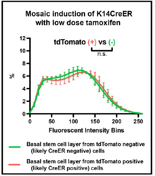
(28) Moldovan, A. & Fraunholz, M.J. In or out: Phagosomal escape of Staphylococcus aureus. Cell Microbiol 21, e12997 (2019).
(29) Slotte, J.P. Biological functions of sphingomyelins. Progress in Lipid Research 52, 424-437 (2013).
(30) Stelzner, K. et al. Intracellular Staphylococcus aureus Perturbs the Host Cell Ca(2+) Homeostasis To Promote Cell Death. mBio 11 (2020).
(31) Kunz, T.C. et al. The Expandables: Cracking the Staphylococcal Cell Wall for Expansion Microscopy. Front Cell Infect Microbiol 11, 644750 (2021).
(32) Sakurai, Y. et al. Ebola virus. Two-pore channels control Ebola virus host cell entry and are drug targets for disease treatment. Science 347, 995-998 (2015).
(33) Grosz, M. et al. Cytoplasmic replication of Staphylococcus aureus upon phagosomal escape triggered by phenol-soluble modulin alpha. Cell Microbiol 16, 451-465 (2014).
(34) Giese, B. et al. Staphylococcal alpha-toxin is not sufficient to mediate escape from phagolysosomes in upper-airway epithelial cells. Infect Immun 77, 3611-3625 (2009).
-
eLife Assessment
This valuable study proposes a novel rapid-entry mechanism of S. aureus that involves the rapid release of calcium from lysosomes. The strength of the paper lies in a very interesting hypothesis; what diminishes enthusiasm is the lack of appropriate methodology, thus making the study incomplete. The methods used are deficient: they are largely reliant on the use of chemical inhibitors and do not adequately support the conclusions.
-
Reviewer #1 (Public review):
Summary:
The manuscript by Rühling et al analyzes the mode of entry of S. aureus into mammalian cells in culture. The authors propose a novel mechanism of rapid entry that involves the release of calcium from lysosomes via NAADP-stimulated activation of TPC1, which in turn causes lysosomal exocytosis; exocytic release of lysosomal acid sphingomyelinase (ASM) is then envisaged to convert exofacial sphingomyelin to ceramide. These events not only induce the rapid entry of the bacteria into the host cells but are also described to alter the fate of the intracellular S. aureus, facilitating escape from the endocytic vacuole to the cytosol.
Strengths:
The proposed mechanism is novel and could have important biological consequences.
Weaknesses:
Unfortunately, the evidence provided is unconvincing and insufficient to document the multiple, complex steps suggested. In fact, there appear to be numerous internal inconsistencies that detract from the validity of the conclusions, which were reached mostly based on the use of pharmacological agents of imperfect specificity.
Firstly, the release of calcium from lysosomes is not demonstrated. Localized changes in the immediate vicinity of lysosomes need to be measured to ascertain that these organelles are the source of cytosolic calcium changes. In fact, 9-phenantrol, which the authors find to be the most potent inhibitor of invasion and hence of the putative calcium changes, is not a blocker of lysosomal calcium release but instead blocks plasmalemmal TRPM4 channels. On the other hand, invasion is seemingly independent of external calcium. These findings are inconsistent with each other and point to non-specific effects of 9-phenantrol. The fact that ionomycin decreases invasion efficiency is taken as additional evidence of the importance of lysosomal calcium release. It is not clear how these observations support involvement of lysosomal calcium release and exocytosis; in fact treatment with the ionophore should itself have induced lysosomal exocytosis and stimulated, rather than inhibited invasion. Yet, manipulations that increase and others that decrease cytosolic calcium both inhibited invasion.
The proposed role of NAADP is based on the effects of "knocking out" TPC1 and on the pharmacological effects of Ned-19. It is noteworthy that TPC2, rather than TPC1, is generally believed to be the primary TPC isoform of lysosomes. Moreover, the gene ablation accomplished in the TPC1 "knockouts" is only partial and rather unsatisfactory. Definitive conclusions about the role of TPC1 can only be reached with proper, full knockouts. Even the pharmacological approach is unconvincing because the high doses of Ned-19 used should have blocked both TPC isoforms and presumably precluded invasion. Instead, invasion is reduced by only ≈50%. A much greater inhibition was reported using 9-phenantrol, the blocker of plasmalemmal calcium channels. How is the selective involvement of lysosomal TPC1 channels justified?
Invoking an elevation of NAADP as the mediator of calcium release requires measurements of the changes in NAADP concentration in response to the bacteria. This was not performed. Instead, the authors analyzed the possible contribution of putative NAADP-generating systems and reported that the most active of these, CD38, was without effect, while the elimination of SARM1, another potential source of NAADP, had a very modest (≈20%) inhibitory effect that may have been due to clonal variation, which was not ruled out. In view of these data, the conclusion that NAADP is involved in the invasion process seems unwarranted.
The involvement of lysosomal secretion is, again, predicated largely on the basis of pharmacological evidence. No direct evidence is provided for the insertion of lysosomal components into the plasma membrane, or for the release of lysosomal contents to the medium. Instead, inhibition of lysosomal exocytosis by vacuolin-1 is the sole source of evidence. However, vacuolin-1 is by no means a specific inhibitor of lysosomal secretion: it is now known to act primarily as a PIKfyve inhibitor and to cause massive distortion of the endocytic compartment, including gross swelling of endolysosomes. The modest (20-25%) inhibition observed when using synaptotagmin 7 knockout cells is similarly not convincing proof of the requirement for lysosomal secretion.
ASM is proposed to play a central role in the rapid invasion process. As above, most of the evidence offered in this regard is pharmacological and often inconsistent between inhibitors or among cell types. Some drugs affect some of the cells, but not others. It is difficult to reach general conclusions regarding the role of ASM. The argument is made even more complex by the authors' use of exogenous sphingomyelinase (beta-toxin). Pretreatment with the toxin decreased invasion efficiency, a seemingly paradoxical result. Incidentally, the effectiveness of the added toxin is never quantified/validated by directly measuring the generation of ceramide or the disappearance of SM.
The use of fluorescent analogs of sphingomyelin and ceramide is not well justified and it is unclear what conclusions can be derived from these observations. Despite the low resolution of the images provided, it appears as if the labeled lipids are largely in endomembrane compartments, where they would presumably be inaccessible to the secreted ASM. Moreover, considering the location of the BODIPY probe, the authors would be unable to distinguish intact sphingomyelin from its breakdown product, ceramide. What can be concluded from these experiments? Incidentally, the authors report only 10% of BODIPY-positive events after 10 min. What are the implications of this finding? That 90% of the invasion events are unrelated to sphingomyelin, ASM, and ceramide?
It is also unclear how the authors can distinguish lysenin entry into ruptured vacuoles from the entry of RFP-CWT, used as a criterion of bacterial escape. Surely the molecular weights of the probes are not sufficiently different to prevent the latter one from traversing the permeabilized membrane until such time that the bacteria escape from the vacuole.
Both SMase inhibitors (Figure 4C) and SMase pretreatment increased bacterial escape from the vacuole. The former should prevent SM hydrolysis and formation of ceramide, while the latter treatment should have the exact opposite effects, yet the end result is the same. What can one conclude regarding the need and role of the SMase products in the escape process?
-
Reviewer #2 (Public review):
Summary:
In this manuscript, Ruhling et al propose a rapid uptake pathway that is dependent on lysosomal exocytosis, lysosomal Ca2+ and acid sphingomyelinase, and further suggest that the intracellular trafficking and fate of the pathogen is dictated by the mode of entry.
The evidence provided is solid, methods used are appropriate and results largely support their conclusions, but can be substantiated further as detailed below. The weakness is a reliance on chemical inhibitors that can be non-specific to delineate critical steps.
Specific comments:
A large number of experiments rely on treatment with chemical inhibitors. While this approach is reasonable, many of the inhibitors employed such as amitriptyline and vacuolin1 have other or non-defined cellular targets and pleiotropic effects cannot be ruled out. Given the centrality of ASM for the manuscript, it will be important to replicate some key results with ASM KO cells.
Most experiments are done in HeLa cells. Given the pathway is projected as generic, it will be important to further characterize cell type specificity for the process. Some evidence for a similar mechanism in other cell types S. aureus infects, perhaps phagocytic cell type, might be good.
I'm a little confused about the role of ASM on the surface. Presumably, it converts SM to ceramide, as the final model suggests. Overexpression of b-toxin results in the near complete absence of SM on phagosomes (having representative images will help appreciate this), but why is phagosomal SM detected at high levels in untreated conditions? If bacteria are engulfed by SM-containing membrane compartments, what role does ASM play on the surface? If surface SM is necessary for phagosomal escape within the cell, do the authors imply that ASM is tuning the surface SM levels to a certain optimal range? Alternatively, can there be additional roles for ASM on the cell surface? Can surface SM levels be visualized (for example, in Figure 4 E, F)?
Related to that, why is ASM activity on the cell surface important? Its role in non-infectious or other contexts can be discussed.
If SM removal is so crucial for uptake, can exocytosis of lysosomes alone provide sufficient ASM for SM removal? How much or to what extent is lysosomal exocytosis enhanced by initial signaling events? Do the authors envisage the early events in their model happening in localized confines of the PM, this can be discussed.
How are inhibitor doses determined? How efficient is the removal of extracellular bacteria at 10 min? It will be good to substantiate the cfu experiments for infectivity with imaging-based methods. Are the roles of TPC1 and TPC2 redundant? If so, why does silencing TPC1 alone result in a decrease in infectivity? For these and other assays, it would be better to show raw values for infectivity. Please show alterations in lysosomal Ca2+ at the doses of inhibitors indicated. Is lysosomal Ca2+ released upon S. aureus binding to the cell surface? Will be good to directly visualize this.
The precise identification of cytosolic vs phagosomal bacteria is not very easy to appreciate. The methods section indicates how this distinction is made, but how do the authors deal with partial overlaps and ambiguities generally associated with such analyses? Please show respective images. The number of events (individual bacteria) for the live cell imaging data should be clearly mentioned.
In the phagosome maturation experiments, what is the proportion of bacteria in Rab5 or Rab7 compartments at each time point? Will the decreased Rab7 association be accompanied by increased Rab5? Showing raw values and images will help appreciate such differences. Given the expertise and tools available in live cell imaging, can the authors trace Rab5 and Rab7 positive compartment times for the same bacteria?
The results with longer-term infection are interesting. Live cell imaging suggests that ASM-inhibited cells show accelerated phagosomal escape that reduces by 6 hpi. Where are the bacteria at this time point ? Presumably, they should have reached lysosomes. The relationship between cytosolic escape, replication, and host cell death is interesting, but the evidence, as presented is correlative for the populations. Given the use of live cell imaging, can the authors show these events in the same cell?
Given the inherent heterogeneity in uptake processes and the use of inhibitors in most experiments, the distinction between ASM-dependent and independent pathways might not be as clear-cut as the authors suggest. Some caution here will be good. Can the authors estimate what fraction of intracellular bacteria are taken up ASM-dependent?
-
-
www.biorxiv.org www.biorxiv.org
-
eLife Assessment
This fundamental study explores how genotypic changes relate to phenotypic stasis or variation within chitons, a molluscan group. Chitons are significant because their ancient body plan has remained largely unchanged for millions of years, yet the paper reveals rapid and large-scale genomic changes. This compelling study is a splendid advance in approximately doubling the number of sequenced chiton genomes, providing what appears to be among the best genome annotations for chiton genomes available to date. The study's key focus is on the genomic rearrangements across five reference-quality genomes of chitons and their implications for understanding evolutionary mechanisms, particularly in comparison to other molluscan clades.
-
Reviewer #1 (Public review):
Summary of Key Findings:
The authors identified 20 ancient molluscan linkage groups (MLGs) that are largely conserved in other molluscan groups but highly dynamic and rearranged in chitons. This contrasts with the stability seen in other animal groups.
Significant chromosome rearrangements, fusions, and duplications were observed in chitons, particularly in the most basal clades like Lepidopleurida, indicating that chitons undergo more extensive genomic changes than expected.
Chitons exhibit extremely high levels of genomic heterozygosity, exceeding that of other molluscan species and even Lepidoptera. This presents challenges for assembling high-quality genomes but also points to genetic diversity as a driver of evolutionary processes.
Partial genome duplications, particularly in Liolophura japonica, extend the knowledge of gene duplication events within the broader Mollusca clade.
The paper speculates that these genomic rearrangements may contribute to maintaining species boundaries in sympatric and parapatric radiations, as observed in certain Acanthochitona species.
Strengths:
The use of high-quality genomic data, including four de novo genome assemblies, provides robust evidence for the conclusions.
The research challenges the common assumption that chitons are evolutionarily conservative, showing that their genomes are highly dynamic despite their morphological stasis.
The study adds to the understanding of how chromosomal rearrangements might contribute to speciation, a concept that can be applied to other taxa.
Limitations:
The paper acknowledges that the limited availability of high-quality genomes across molluscs may restrict the scope of comparative analyses. More genomic data from other molluscan groups could strengthen the conclusions.
The role of high heterozygosity in chitons is highlighted, but more information is needed to clarify how this affects genome assembly and evolutionary outcomes.
Implications for Future Research:
The research raises important questions about the relationship between genomic instability and phenotypic stasis, which can inform studies in other animal groups.
The findings call for a re-evaluation of how we define and measure biodiversity, particularly in "neglected" clades like chitons. Further studies could focus on linking the observed genomic changes to specific adaptive traits or ecological niches.
-
Reviewer #2 (Public review):
Summary:
The authors provide four new annotated genomes for an important taxon within Mollusca known as Polyplacophora (chitons). They provide an impressive analysis showing syntenic relationships between the chromosomes of these four genomes but also other available chiton genome sequences and analysis of 20 molluscan linkage groups to expand this analysis across Mollusca.
Strengths:
The authors have selected particular chiton species for genome sequencing and annotation that expand what is known about genomes across portions of chiton phylogenetic diversity lacking genome sequences. The manuscript is well-written and illustrated in a concise manner. The figures are mostly clear, allowing a reader to visually compare the syntenic relationships of chromosomes, especially within chitons. Their phylogenetic analysis provides a simple manner to map important events in molluscan genome evolution. This study greatly expands what is known about molluscan and chiton comparative genomics.
Weaknesses:
I am not especially convinced that chitons have experienced more substantial genomic rearrangements or other genomic events than other molluscan classes, and for this reason, I did not personally find the title compelling: "Still waters run deep: Large scale genome rearrangements in the evolution of morphologically conservative Polyplacophora." Are the documented events "large scale genomic rearrangements"? It seems that mostly they found two cases of chromosome fusion, plus one apparent case of whole genome duplication. What do they mean by "Still waters run deep"? I have no idea. I guess they consider chitons to be morphologically conservative in their appearance and lifestyle so they are calling attention to this apparent paradox. However, most chiton genomes seem to be relatively conserved, but there are unexpected chromosome fusion events within a particular genus, Acanthochitona. Likewise, they found a large-scale gene duplication event in Acanthopleurinae, a different subfamily of chitons, which is quite interesting but these seem to be geologically recent events that do not especially represent the general pattern of genome evolution across this ancient molluscan taxon.
-
-
www.biorxiv.org www.biorxiv.org
-
eLife Assessment
This important study reports new insights into the roles of a long noncoding RNA, lnc-FANCI-2, in the progression of cervical cancer induced by a type of human papillomavirus. Through a blend of cell biological, biochemical, and genetic analyses of RNA and protein expression, protein-protein interaction, cell signaling, and cell morphology, the authors provide convincing evidence that lnc-FANCI-2 affects cervical cancer outcome by regulating the RAG signaling pathway. These findings will be of interest to scientists in the fields of cervical cancer, long noncoding RNA, and cell signaling.
-
Reviewer #1 (Public review):
Summary:
The authors attempted to dissect the function of a long non-coding RNA, lnc-FANCI-2, in cervical cancer. They profiled lnc-FANCI-2 in different cell lines and tissues, generated knockout cell lines, and characterized the gene using multiple assays.
Strengths:
A large body of experimental data has been presented and can serve as a useful resource for the scientific community, including transcriptomics and proteomics datasets. The reported results also span different parts of the regulatory network and open up multiple avenues for future research.
Weaknesses:
The write-up is somewhat unfocused and lacks deep mechanistic insights in some places.
-
Reviewer #2 (Public review):
The study by Liu et al provides a functional analysis of lnc-FANCI-2 in cervical carcinogenesis, building on their previous discovery of FANCI-2 being upregulated in cervical cancer by HPV E7.
The authors conducted a comprehensive investigation by knocking out (KO) FANCI-2 in CaSki cells and assessing viral gene expression, cellular morphology, altered protein expression and secretion, altered RNA expression through RNA sequencing (verification of which by RT-PCR is well appreciated), protein binding, etc. Verification experiments by RT-PCR, Western blot, etc are notable strengths of the study.
The KO and KD were related to increased Ras signaling and EMT and reduced IFN-y/a responses.
Although the large amount of data is well acknowledged, it is a limitation that most data come from CaSki cells, in which FANCI-2 localization is different from SiHa cells and cancer tissues (Figure 1). The cytoplasmic versus nuclear localization is somewhat puzzling.
-
Reviewer #3 (Public review):
Summary:
A long noncoding RNA, lnc-FANCI-2, was reported to be regulated by HPV E7 oncoprotein and a cell transcription factor, YY1 by this group. The current study focuses on the function of lnc-FANCI-2 in HPV-16 positive cervical cancer is to intrinsically regulate RAS signaling, thereby facilitating our further understanding of additional cellular alterations during HPV oncogenesis. The authors used advanced technical approaches such as KO, transcriptome and (IRPCRP) and LC- MS/MS analyses in the current study and concluded that KO Inc-FANCI-2 significantly increases RAS signaling, especially phosphorylation of Akt and Erk1/2.
Strengths:
(1) HPV E6E7 are required for full immortalization and maintenance of the malignant phenotype of cervical cancer, but they are NOT sufficient for full transformation and tumorigenesis. This study helps further understanding of other cellular alterations in HPV oncogenesis.
(2) lnc-FANCI-2 is upregulated in cervical lesion progression from CIN1, CIN2-3 to cervical cancer, cancer cell lines, and HPV transduced cell lines.
(3) Viral E7 of high-risk HPVs and host transcription factor YY1 are two major factors promoting lnc-FANCI-2 expression.
(4) Proteomic profiling of cytosolic and secreted proteins showed inhibition of MCAM, PODXL2, and ECM1 and increased levels of ADAM8 and TIMP2 in KO cells.
(5) RNA-seq analyses revealed that KO cells exhibited significantly increased RAS signaling but decreased IFN pathways.
(6) Increased phosphorylated Akt and Erk1/2, IGFBP3, MCAM, VIM, and CCND2 (cyclin D2) and decreased RAC3 were observed in KO cells.
Weaknesses:
(1) The authors observed the increased Inc-FANCI-2 in HPV 16 and 18 transduced cells, and other cervical cancer tissues as well, HPV-18 positive HeLa cells exhibited different expressions of Inc-FANCI-2.
(2) Previous studies and data in the current showed a steadily increased Inc-FANCI-2 during cancer progression, however, the authors did not observe significant changes in cell behaviors (both morphology and proliferation) in KO Inc-FANCI-2.
(3) The authors observed the significant changes of RAS signaling (downstream) in KO cells, but they provided limited interpretations of how these results contributed to full transformation or tumorigenesis in HPV-positive cancer.
-
-
www.biorxiv.org www.biorxiv.org
-
eLife Assessment
In this potentially important study, the authors employed advanced computational techniques to explore a detailed atomistic description of the mechanism and energetics of substrate translocation in the MelB transporter. The overall approach is solid and reveals the coupling between sodium binding and melibiose transport through a series of conformational transitions, and the results for a mutant are also in qualitative agreement with the experiment, providing further support to the computational analyses. Nevertheless, the level of evidence is considered incomplete since there are concerns regarding the convergence and initial guess of the string calculations, leaving doubts that the computed pathway does not reflect the most energetically favorable mechanism.
-
Reviewer #1 (Public review):
Summary:
Liang and Guan have studied the transport mechanism of Melbiose transporter MelB using the string method in collective variables and replica-exchange umbrella sampling simulations. The authors study the mechanism of substrate binding to the outward-facing state, conformational change of the transporter from outward-facing to inward-facing, and substrate unbinding from inward-facing state. In their analysis, they also highlight the effects of mutant D59C and the effect of sodium binding on the substrate transport process.
Strengths:
The authors employ a combination of string method and replica-exchange umbrella sampling simulation techniques to provide a complete map of the free energy landscape for sodium-coupled melibiose transport in MelB.
Weaknesses:
(1) Free energy barriers appear to be very high for a substrate transport process. In Figure 3, the transitions from IF (Inward facing) to OF (Outward facing) state appear to have a barrier of 12 kcal/mol. Other systems with mutant or sodium unbound have even higher barriers. This does not seem consistent with previous studies where transport mechanisms of transporters have been explored using molecular dynamics.
(2) Figure 2b: The PMF between images 20-30 shows the conformation change from OF to IF, where the occluded (OC) state is the highest barrier for transition. However, OC state is usually a stable conformation and should be in a local minimum. There should be free energy barriers between OF and OC and in between OC and IF.
(3) String method pathway is usually not the only transport pathway and alternate lower energy pathways should be explored. The free energy surface looks like it has not deviated from the string pathway. Longer simulations can help in the exploration of lower free energy pathways.
(4) The conformational change in transporters from OF to IF state is a complicated multi-step process. First, only 10 images in the string pathway are used to capture the transition from OF to IF state. I am not sure is this number is enough to capture the process. Second, the authors have used geodesic interpolation algorithm to generate the intermediate images. However, looking at Figure 3B, it looks like the transition pathway has not captured the occluded (OC) conformation, where the transport tunnel is closed at both the ends. Transporters typically follow a stepwise conformational change mechanism where OF state transitions to OC and then to IF state. It appears that the interpolation algorithm has created a hourglass-like state, where IF gates are opening and OF gates are closing simultaneously thereby creating a state where the transport tunnel is open on both sides of the membrane. These states are usually associated with high energy. References 30-42 cited in the manuscript reveal a distinct OC state for different transporters.
-
Reviewer #2 (Public review):
Summary:
The manuscript by Liang and Guan provides an impressive attempt to characterize the conformational free energy landscape of a melibiose permease (MelB), a symporter member of major facilitator superfamily (MFS) of transporters. Although similar studies have been conducted previously for other members of MFS, each member or subfamily has its own unique features that make the employment of such methods quite challenging. While the methodology is indeed impressive, characterizing the coupling between large-scale conformational changes and substrate binding in membrane transporters is quite challenging and requires a sophisticated methodology. The conclusions obtained from the three sets of path-optimization and free energy calculations done by the authors are generally supported by the provided data and certainly add to our understanding of how sodium binding facilitates the transport of melibiose in MelB. However, the data is not generated reliably which questions the relevance of the conclusions as well. I particularly have some concerns regarding the implementation of the methodology that I will discuss below.
(1) In enhanced sampling techniques, often much attention is given to the sampling algorithm. Although the sampling algorithm is quite important and this manuscript has chosen an excellent pair: string method with swarms of trajectories (SMwST) and replica-exchange umbrella sampling (REUS) for this task, there are other important factors that must be taken into account. More specifically, the collective variables used and the preparation of initial conformations for sampling. I have objectives for both of these (particularly the latter) that I detail below. Overall, I am not confident that the free energy profiles generated (summarized in Figure 5) are reliable, and unfortunately, much of the data presented in this manuscript heavily relies on these free energy profiles.
(2) The authors state that they have had an advantage over other similar studies in that they had two endpoints of the string to work from experimental data. I agree that this is an advantage. However, this could lead to some dangerous flaws in the methodology if not appropriately taken into account. Proteins such as membrane transporters have many slow degrees of freedom that can be fully captured within tens of nanoseconds (90 ns was the simulation time used here for the REUS). Biased sampling allows us to overcome this challenge to some extent, but it is virtually impossible to take into account all slow degrees of freedom in the enhanced sampling protocol (e.g., the collective variables used here do not represent anything related to sidechain dynamics). Therefore, if one mixes initial conformations that form different initial structures (e.g., an OF state and an IF state from two different PDB files), it is very likely that despite all equilibration and relaxation during SMwST and REUS simulations, the conformations that come from different sources never truly mix. This is dangerous in that it is quite difficult to detect such inconsistencies and from a theoretical point of view it makes the free energy calculations impossible. Methods such as WHAM and its various offshoots all rely on overlap between neighboring windows to calculate the free energy difference between two windows and the overlap should be in all dimensions and not just the ones that we use for biasing. This is related to well-known issues such as hidden barriers and metastability. If one uses two different structures to generate the initial conformations, then the authors need to show their sampling has been long enough to allow the two sets of conformations to mix and overlap in all dimensions, which is a difficult task to do.
(3) I also have concerns regarding the choice of collective variables. The authors have split the residues in each transmembrane helix into the cyto- and periplasmic sides. Then they have calculated the mass center distance between the cytoplasmic sides of certain pairs of helices and have also done the same for the periplasmic side. Given the shape of a helix, this does not seem to be an ideal choice since rather than the rotational motion of the helix, this captures more the translational motion of the helix. However, the transmembrane helices are more likely to undergo rotational motion than the translational one.
(4) Convergence: String method convergence data does not show strong evidence for convergence (Figure S2) in my opinion. REUS convergence is also not discussed. No information is provided on the exchange rate or overlap between the windows.
-
Reviewer #3 (Public review):
The paper from Liang and Guan details the calculation of the potential mean force for the transition between two key states of the melibiose (Mel) transporter MelB. The authors used the string method along with replica-exchange umbrella sampling to model the transition between the outward and inward-facing Mel-free states, including the binding and subsequent release of Mel. They find a barrier of ~6.8 kcal/mol and an overall free-energy difference of ~6.4 kcal/mol. They also investigate the same process without the co-transported Na+, finding a higher barrier, while in the D59C mutant, the barrier is nearly eliminated.
I found this to be an interesting and technically competent paper. I was disappointed actually to see that the authors didn't try to complete the cycle. I realize this is beyond the scope of the study as presented.
The results are in qualitative agreement with expectations from experiments. Could the authors try to make this comparison more quantitative? For example, by determining the diffusivity along the path, the authors could estimate transition rates.
Relatedly, could the authors comment on how typical concentration gradients of Mel and Na+ would affect these numbers?
-
Author response:
Reviewer 1:
(1) Free energy barriers appear to be very high for a substrate transport process. In Figure 3, the transitions from IF (Inward facing) to OF (Outward facing) state appear to have a barrier of 12 kcal/mol. Other systems with mutant or sodium unbound have even higher barriers. This does not seem consistent with previous studies where transport mechanisms of transporters have been explored using molecular dynamics.
First, in Figure 3, the transition from IF to OF state doesn’t have a barrier of 12 kcal/mol. The IFF to OFB transition is almost barrierless, and from OFB to OFF is ~5 kcal/mol, which is also evident in Figure 2.
If the reviewer was referring to the transition from OFB to IFB states, the barrier is 6.8 kcal/mol (Na+ bound state), and the rate-limiting barrier in the entire sugar transport process (Na+ bound state) is 8.4 kcal/mol, as indicated in Figure 2 and Table 1, which is much lower than the 12 kcal/mol barrier the reviewer mentioned. When the Na+ is unbound, the barrier can be as high as 12 kcal/mol, but it is this high barrier that leads to our conclusion that the Na+ binding is essential for sugar transport, and the 12 kcal/mol barrier indicates an energetically unfavorable sugar translocation process when the Na+ is unbound, which is unlikely to be the major translocation process in nature.
Even for the 12 kcal/mol barrier reported for the Na+ unbound state, it is still not too high considering the experimentally measured MelB sugar active transport rate, which is estimated to be on the order of 10 to 100 s-1. This range of transport rate is typical for similar MFS transporters such as the lactose permease (LacY), which has an active transport rate of 20 s-1. The free energy barrier associated with the active transport is thus on the order of ~15-16 kcal/mol based on transition state theory assuming kBT/h as the prefactor. This experimentally estimated barrier is higher than all of our calculated barriers. Our calculated barrier for the sugar translocation with Na+ bound is 8.4 kcal/mol, which means an additional ~7-8 kcal/mol barrier is contributed by the Na+ release process after sugar release in the IFF state. This is a reasonable estimation of the Na+ unbinding barrier.
Therefore, whether the calculated barrier is too high depends on the experimental kinetics measurements, which are often challenging to perform. Based on the existing experimental data, the MFS transporters are
usually relatively slow in their active transport cycle. The calculated barrier thus falls within the reasonable range considering the experimentally measured active transport rates.
(2) Figure 2b: The PMF between images 20-30 shows the conformation change from OF to IF, where the occluded (OC) state is the highest barrier for transition. However, OC state is usually a stable conformation and should be in a local minimum. There should be free energy barriers between OF and OC and in between OC and IF.
First, the occluded state (OCB) is not between images 20-30, it is between images 10 to 20. Second, there is no solid evidence that the OCB state is a stable conformation and a local minimum. Existing experimental structures of MFS transporters seldom have the fully occluded state resolved.
(3) String method pathway is usually not the only transport pathway and alternate lower energy pathways should be explored. The free energy surface looks like it has not deviated from the string pathway. Longer simulations can help in the exploration of lower free energy pathways.
We agree with the reviewer that the string method pathway is usually not the only transport pathway and alternate lower energy pathways could exist. However, we also note that even if the fully occluded state is a local minimum and our free energy pathway does visit this missing local minimum after improved sampling, the overall free energy barrier will not be lowered from our current calculated value. This is because the current rate-limiting barrier arises from the transition from the OFB state to the IFF state, and the barrier top corresponds to the sugar molecule passing through the most constricted region in the cytoplasmic region, i.e., the IFC intermediate state visited after the IFB state is reached. Therefore, the free energy difference between the OFB state and the IFC state will not be changed by another hypothetical local minimum between the OFB and IFB states, i.e., the occluded OCB state. In other words, a hypothetical local minimum corresponding to the occluded state, even if it exists, will not decrease the overall rate-limiting barrier and may even increase it further, depending on the depth of the local minimum and the additional barriers of entering and escaping from this new minimum.
(4) The conformational change in transporters from OF to IF state is a complicated multi-step process. First, only 10 images in the string pathway are used to capture the transition from OF to IF state. I am not sure is this number is enough to capture the process. Second, the authors have used geodesic interpolation algorithm to generate the intermediate images. However, looking at Figure 3B, it looks like the transition pathway has not captured the occluded (OC) conformation, where the transport tunnel is closed at both the ends. Transporters typically follow a stepwise conformational change mechanism where OF state transitions to OC and then to IF state. It appears that the interpolation algorithm has created a hourglasslike state, where IF gates are opening and OF gates are closing simultaneously thereby creating a state where the transport tunnel is open on both sides of the membrane. These states are usually associated with high energy. References 30-42 cited in the manuscript reveal a distinct OC state for different transporters.
In our simulations, even with 10 initial images representing the OF to IF conformational transition, the occluded state is sampled in the final string pathway. There is an ensemble of snapshots where the extracellular and intracellular gates are both relatively narrower than the OF and IF states, preventing the sugar from leaking into either side of the bulk solution. In contrast to the reviewer’s guess, we never observed an hourglass-like state in our simulation where both gates are open. Figure 3B is a visual representation of the backbone structure of the OCB state without explicitly showing the actual radius of the gating region, which also depends on the side chain conformations. Thus, Figure 3B alone cannot be used to conclude that we are dominantly sampling an hourglass-like intermediate conformation instead of the occluded state, as mentioned by the reviewer.
Moreover, not all references in 30-42 have sampled the occluded state since many of them did not even simulate the substrate translocation process at all. For the ones that did sample substrate translocation processes, only two of them were studying the cation-coupled MFS family symporter (ref 38, 40) and they didn’t provide the PMF for the entire translocation process. There is no strong evidence for a stable minimum corresponding to a fully occluded state in these two studies. In fact, different types of transporters with different coupling cations may exhibit different stability of the fully occluded state. For example, the fully occluded state has been experimentally observed for some MFS transporters, such as multidrug transporter EmrD, but not for others, such as lactose permease LacY. Thus, it is not generally true that a stable, fully-occluded state exists in all transporters, and it highly depends on the specific type of transporter and the coupling ion under study.
Reviewer 2:
The manuscript by Liang and Guan provides an impressive attempt to characterize the conformational free energy landscape of a melibiose permease (MelB), a symporter member of major facilitator superfamily (MFS) of transporters. Although similar studies have been conducted previously for other members of MFS, each member or subfamily has its own unique features that make the employment of such methods quite challenging. While the methodology is indeed impressive, characterizing the coupling between large-scale conformational changes and substrate binding in membrane transporters is quite challenging and requires a sophisticated methodology. The conclusions obtained from the three sets of path-optimization and free energy calculations done by the authors are generally supported by the provided data and certainly add to our understanding of how sodium binding facilitates the transport of melibiose in MelB. However, the data is not generated reliably which questions the relevance of the conclusions as well. I particularly have some concerns regarding the implementation of the methodology that I will discuss below.
(1) In enhanced sampling techniques, often much attention is given to the sampling algorithm. Although the sampling algorithm is quite important and this manuscript has chosen an excellent pair: string method with swarms of trajectories (SMwST) and replica-exchange umbrella sampling (REUS) for this task, there are other important factors that must be taken into account. More specifically, the collective variables used and the preparation of initial conformations for sampling. I have objectives for both of these (particularly the latter) that I detail below. Overall, I am not confident that the free energy profiles generated (summarized in Figure 5) are reliable, and unfortunately, much of the data presented in this manuscript heavily relies on these free energy profiles.
Since comments (1) and (2) from this review are related, please see our response to (2) below.
(2) The authors state that they have had an advantage over other similar studies in that they had two endpoints of the string to work from experimental data. I agree that this is an advantage. However, this could lead to some dangerous flaws in the methodology if not appropriately taken into account. Proteins such as membrane transporters have many slow degrees of freedom that can be fully captured within tens of nanoseconds (90 ns was the simulation time used here for the REUS). Biased sampling allows us to overcome this challenge to some extent, but it is virtually impossible to take into account all slow degrees of freedom in the enhanced sampling protocol (e.g., the collective variables used here do not represent anything related to sidechain dynamics). Therefore, if one mixes initial conformations that form different initial structures (e.g., an OF state and an IF state from two different PDB files), it is very likely that despite all equilibration and relaxation during SMwST and REUS simulations, the conformations that come from different sources never truly mix. This is dangerous in that it is quite difficult to detect such inconsistencies and from a theoretical point of view it makes the free energy calculations impossible. Methods such as WHAM and its various offshoots all rely on overlap between neighboring windows to calculate the free energy difference between two windows and the overlap should be in all dimensions and not just the ones that we use for biasing. This is related to well-known issues such as hidden barriers and metastability. If one uses two different structures to generate the initial conformations, then the authors need to show their sampling has been long enough to allow the two sets of conformations to mix and overlap in all dimensions, which is a difficult task to do.
We partly agree with the reviewer in that it is challenging to investigate whether the structures generated from the two different initial structures are sufficiently mixed in terms of orthogonal degrees of freedom outside the CV space during our string method and REUS simulations. We acknowledge that our simulations are within 100 ns for each REUS window, and there could be some slow degrees of freedom that are not fully sampled within this timescale. However, the conjectures and concerns raised by the reviewer are somewhat subjective in that they are almost impossible to be completely disproven. In a sense, these concerns are essentially the same as the general suspicion that the biomolecular simulation results are not completely converged, which cannot be fully ruled out for relatively complex biomolecular systems in any computational study involving MD simulations. We also note that comparison among the PMFs of different cation bound/unbound states will have some error cancellation effects because of the consistent use of the same sampling methods for all three systems. Our main conclusions regarding the cooperative binding and transport of the two substrates lie in such comparison of the PMFs and additionally on the unbiased MD simulations. Thus, although there could be insufficient sampling, our key conclusions based on the relative comparison between the PMFs are more robust and less likely to suffer from insufficient sampling.
(3) I also have concerns regarding the choice of collective variables. The authors have split the residues in each transmembrane helix into the cyto- and periplasmic sides. Then they have calculated the mass center distance between the cytoplasmic sides of certain pairs of helices and have also done the same for the periplasmic side. Given the shape of a helix, this does not seem to be an ideal choice since rather than the rotational motion of the helix, this captures more the translational motion of the helix. However, the transmembrane helices are more likely to undergo rotational motion than the translational one.
Our choice of CVs not only captures the translational motion but also the rotational motion of the helix. Consider a pair of helices. If there is a relative rotation in the angle between the two helices, causing the extracellular halves of the two helices to get closer and the intracellular halves to be more separated, this rotational motion can be captured as the decrease of one CV describing the extracellular distance and increase in the other CV describing the intracellular distance between the two helices. Reversely, if one of the two CVs is forced to increase and the other one forced to decrease, it can, in principle, bias the relative rotation of the two helices with respect to each other. Indeed, comparing Figure 3 with Figure S4, the reorientation of the helices with respect to the membrane normal (Fig. S4) is accompanied by the simultaneous decrease and increase in the pairwise distances between different segments of the helices. Therefore, our choice of CVs in the string method and REUS are not biased against the rotation of the helices, as the reviewer assumed.
(4) Convergence: String method convergence data does not show strong evidence for convergence (Figure S2) in my opinion. REUS convergence is also not discussed. No information is provided on the exchange rate or overlap between the windows.
The convergence of string method, REUS, the exchange rate and overlap between windows will be discussed in the reviewed manuscript.
Reviewer 3:
The paper from Liang and Guan details the calculation of the potential mean force for the transition between two key states of the melibiose (Mel) transporter MelB. The authors used the string method along with replica-exchange umbrella sampling to model the transition between the outward and inwardfacing Mel-free states, including the binding and subsequent release of Mel. They find a barrier of ~6.8 kcal/mol and an overall free-energy difference of ~6.4 kcal/mol. They also investigate the same process without the co-transported Na+, finding a higher barrier, while in the D59C mutant, the barrier is nearly eliminated.
For Na+ bound state, the rate-limiting barrier is 8.4 kcal/mol instead of 6.8 kcal/mol. The overall free energy difference is 3.7 kcal/mol instead of 6.4 kcal/mol. These numbers need to be corrected in the public review.
I found this to be an interesting and technically competent paper. I was disappointed actually to see that the authors didn't try to complete the cycle. I realize this is beyond the scope of the study as presented.
We agree with the reviewer that characterizing the complete cycle is our eventual goal. However, in order to characterize the complete cycle of the transporter, the free energy landscapes of the Na+ binding and unbinding process in the sugar-bound and unbound states, as well as the OF to IF conformational transition in the apo state. These additional calculations are expensive, and the amount of work devoted to these new calculations is estimated to be at least the same as the current study. Therefore, we prefer to carry out and analyze these new simulations in a future study.
The results are in qualitative agreement with expectations from experiments. Could the authors try to make this comparison more quantitative? For example, by determining the diffusivity along the path, the authors could estimate transition rates.
In our revised manuscript, we will determine the diffusivity along the path and estimate transition rates.
Relatedly, could the authors comment on how typical concentration gradients of Mel and Na+ would affect these numbers?
The concentration gradient of Mel and Na+ can be varied in different experimental setups. In a typical active transport essay, the Na+ has a higher concentration outside the cell, and the melibiose has a higher concentration inside the cell. In the steady state, depending on the experiment setup, the extracellular Na+ concentration is in the range of 10-20 mM, and the intracellular concentration is self-balanced in the range of 3-4 mM due to the presence of other ion channels and pumps. In addition to the Na+ concentration gradient, there is also a transmembrane voltage potential of -200 mV (the intracellular side being more negative than the extracellular side), which facilitates the Na+ release into the intracellular side. In the steady state, the extracellular concentration of melibiose is ~0.4 mM, and the intracellular concentration is at least 1000 times the extracellular concentration, greater than 0.4 M. In this scenario, the free energy change of intracellular melibiose translocation will be increased by about ~5 kcal/mol at 300K temperature, leading to a total ∆𝐺 of ~8 kcal/mol. The total barrier for the melibiose translocation is expected to be increased by less than 5 kcal/mol. However, the increase in ∆𝐺 for intracellular melibiose translocation will be compensated by a decrease in ∆𝐺 of similar magnitude ( ~5 kcal/mol) for intracellular Na+ translocation. In a typical sugar self-exchange essay, there is no net gradient in the melibiose or Na+ across the membrane, and the overall free energy changes we calculated apply to this situation.
-
-
www.biorxiv.org www.biorxiv.org
-
eLife Assessment
This fundamental work provides new mechanistic insight in regulation of PDGF signaling through splicing controls. The evidence is compelling to demonstrate functional involvement of Srsf3, an RNA binding protein to this new and interesting mechanism. The work will be of broad interest to developmental biologists in general and molecular biologists/biochemists in the field of growth factor signaling and RNA processing.
-
Reviewer #1 (Public review):
In their manuscript "PDGFRRa signaling regulates Srsf3 transcript binding to affect PI3K signaling and endosomal trafficking" Forman and colleagues use iMEPM cells to characterize the effects of PDGF signaling on alternative splicing. They first perform RNA-seq using a one-hour stimulation with Pdgf-AA in control and Srsf3 knockdown cells. While Srsf3 manipulation results in a sizeable number of DE genes, PDGF does not. They then turn to examine alternative splicing, due to findings from this lab. They find that both PDGF and Srsf3 contribute much more to splicing than transcription. They find that the vast majority of PDGF-mediated alternative splicing depends upon Srsf3 activity and that skipped exons are the most common events with PDGF stimulation typically promoting exon skipping in the presence of Srsf3. They used eCLIP to identify RNA regions bound to Srsf3. Under both PDGF conditions, the majority of peaks were in exons with +PDGF having a substantially greater number of these peaks. Interestingly, they find differential enrichment of sequence motifs and GC content in stimulated versus unstimulated cells. They examine 2 transcripts encoding PI3K pathway (enriched in their GO analysis) members: Becn1 and Wdr81. They then go on to examine PDGFRRa and Rab5, an endosomal marker, colocalization. They propose a model in which Srsf3 functions downstream of PDGFRRa signaling to, in part, regulate PDGFRa trafficking to the endosome. The findings are novel and shed light on the mechanisms of PDGF signaling and will be broadly of interest. This lab previously identified the importance of PDGF naling on alternative splicing. The combination of RNA-seq and eCLIP is an exceptional way to comprehensively analyze this effect. The results will be of great utility to those studying PDGF signaling or neural crest biology.
Comments on the revised version:
The authors have fully addressed my previous comments and I have no further concerns.
-
Reviewer #2 (Public review):
Summary:
This manuscript builds upon the work of a previous study published by the group (Dennison, 2021) to further elucidate the coregulatory axis of Srsf3 and PDGFRa on craniofacial development. The authors in this study investigated the molecular mechanisms by which PDGFRa signaling activates the RNA-binding protein Srsf3 to regulate alternative splicing (AS) and gene expression (GE) necessary for craniofacial development. PDGFRa signaling-mediated Srsf3 phosphorylation drives its translocation into the nucleus and affect binding affinity to different proteins and RNA, but the exact molecular mechanisms were not known. The authors performed RNA sequencing on immortalized mouse embryonic mesenchyme (MEPM) cells treated with shRNA targeting 3' UTR of Srsf3 or scramble shRNA (to probe AS and DE events that are Srsf3 dependent) and with and without PDGF-AA ligand treatment (to probe AS and DE events that are PDGFRa signaling dependent). They found that PDGFRa signaling has more effect on AS than on DE. A matching eCLIP-seq experiment was performed to investigate how Srsf3 binding sites change with and without PDGFRa signaling.
Strengths:
(1) The work builds well upon the previous data and the authors employ a variety of appropriate techniques to answer their research questions.
(2) The authors show that Srsf3 binding pattern within the transcript as well as binding motifs change significantly upon PDGFRa signaling, providing a mechanistic explanation for the significant changes in AS.
(3) By combining RNA-seq and eCLIP datasets together, the authors identified a list of genes that are directly bound by Srsf3 and undergo changes in GE and/or AS. Two examples are Becn1 and Wdr81, which are involved in early endosomal trafficking.
Weaknesses:
(1) The authors identify two genes whose AS are directly regulated by Srsf3 and involved in endosomal trafficking; however, they do not validate the differential AS results and whether changes in these genes can affect endosomal trafficking. In Figure 6, they show that PDGFRa signaling is involved in endosome size and Rab5 colocalization, but do not show how Srsf3 and the two genes are involved.
(2) The proposed model does not account for other proteins mediating the activation of Srsf3 after Akt phosphorylation. How do we know this is a direct effect (and not secondary or tertiary effect)?
This is a thoroughly revised manuscript. I would like to congratulate the authors to have invested a lot of time, resources, new data, and a more refined discussion to make this a compelling piece of work. I have no further concerns.
-
Author response:
The following is the authors’ response to the original reviews.
Public Reviews:
Reviewer #1 (Public Review):
In their manuscript "PDGFRRa signaling regulates Srsf3 transcript binding to affect PI3K signaling and endosomal trafficking" Forman and colleagues use iMEPM cells to characterize the effects of PDGF signaling on alternative splicing. They first perform RNA-seq using a one-hour stimulation with Pdgf-AA in control and Srsf3 knockdown cells. While Srsf3 manipulation results in a sizeable number of DE genes, PDGF does not. They then turn to examine alternative splicing, due to findings from this lab. They find that both PDGF and Srsf3 contribute much more to splicing than transcription. They find that the vast majority of PDGF-mediated alternative splicing depends upon Srsf3 activity and that skipped exons are the most common events with PDGF stimulation typically promoting exon skipping in the presence of Srsf3. They used eCLIP to identify RNA regions bound to Srsf3. Under both PDGF conditions, the majority of peaks were in exons with +PDGF having a substantially greater number of these peaks. Interestingly, they find differential enrichment of sequence motifs and GC content in stimulated versus unstimulated cells. They examine 2 transcripts encoding PI3K pathway (enriched in their
GO analysis) members: Becn1 and Wdr81. They then go on to examine PDGFRRa and Rab5, an endosomal marker, colocalization. They propose a model in which Srsf3 functions downstream of PDGFRRa signaling to, in part, regulate PDGFRa trafficking to the endosome. The findings are novel and shed light on the mechanisms of PDGF signaling and will be broadly of interest. This lab previously identified the importance of PDGF naling on alternative splicing. The combination of RNA-seq and eCLIP is an exceptional way to comprehensively analyze this effect. The results will be of great utility to those studying PDGF signaling or neural crest biology. There are some concerns that should be considered, however.
We thank the Reviewer for these supportive comments.
(1) It took some time to make sense of the number of DE genes across the results section and Figure 1. The authors give the total number of DE genes across Srsf3 control and loss conditions as 1,629 with 1,042 of them overlapping across Pdgf treatment. If the authors would add verbiage to the point that this leaves 1,108 unique genes in the dataset, then the numbers in Figure 1D would instantly make sense. The same applies to PDGF in Figure 1F and the Venn diagrams in Figure 2.
We have edited the relevant sentence for Figure 1D as follows: “There was extensive overlap (521 out of 1,108; 47.0%) of Srsf3-dependent DE genes across ligand treatment conditions, resulting in a total of 1,108 unique genes within both datasets (Fig. 1C,D; Fig. S1A).” Similarly, we edited the relevant sentence for Figure 1F as follows: “There was limited overlap (4 out of 47; 8.51%) of PDGF-AA-dependent DE genes across Srsf3 conditions, resulting in a total of 47 unique genes within both datasets (Fig. 1E,F; Fig. S1B).” We edited the relevant sentence for Figure 2B as follows: “There was limited overlap (203 out of 1,705; 11.9%) of Srsf3-dependent alternatively-spliced transcripts across ligand treatment conditions, resulting in a total of 1,705 unique events within both datasets (Fig. 2A,B).” Finally, we edited the relevant sentence for Figure 2D as follows: “There was negligible overlap (9 out of 622; 1.45%) of PDGF-AA-dependent alternatively-spliced transcripts across Srsf3 conditions, resulting in a total of 622 unique events within both datasets (Fig. 2C,D).”
(2) The percentage of skipped exons in the +DPSI on the righthand side of Figure 2F is not readable.
We have moved the label for the percentage of skipped exon events with a +DPSI for the -PDGF-AA vs +PDGF-AA (scramble) alternatively-spliced transcripts in Figure 2E so that it is legible.
(3) It would be useful to have more information regarding the motif enrichment in Figure 3. What is the extent of enrichment? The authors should also provide a more complete list of enriched motifs, perhaps as a supplement.
We have added P values beneath the motifs in Figure 3F and 3G. Further, we have added a new Supplementary Figure, Figure S5, that lists the occurrence of the top 10 most enriched motifs in the unstimulated and, separately, stimulated samples in the eCLIP dataset and in a control dataset, as well as their P values.
(4) It is unclear what subset of transcripts represent the "overlapping datasets" on lines 280-315. The authors state that there are 149 unique overlapping transcripts, but the Venn diagram shows 270. Also, it seems that the most interesting transcripts are the 233 that show alternative splicing and are bound by Srsf3. Would the results shown in Figure 5 change if the authors focused on these transcripts?
The Reviewer is correct that 233 of the alternatively-spliced transcripts had an Srsf3 eCLIP peak, as indicated in Figure 5A. However, several of these eCLIP peaks were a large distance from an alternatively-spliced element in the rMATS datasets, indicating that Srsf3 binding may not be contributing to the splicing outcomes in these cases. Instead, we correlated the eCLIP peaks with AS events by identifying transcripts in which Srsf3 bound within an alternatively-spliced exon or within 250 bp of the neighboring introns. We have added additional text clarifying this point in the Results: “We next sought to identify high-confidence transcripts for which Srsf3 binding had an increased likelihood of contributing to AS. Previous studies revealed enrichment of functional RBP motifs near alternatively-spliced exons (Yee et al., 2019). As such, we correlated the eCLIP peaks with AS events across all four treatment comparisons by identifying transcripts in which Srsf3 bound within an alternatively-spliced exon or within 250 bp of the neighboring introns (Tables S12-S15).” Further, we have relabeled Figure 5B as “Highconfidence, overlapping datasets biological process GO terms”.
(5) In general, there is little validation of the sequencing results, performing qPCR on Arhgap12 and Cep55. The authors should additionally validate the PI3K pathway members that they analyze. Related, is Becn1 expression downregulated in the absence of Srsf3, as would be predicted if it is undergoing NMD?
We have added two new figure panels, Figure 5F-5G, assessing Wdr81 AS and Wdr81 protein sizes, as this gene has previously been implicated in craniofacial development. We have added the following text to the Results section: “Finally, as Wdr81 protein levels are predicted to regulate RTK trafficking between early and late endosomes, we confirmed the differential AS of Wdr81 transcripts between unstimulated scramble cells and scramble cells treated with PDGFAA ligand for 1 hour by qPCR using primers within constitutively-expressed exons flanking alternatively-spliced exon 9. This analysis revealed a decreased PSI for Wdr81 in each of three biological replicates upon PDGF-AA ligand treatment (Fig. 5F). Relatedly, we assessed the ratio of larger isoforms of Wdr81 protein (containing the WD3 domain) to smaller isoforms (missing the WD3 domain) via western blotting. Consistent with our RNA-seq and qPCR results, PDGFAA stimulation for 24 hours in the presence of Srsf3 led to an increase in smaller Wdr81 protein isoforms (Fig. 5G).”
(6) What is the alternative splicing event for Acap3?
We have added the following text to the Results section and updated Figure 5E with Acap3 eCLIP peak visualization and the predicted alternative splicing outcome: “Finally, Acap3 is a GTPase-activating protein (GAP) for the small GTPase Arf6, converting Arf6 to an inactive, GDP-bound state (Miura et al., 2016). Arf6 localizes to the plasma membrane and endosomes, and has been shown to regulate endocytic membrane trafficking by increasing PI(4,5)P2 levels at the cell periphery (D’Souza-Schorey and Chavrier, 2006). Further, constitutive activation of Arf6 leads to upregulation of the gene encoding the p85 regulatory subunit of PI3K and increased activity of both PI3K and AKT (Yoo et al., 2019)… Srsf3 binding was additionally increased in Acap3 exon 19 upon PDGF-AA stimulation, at an enriched motif within the highconfidence, overlapping datasets, and we observed a corresponding increase in excision of adjacent intron 19 (Fig. 5D,E). As Acap3 intron 19 contains a PTC, this event is predicted to result in more transcripts encoding full-length protein (Fig. 5E).”
(7) The insets in Figure 6 C"-H" are useful but difficult to see due to their small size. Perhaps these could be made as their own figure panels.
We have increased the size of the previous insets in new Figure 6 panels C’’’-H’’’.
(8) In Figure 6A, it is not clear which groups have statistically significant differences. A clearer visualization system should be used.
We have added bracket shapes to Figure 6A indicating the statistically significant differences between scramble 0 minutes and scramble 60 minutes, and between scramble 60 minutes and shSrsf3 60 minutes.
(9) Similarly in Figure 6B, is 15 vs 60 minutes in the shSrsf3 group the only significant difference? Is there a difference between scramble and shSrsf3 at 15 minutes? Is there a difference between 0 and 15 minutes for either group?
We have added a bracket shape to Figure 6B indicating the statistically significant difference between shSrsf3 at 15 minutes and shSrsf3 at 60 minutes. No other pairwise comparisons between treatments or timepoints were statistically significantly different.
Reviewer #2 (Public Review):
Summary:
This manuscript builds upon the work of a previous study published by the group (Dennison, 2021) to further elucidate the coregulatory axis of Srsf3 and PDGFRa on craniofacial development. The authors in this study investigated the molecular mechanisms by which PDGFRa signaling activates the RNA-binding protein Srsf3 to regulate alternative splicing (AS) and gene expression (GE) necessary for craniofacial development. PDGFRa signaling-mediated Srsf3 phosphorylation drives its translocation into the nucleus and affects binding affinity to different proteins and RNA, but the exact molecular mechanisms were not known. The authors performed RNA sequencing on immortalized mouse embryonic mesenchyme (MEPM) cells treated with shRNA targeting 3' UTR of Srsf3 or scramble shRNA (to probe AS and DE events that are Srsf3 dependent) and with and without PDGF-AA ligand treatment (to probe AS and DE events that are PDGFRa signaling dependent). They found that PDGFRa signaling has more effect on AS than on DE. A matching eCLIP-seq experiment was performed to investigate how Srsf3 binding sites change with and without PDGFRa signaling.
Strengths:
(1) The work builds well upon the previous data and the authors employ a variety of appropriate techniques to answer their research questions.
(2) The authors show that Srsf3 binding pattern within the transcript as well as binding motifs change significantly upon PDGFRa signaling, providing a mechanistic explanation for the significant changes in AS.
(3) By combining RNA-seq and eCLIP datasets together, the authors identified a list of genes that are directly bound by Srsf3 and undergo changes in GE and/or AS. Two examples are Becn1 and Wdr81, which are involved in early endosomal trafficking. We thank the Reviewer for these supportive comments.
Weaknesses:
(1) The authors identify two genes whose AS are directly regulated by Srsf3 and involved in endosomal trafficking; however, they do not validate the differential AS results and whether changes in these genes can affect endosomal trafficking. In Figure 6, they show that PDGFRa signaling is involved in endosome size and Rab5 colocalization, but do not show how Srsf3 and the two genes are involved.
We have added two new figure panels, Figure 5F-5G, assessing Wdr81 AS and Wdr81 protein sizes, as this gene has previously been implicated in craniofacial development. We have added the following text to the Results section: “Finally, as Wdr81 protein levels are predicted to regulate RTK trafficking between early and late endosomes, we confirmed the differential AS of Wdr81 transcripts between unstimulated scramble cells and scramble cells treated with PDGFAA ligand for 1 hour by qPCR using primers within constitutively-expressed exons flanking alternatively-spliced exon 9. This analysis revealed a decreased PSI for Wdr81 in each of three biological replicates upon PDGF-AA ligand treatment (Fig. 5F). Relatedly, we assessed the ratio of larger isoforms of Wdr81 protein (containing the WD3 domain) to smaller isoforms (missing the WD3 domain) via western blotting. Consistent with our RNA-seq and qPCR results, PDGFAA stimulation for 24 hours in the presence of Srsf3 led to an increase in smaller Wdr81 protein isoforms (Fig. 5G).” The experiments in Figure 6 compare early endosome size, PDGFRa localization in early endosomes and phospho-Akt levels in response to PDGF-AA stimulation in scramble versus shSrsf3 cells, demonstrating that Srsf3-mediated PDGFRa signaling leads to enlarged early endosomes, retention of PDGFRa in early endosomes and increased downstream phospho-Akt signaling. Though we agree with the Reviewer that functionally linking the AS events to the endosomal phenotype would strengthen our conclusions, these are technically challenging experiments for several reasons. First, this approach has typically relied on tiling oligos against a region of interest to find the optimal sequence. We identified several transcripts that are bound by Srsf3 and undergo alternative splicing upon PDGFRa signaling to potentially contribute to the regulation of PI3K signaling and early endosomal trafficking. We do not expect that these effects are mediated by a single transcript but may instead by mediated by a combination of alternative splicing changes. As such, these experiments would require us to identify and validate multiple splice-switching antisense oligonucleotides (ASOs). Second, ASOs designed against a specific target may not lead to alternative splicing of that target, even in cases of high predicted binding affinities (Scharner et al., 2020, Nucleic Acid Res 48(2), 802816). Third, ASOs have been shown to result in off-target mis-splicing effects, which are hard to predict (Scharner et al., 2020, Nucleic Acid Res 48(2), 802-816). The design of functional ASOs is thus a long-standing challenge in the field, and likely beyond the scope of this manuscript. We have added the following text to the Discussion to highlight this potential future direction: “In the future, it will be worthwhile to attempt to functionally link the AS of transcripts such as Becn1, Wdr81 and/or Acap3 to the endosomal trafficking changes observed above using spliceswitching antisense oligonucleotides (ASOs).”
(2) The proposed model does not account for other proteins mediating the activation of Srsf3 after Akt phosphorylation. How do we know this is a direct effect (and not a secondary or tertiary effect)?
This point is introduced in the Discussion: “Whether phosphorylation of Srsf3 directly influences its binding to target RNAs or acts to modulate Srsf3 protein-protein interactions which then contribute to differential RNA binding remains to be determined, though findings from Schmok et al., 2024 may argue for the latter mechanism. Studies identifying proteins that differentially interact with Srsf3 in response to PDGF-AA ligand stimulation are ongoing and will shed light on these mechanisms…. Again, this shift could be due to loss of RNA binding owing to electrostatic repulsion and/or changes in ribonucleoprotein composition and will be the subject of future studies.” We have added a potential change in Srsf3 protein-protein interactions upon Akt phosphorylation in the model in Figure 6J.
Reviewer #2 (Recommendations For The Authors):
Suggestions:
(1) It would strengthen the paper and improve the connection with the other sections of the paper if the authors show:
a) validation of PDGFRa signaling leading to AS of Becn1 and Wdr81 and corresponding changes in protein, and
We have added two new figure panels, Figure 5F-5G, assessing Wdr81 AS and Wdr81 protein sizes, as this gene has previously been implicated in craniofacial development. We have added the following text to the Results section: “Finally, as Wdr81 protein levels are predicted to regulate RTK trafficking between early and late endosomes, we confirmed the differential AS of Wdr81 transcripts between unstimulated scramble cells and scramble cells treated with PDGFAA ligand for 1 hour by qPCR using primers within constitutively-expressed exons flanking alternatively-spliced exon 9. This analysis revealed a decreased PSI for Wdr81 in each of three biological replicates upon PDGF-AA ligand treatment (Fig. 5F). Relatedly, we assessed the ratio of larger isoforms of Wdr81 protein (containing the WD3 domain) to smaller isoforms (missing the WD3 domain) via western blotting. Consistent with our RNA-seq and qPCR results, PDGFAA stimulation for 24 hours in the presence of Srsf3 led to an increase in smaller Wdr81 protein isoforms (Fig. 5G).”
b) functionally link the AS event(s) to endosomal phenotype using ASOs, etc.
Though we agree with the Reviewer that such results would strengthen our conclusions, these are technically challenging experiments for several reasons. First, this approach has typically relied on tiling oligos against a region of interest to find the optimal sequence. We identified several transcripts that are bound by Srsf3 and undergo alternative splicing upon PDGFRa signaling to potentially contribute to the regulation of PI3K signaling and early endosomal trafficking. We do not expect that these effects are mediated by a single transcript but may instead by mediated by a combination of alternative splicing changes. As such, these experiments would require us to identify and validate multiple splice-switching antisense oligonucleotides (ASOs). Second, ASOs designed against a specific target may not lead to alternative splicing of that target, even in cases of high predicted binding affinities (Scharner et al., 2020, Nucleic Acid Res 48(2), 802-816). Third, ASOs have been shown to result in off-target mis-splicing effects, which are hard to predict (Scharner et al., 2020, Nucleic Acid Res 48(2), 802-816). The design of functional ASOs is thus a long-standing challenge in the field, and likely beyond the scope of this manuscript. We have added the following text to the Discussion to highlight this potential future direction: “In the future, it will be worthwhile to attempt to functionally link the AS of transcripts such as Becn1, Wdr81 and/or Acap3 to the endosomal trafficking changes observed above using splice-switching antisense oligonucleotides (ASOs).”
(2) The Venn diagram in Figure 5A and the description of the analysis the authors did to combine the RNA-seq and eCLIP-seq data are a little confusing. The authors say that they correlated eCLIP peaks with GE or AS events across all four treatment comparisons. The purpose of looking at both datasets was to find genes that are directly bound by Srsf3 and also have significantly affected GE and/or AS. Therefore, the data with and without PDGF-AA should be considered separately. For example, eCLIP peaks in the PDGF-AA condition can be correlated to Srsf3-dependent AS differences (comparing shSrsf3 and scramble) in the -PDGF-AA condition, and eCLIP peaks in the +PDGF-AA condition can be correlated to Srsf3-dependent AS differences in the +PDGF-AA condition. In the Venn diagram and the description, it seems like all comparisons were combined and it is not clear how the data were analyzed.
As indicated in Figure 5A, 233 of the alternatively-spliced transcripts uniquely found in one of the four treatment comparisons had an Srsf3 eCLIP peak. However, several of these eCLIP peaks were a large distance from an alternatively-spliced element in the rMATS datasets, indicating that Srsf3 binding may not be contributing to the splicing outcomes in these cases. Instead, we correlated the eCLIP peaks with AS events by identifying transcripts in which Srsf3 bound within an alternatively-spliced exon or within 250 bp of the neighboring introns. We have added additional text clarifying this point in the Results: “We next sought to identify highconfidence transcripts for which Srsf3 binding had an increased likelihood of contributing to AS.
Previous studies revealed enrichment of functional RBP motifs near alternatively-spliced exons (Yee et al., 2019). As such, we correlated the eCLIP peaks with AS events across all four treatment comparisons by identifying transcripts in which Srsf3 bound within an alternativelyspliced exon or within 250 bp of the neighboring introns (Tables S12-S15).” Further, we have relabeled Figure 5B as “High-confidence, overlapping datasets biological process GO terms”. We respectfully disagree with the Reviewer’s suggested comparisons. A comparison of the PDGF-AA eCLIP data with the scramble vs shSrsf3 (-PDGF-AA) data from the list of highconfidence transcripts resulted in only 7 transcripts. Similarly, a comparison of the +PDGF-AA eCLIP data with the scramble vs shSrsf3 (+PDGF-AA) data from the list of high-confidence transcripts resulted in only 14 transcripts. Separate gene ontology analyses of these lists of 7 and 14 transcripts revealed 21 and 40 significant terms for biological process, respectively, the majority of which encompassed one, and never more than two, transcripts. Had we separately examined the -PDGF-AA and +PDGF-AA data, we would not have detected the changes in Becn1, Wdr81 and Acap3 in Figure 5E.
-
-
www.medrxiv.org www.medrxiv.org
-
eLife Assessment
This valuable manuscript presents a spatiotemporal genetic analysis of malaria-infected individuals from four villages in The Gambia, covering the period between December 2014 and May 2017. Overall, laboratory and data analyses are solid, although details of the methods are lacking. This study offers evidence to advance the understanding of malaria epidemiology in sub-Saharan Africa, but would benefit from additional analysis to strengthen the findings.
-
Reviewer #1 (Public review):
Summary:
The manuscript titled "Household clustering and seasonal genetic variation of Plasmodium falciparum at the community-level in The Gambia" presents a valuable genetic spatio-temporal analysis of malaria-infected individuals from four villages in The Gambia, covering the period between December 2014 and May 2017. The majority of samples were analyzed using a SNP barcode with the Spotmalaria panel, with a subset validated through WGS. Identity-by-descent (IBD) was calculated as a measure of genetic relatedness and spatio-temporal patterns of the proportion of highly related infections were investigated. Related clusters were detected at the household level, but only within a short time period.
Strengths:
This study offers a valuable dataset, particularly due to its longitudinal design and the inclusion of asymptomatic cases. The laboratory analysis using the Spotmalaria platform combined and supplemented with WGS is solid, and the authors show a linear correlation between the IBD values determined with both methods, although other studies have reported that at least 200 SNPs are required for IBD analysis. Data-analysis pipelines were created for (1) variant filtering for WGS and subsequent IBD analysis, and (2) creating a consensus barcode from the spot malaria panel and WGS data and subsequent SNP filtering and IBD analysis.
Weaknesses:
Further refining the data could enhance its impact on both the scientific community and malaria control efforts in The Gambia.
(1) The manuscript would benefit from improved clarity and better explanation of results to help readers follow more easily. Despite familiarity with genotyping, WGS, and IBD analysis, I found myself needing to reread sections. While the figures are generally clear and well-presented, the text could be more digestible. The aims and objectives need clearer articulation, especially regarding the rationale for using both SNP barcode and WGS (is it to validate the approach with the barcode, or is it to have less missing data?). In several analyses, the purpose is not immediately obvious and could be clarified.
(2) Some key results are only mentioned briefly in the text without corresponding figures or tables in the main manuscript, referring only to supplementary figures, which are usually meant for additional detail, but not main results. For example, data on drug resistance markers should be included in a table or figure in the main manuscript.
(3) The study uses samples from 2 different studies. While these are conducted in the same villages, their study design is not the same, which should be addressed in the interpretation and discussion of the results. Between Dec 2014 and Sept 2016, sampling was conducted only in 2 villages and at less frequent intervals than between Oct 2016 to May 2017. The authors should assess how this might have impacted their temporal analysis and conclusions drawn. In addition, it should be clarified why and for exactly in which analysis the samples from Dec 2016 - May 2017 were excluded as this is a large proportion of your samples.
(4) Based on which criteria were samples selected for WGS? Did the spatiotemporal spread of the WGS samples match the rest of the genotyped samples? I.e. were random samples selected from all times and places, or was it samples from specific times/places selected for WGS?
(5) The manuscript would benefit from additional detail in the methods section.
(6) Since the authors only do the genotype replacement and build consensus barcode for 199 samples, there is a bias between the samples with consensus barcode and those with only the genotyping barcode. How did this impact the analysis?
(7) The linear correlation between IBD-values of barcode vs genome is clear. However, since you do not use absolute values of IBD, but a classification of related (>=0.5 IBD) vs. unrelated (<0.5), it would be good to assess the agreement of this classification between the 2 barcodes. In Figure S6 there seem to be quite some samples that would be classified as unrelated by the consensus barcode, while they have IBD>0.5 in the Genome-IBD; in other words, the barcode seems to be underestimating relatedness.<br /> a. How sensitive is this correlation to the nr of SNPs in the barcode?
(8) With the sole focus on IBD, a measure of genetic relatedness, some of the conclusions from the results are speculative.<br /> a. Why not include other measures such as genetic diversity, which relates to allele frequency analysis at the population level (using, for example, nucleotide diversity)? IBD and the proportion of highly related pairs are not a measure of genetic diversity. Please revise the manuscript and figures accordingly.<br /> b. Additionally, define what you mean by "recombinatorial genetic diversity" and explain how it relates to IBD and individual-level relatedness.<br /> c. Recombination is one potential factor contributing to the loss of relatedness over time. There are several other factors that could contribute, such as mobility/gene flow, or study-specific limitations such as low numbers of samples in the low transmission season and many months apart from the high transmission samples.<br /> d. By including other measures such as linkage disequilibrium you could further support the statements related to recombination driving the loss of relatedness.
(9) While the authors conclude there is no seasonal pattern in the drug-resistant markers, one can observe a big fluctuation in the dhps haplotypes, which go down from 75% to 20% and then up and down again later. The authors should investigate this in more detail, as dhps is related to SP resistance, which could be important for seasonal malaria chemoprofylaxis, especially since the mutations in dhfr seem near-fixed in the population, indicating high levels of SP resistance at some of the time points.
(10) I recommend that raw data from genotyping and WGS should be deposited in a public repository.
-
Reviewer #2 (Public review):
Summary:
Malaria transmission in the Gambia is highly seasonal, whereby periods of intense transmission at the beginning of the rainy season are interspersed by long periods of low to no transmission. This raises several questions about how this transmission pattern impacts the spatiotemporal distribution of circulating parasite strains. Knowledge of these dynamics may allow the identification of key units for targeted control strategies, the evaluation of the effect of selection/drift on parasite phenotypes (e.g., the emergence or loss of drug resistance genotypes), and analyze, through the parasites' genetic nature, the duration of chronic infections persisting during the dry season. Using a combination of barcodes and whole genome analysis, the authors try to answer these questions by making clever use of the different recombination rates, as measured through the proportion of genomes with identity-by-descent (IBD), to investigate the spatiotemporal relatedness of parasite strains at different spatial (i.e., individual, household, village, and region) and temporal (i.e., high, low, and the corresponding the transitions) levels. The authors show that a large fraction of infections are polygenomic and stable over time, resulting in high recombinational diversity (Figure 2). Since the number of recombination events is expected to increase with time or with the number of mosquito bites, IBD allows them to investigate the connectivity between spatial levels and to measure the fraction of effective recombinational events over time. The authors demonstrate the epidemiological connectivity between villages by showing the presence of related genotypes, a higher probability of finding similar genotypes within the same household, and how parasite-relatedness gradually disappears over time (Figure 3). Moreover, they show that transmission intensity increases during the transition from dry to wet seasons (Figure 4). If there is no drug selection during the dry season and if resistance incurs a fitness cost it is possible that alleles associated with drug resistance may change in frequency. The authors looked at the frequencies of six drug-resistance haplotypes (aat1, crt, dhfr, dhps, kelch13, and mdr1), and found no evidence of changes in allele frequencies associated with seasonality. They also find chronic infections lasting from one month to one and a half years with no dependence on age or gender.
The use of genomic information and IBD analytic tools provides the Control Program with important metrics for malaria control policies, for example, identifying target populations for malaria control and evaluation of malaria control programs.
Strength:
The authors use a combination of high-quality barcodes (425 barcodes representing 101 bi-allelic SNPs) and 199 high-quality genome sequences to infer the fraction of the genome with shared Identity by Descent (IBD) (i.e. a metric of recombination rate) over several time points covering two years. The barcode and whole genome sequence combination allows full use of a large dataset, and to confidently infer the relatedness of parasite isolates at various spatiotemporal scales.
-
Reviewer #3 (Public review):
This study aimed to investigate the impact of seasonality on the malaria parasite population genetic. To achieve this, the researchers conducted a longitudinal study in a region characterized by seasonal malaria transmission. Over a 2.5-year period, blood samples were collected from 1,516 participants residing in four villages in the Upper River Region of The Gambia and tested the samples for malaria parasite positivity. The parasites from the positive samples were genotyped using a genetic barcode and/or whole genome sequencing, followed by a genetic relatedness analysis.
The study identified three key findings:
(1) The parasite population continuously recombines, with no single genotype dominating, in contrast to viral populations;
(2) The relatedness of parasites is influenced by both spatial and temporal distances; and
(3) The lowest genetic relatedness among parasites occurs during the transition from low to high transmission seasons. The authors suggest that this latter finding reflects the increased recombination associated with sexual reproduction in mosquitoes.
The results section is well-structured, and the figures are clear and self-explanatory. The methods are adequately described, providing a solid foundation for the findings. While there are no unexpected results, it is reassuring to see the anticipated outcomes supported by actual data. The conclusions are generally well-supported; however, the discussion on the burden of asymptomatic infections falls outside the scope of the data, as no specific analysis was conducted on this aspect and was not stated as part of the aims of the study. Nonetheless, the recommendation to target asymptomatic infections is logical and relevant.
-
-
www.biorxiv.org www.biorxiv.org
-
eLife Assessment
This manuscript describes a novel magnetic steering technique to target human adipose derived mesenchymal stem cells (hAMSC) or induce pluripotent stem cells to the TM (iPSC-TM). The authors demonstrate the valuable findings that delivery of the stem cells compared to baseline lowered IOP, increased outflow facility, and increased TM cellularity. Although the methods, data, and analysis are solid, there is an overall weakness in the experimental controls, and questions around the transgenic mouse model. If these issues are addressed, the manuscript will be significantly improved.
-
Reviewer #1 (Public review):
Summary:
This manuscript describes a novel magnetic steering technique to target human adipose derived mesenchymal stem cells (hAMSC) or induce pluripotent stem cells to the TM (iPSC-TM). The authors show that delivery of the stem cells lowered IOP, increased outflow facility, and increased TM cellularity.
Strengths:
The technique is novel and shows promise as a novel therapeutic to lower IOP in glaucoma. hAMSC are able to lower IOP below the baseline as well as increase outflow facility above baseline with no tumorigenicity. These data will have a positive impact on the field and will guide further research using hAMSC in glaucoma models.
Weaknesses:
The transgenic mouse model of glaucoma the authors used did not show ocular hypertensive phenotypes at 6-7 months of age as previously reported. Therefore, if there is no pathology in these animals the authors did not show a restoration of function, but rather a decrease in pressure below normal IOP.
-
Reviewer #2 (Public review):
Summary:
This observational study investigates the efficacy of intracameral injected human stem cells as a means to re-functionalize the trabecular meshwork for the restoration of intraocular pressure homeostasis. Using a murine model of glaucoma, human adipose-derived mesenchymal stem cells are shown to be biologically safer and functionally superior at eliciting a sustained reduction in intraocular pressure (IOP). The authors conclude that the use of human adipose-derived mesenchymal stem cells has the potential for long-term treatment of ocular hypertension in glaucoma.
Strengths:
A noted strength is the use of a magnetic steering technique to direct injected stem cells to the iridocorneal angle. An additional strength is the comparison of efficacy between two distinct sources of stem cells: human adipose-derived mesenchymal vs. induced pluripotent cell derivatives. Utilizing both in vivo and ex vivo methodology coupled with histological evidence of introduced stem cell localization provides a consistent and compelling argument for a sustainable impact exogenous stem cells may have on the re-functionalization of a pathologically compromised TM.
Weaknesses:
A noted weakness of the study, as pointed out by the authors, includes the unanticipated failure of the genetic model to develop glaucoma-related pathology (elevated IOP, TM cell changes). While this is most unfortunate, it does temper the conclusion that exogenous human adipose derived mesenchymal stem cells may restore TM cell function. Given that TM cell function was not altered in their genetic model, it is difficult to say with any certainty that the introduced stem cells would be capable of restoring pathologically altered TM function. A restoration effect remains to be seen. Another noted complication to these findings is the observation that sham intracameral-injected saline control animals all showed elevated IOP and reduced outflow facility, compared to WT or Tg untreated animals, which allowed for more robust statistically significant outcomes. Additional comments/concerns that the authors may wish to address are elaborated in the Private Review section.
-
Reviewer #3 (Public review):
Summary:
The purpose of the current manuscript was to investigate a magnetic cell steering technique for efficiency and tissue-specific targeting, using two types of stem cells, in a mouse model of glaucoma. As the authors point out, trabecular meshwork (TM) cell therapy is an active area of research for treating elevated intraocular pressure as observed in glaucoma. Thus, further studies determining the ideal cell choice for TM cell therapy is warranted. The experimental protocol of the manuscript involved the injection of either human adipose derived mesenchymal stem cells (hAMSCs) or induced pluripotent cell derivatives (iPSC-TM cells) into a previously reported mouse glaucoma model, the transgenic MYOCY437H mice and wild-type littermates followed by the magnetic cell steering. Numerous outcome measures were assessed and quantified including IOP, outflow facility, TM cellularity, retention of stem cells, and the inner wall BM of Schlemm's canal.
Strengths:
All of these analyses were carefully carried out and appropriate statistical methods were employed. The study has clearly shown that the hAMSCs are the cells of choice over the iPSC-TM cells, the latter of which caused tumors in the anterior chamber. The hAMSCs were shown to be retained in the anterior segment over time and this resulted in increased cellular density in the TM region and a reduction in IOP and outflow facility. These are all interesting findings and there is substantial data to support it.
Weaknesses:
However, where the study falls short is in the MYOCY437H mouse model of glaucoma that was employed. The authors clearly state that a major limitation of the study is that this model, in their hands, did not exhibit glaucomatous features as previously reported, such as a significant increase in IOP, which was part of the overall purpose of the study. The authors state that it is possible that "the transgene was silenced in the original breeders". The authors did not show PCR, western blot, or immuno of angle tissue of the tg to determine transgenic expression (increased expression of MYOC was shown in the angle tissue of the transgenics in the original paper by Zode et al, 2011). This should be investigated given that these mice were rederived. Thus, it is clearly possible that these are not transgenic mice. If indeed they are transgenics, the authors may want to consider the fact that in the Zode paper, the most significant IOP elevation in the mutant mice was observed at night and thus this could be examined by the authors. Other glaucomatous features of these mice could also have been investigated such as loss of RGCs, to further determine their transgenic phenotype. Finally, while increased cellular density in the TM region was observed, proliferative markers could be employed to determine if the transplanted cells are proliferating.
-
-
www.biorxiv.org www.biorxiv.org
-
eLife Assessment
In this potentially valuable study, the authors employed in vivo experiments and theoretical modeling to study the growth dynamics of nuclear condensates. They observed that condensates can exhibit distinct growth modes, as dictated by the competition between condensate surface tension and local elasticity of chromatin. While the theoretical model appears to capture the experimental observations, the level of evidence supporting the proposed growth mechanism is incomplete due to, among other limitations, the multiple fitting parameters and poorly justified Neo-Hookean elasticity.
-
Reviewer #1 (Public review):
Summary:
The manuscript "Interplay of condensate material properties and chromatin heterogeneity governs nuclear condensate ripening" presents experiments and theory to explain the dynamic behavior of nuclear condensates. The authors present experimental data that shows the size of multiple artificially induced condensates as a function of time for various conditions. They identify different dynamic regimes, which all differ from traditional Ostwald ripening. By careful analysis and comparison with a quantitative model, the authors conclude that the elastic effects of the chromatin are relevant and the interplay between (heterogeneous) elasticity and surface tension governs the droplets' behavior. However, since they apply a simple model to a complex system, I think that the work is sometimes prone to over-interpretation, which I detail below. In summary, since droplet growth in a heterogeneous, elastic environment is unavoidable for condensates, this work achieves an important step toward understanding this complex setting. The work will likely stimulate more experiments (using different methods or alternative settings) as well as theory (accounting for additional effects, like spatial correlations).
Strengths:
A particularly strong point of the work is the tight integration between experiment and theory. Both parts are explained well at an appropriate level with more details in the methods section and the supplementary information. I cannot comment much on the experiments, but they seem convincing to me and the authors quantify the relevant parameters. Concerning the theory, they derive a model at the appropriate level of description. The analysis of the model is performed and explained well. Even though spatial correlations are not taken into account, the model will serve as a useful basis for developing more complicated models in the future. It is also worth mentioning that the clear classification into different growth regimes is helpful since such results, with qualitative predictions for parameter dependencies, likely also hold in more complex scenarios.
Weaknesses:
I think that the manuscript would profit from more precise definitions and explanations in multiple points, as detailed below. Clearly, not all these points can be fully incorporated in a model at this point, but I think it would be helpful to mention weaknesses in the manuscript and to discuss the results a bit more carefully.
(1) The viscosity analysis likely over-interprets the data. First, the FRAP curves do not show clear exponential behavior. For Figure 1C, there are at least two time scales and it is not clear to me why the shorter time scale right after bleaching is not analyzed. If the measured time scale were based on the early recovery, the differences between the two cases would likely be very small. For Figure 1D, the recovery is marginal, so it is not clear how reliable the measurements are. More generally, the analysis was performed on condensates of very different sizes, which can surely affect the measurements; see https://doi.org/10.7554/eLife.68620 for many details on using FRAP to analyze condensate dynamics. Second, the relaxation dynamics are likely not purely diffusive in a viscous environment since many condensates show elastic properties (https://doi.org/10.1126/science.aaw4951). I could very well imagine that the measured recovery time is related to the viscoelastic time scale. Third, the assumption of the Stokes-Einstein-Sutherland equation to relate diffusivity and viscosity is questionable because of viscoelasticity and the fact that the material is clearly interacting, so free diffusion is probably not expected.
(2) A large part of the paper is spent on the difference between different dynamic regimes, which are called "fusion", "ripening", and "diffusion-based" (with slightly different wording in different parts). First, I would welcome consistent language, e.g., using either fusion or coalescence. Second, I would welcome an early, unambiguous definition of the regimes. A definition is given at the end of page 2, but this definition is not clear to me: Does the definition pertain to entire experiments (e.g., is something called "fusion" if any condensates fuse at any time in the experiment?), or are these labels used for different parts of the experiment (e.g., would the data in Figure 1H first be classified as "ripening" and then "diffusion-based")? More generally, the categorization seems to depend on the observed system size (or condensate count) and time scale. Third, I find the definition of the ripening time a bit strange since it is clearly correlated with droplet size. Is this dependency carefully analyzed in the subsequent parts?
(3) The effect of the elastic properties of the chromatin is described by a Neo-Hookean model, but the strains R/\xi used in the theory are of the order of 100, which is huge. At such high strains, the Neo-Hookean model essentially has a constant pressure 5E/6, so the mesh size \xi does not matter. It is not clear to me whether chromatin actually exhibits such behavior, and I find it curious that the authors varied the stiffness E but not the mesh size \xi when explaining the experiments in the last section although likely both parameters are affected by the experimental perturbations. In any case, https://doi.org/10.1073/pnas.2102014118 shows that non-linear elastic effects related to breakage and cavitation could set in, which might also be relevant to the problem described here. In particular, the nucleation barrier discussed in the later part of the present manuscript might actually be a cavitation barrier due to elastic confinement. In any case, I would welcome a more thorough discussion of these aspects (in particular the large strains).
(4) The description of nucleation on page 7 is sloppy and might be misleading. First, at first reading I understood the text as if droplets of any radius could nucleate with probability p_nuc related to Eq. 7. This must be wrong since large droplets have ΔG<0 implying p_nuc > 1. Most likely, the nucleation rate only pertains to the critical radius (which is what might be meant by R_0, but it is unclear from the description). In this case, the critical radius and its dependence on parameters should probably be discussed. It might also help to give the value of the supersaturation S in terms of the involved concentrations, and it should be clarified whether P_E depends on R_0 or not (this might also relate to the cavitation barrier raised in point 3 above). Secondly, it is a bit problematic that E is sampled from a normal distribution, which allows for negative stiffnesses! More importantly, the exact sampling protocol is important since sampling more frequently (in the simulations) leads to a larger chance of hitting a soft surrounding, which facilitates nucleation. I could not find any details on the sampling in the numerical simulations, but I am convinced that it is a crucial aspect. I did find a graphical representation of the situation in Figure S4A, but I think it is misleading since there is no explicit space in the model and stiffnesses are not correlated.
-
Reviewer #2 (Public review):
Summary:
The authors used a chemical linker to induce phase separation in U2OS cell nuclei with two different proteins, a coiled-coil protein (Mad1) and a disordered domain (from LAF-1), whose condensates were purported to have different material properties. First, they performed Fluorescence Recovery After Photobleaching (FRAP) and estimated the viscosity via the Stokes-Einstein equation. Combined with droplet fusion assays, this yielded an estimate of the surface tension, wherein the disordered condensates were found to have 130 times higher surface tension than the coiled-coil condensates. Confocal fluorescence microscopy was used to follow condensates over time, enabling classification of growth events as either fusion-, ripening-, or diffusion-based, and subsequent comparison of the relative abundances of these growth events between the two condensate types. Coiled-coil condensates grew primarily by diffusive processes, whereas disordered condensates grew primarily by ripening processes. The coarsening rates were described by growth exponents extracted from power-law fits of average normalized condensate radius over time. In both cases, these growth exponents were smaller than those predicted by theory, leading the authors to propose that nuclear condensate growth is generally suppressed by chromatin mechanics, as found in previous studies albeit with different exponents. The authors developed a theory to understand how the extent of this effect may depend on condensate material properties like surface tension. Treating chromatin as a neo-Hookean elastic solid, the authors assume a form of mechanical pressure that plateaus with increasing condensate size, and the resulting theory is used to analyze the observed condensate growth dynamics. A linearized extension of the theory is used to distinguish between suppressed, elastic, and Ostwald ripening. Finally, the authors consider the impact of different chromatin environments on condensate growth patterns and dynamics, which is achieved experimentally with another cell type (HeLa) and with a drug that decondenses chromatin (TSA). They find that condensate growth patterns are not significantly changed in either condensate type, but that the number of condensates nucleated and their related growth exponent are more sensitive to variations in chromatin stiffness in the coiled-coil system due to its low surface tension.
Strengths:
This work provides evidence that nuclear condensates can coarsen not only by fusion but also by continuous diffusive growth processes, predominant in coiled-coil condensates, and ripening, predominant in disordered condensates. Across these different condensate types and coarsening mechanisms, the authors find growth exponents lower than theoretical expectations, reinforcing the notion that elastic media can suppress condensate growth in the nucleus. Combined with theory, these observed differences in growth patterns and rates are argued to originate from differences in material properties, namely, surface tension relative to local chromatin stiffness. The authors further suggest that the few ripening events that are seen in coiled-coil condensates may be elastic in nature due to gradients in chromatin stiffness as opposed to Ostwald ripening. If this assertion proves to be robust, it would mark an early observation of elastic ripening in living cells.
Weaknesses:
(1) The assertion that nuclear condensates experience an external pressure from the chromatin network implies that chromatin should be excluded from the condensates (Nott et al., Molecular Cell (2015); Shin et al., Cell (2018)). This has not been shown or discussed here. While Movie 1 suggests the coiled-coil condensates may exclude chromatin, Movie 2 suggests the disordered condensates do not. LAF-1, as an RNA helicase, interacts with RNA, and RNA can be associated with chromatin in the nucleus. RNA can also modulate droplet viscosity. The authors' analysis of the disordered condensate data only makes sense if these condensates exclude chromatin, which they have not demonstrated, and which appears not to be the case.
(2) Critical physical parameters like viscosity and surface tension have not been directly measured but rather are estimated indirectly using FRAP and the Stokes-Einstein equation. While not uncommon in the field, this approach is flawed as droplet viscosity is not simply determined by the size of the composing particles. Rather, in polymeric systems, viscosity strongly depends on the local protein concentration and intermolecular interactions (Rubinstein & Semenov Macromolecules (2001)). This unjustified approach propagates to the surface tension estimate since only the ratio of viscosity to surface tension is explicitly measured. Since the paper's conclusions strongly hinge on the magnitude of the surface tension, a more accurate estimate or direct measurement of this salient material property is called for.
(3) The phase diagram of growth modes very much depends on the assumption of neo-Hookean elasticity of the chromatin network. This assumption is poorly justified and calls into question the general conclusions about possible growth phases. The authors need to either provide evidence for neo-Hookean elasticity, or, alternatively, consider a model in which strain stiffening or thinning continues as droplets grow, which would likely lead to very different conclusions, and acknowledge this uncertainty.
(4) There is limited data for the elastic ripening claim. In Figure 3E, only one data point resides in the elastic ripening (δ < 0) range, with a few data points very close to zero.
(5) The authors claim that "our work shows that the elastic chromatin network can stabilize condensates against Ostwald ripening but only when condensate surface tension is low." This claim also depends on the details of the chosen neo-Hookean model of chromatic elasticity, and it is not studied here whether these results are robust to other models.
(6) It is also not clear how the total number of Mad1 proteins and LAF-1 disordered regions change while the condensates evolve with time. As the experiments span longer than 6 hours, continued protein production could lead to altered condensate coarsening dynamics. For example, continued production of Mad1 can lead to the growth of all Mad1 condensates, mimicking the diffusive growth process.
-
Author response:
We appreciate the reviewer’s recognition of the strengths of our work as well as their constructive critiques and insightful suggestions for improvement. In this provisional response, we outline how we plan to address the reviewer’s comments in the revised manuscript.
(1) Viscosity and surface tension are not accurately measured.
We thank the reviewers for bringing up this important point. We are aware that FRAP is not the best method to accurately measure condensate viscoelasticity due to the problems the reviewers and others in the field have pointed out. More accurate methods of measuring fluorescent protein mobility, such as single-molecule tracking or fluorescence correlation spectroscopy, can be used; however, they cannot accurately reflect the time scale dependence of viscoelasticity in the condensate either. Other methods such as rheology and micropipette aspiration that have been used to measure condensate viscoelasticity in vitro are not accessible in living cells yet. Similarly, there is no readily available method to directly measure the surface tension of condensates in live cells. Therefore, we used FRAP and fusion assays to estimate the ratio of surface tension between the two condensates. This ratio was then used to determine the surface tension of the coiled coil condensates in the model after estimating the surface tension for disordered condensate from in vitro measurements (https://doi.org/10.1016/j.bpr.2021.100011). In the revision, we will adjust our FRAP fitting and use condensates with similar sizes to make our FRAP data more accurate. However, based on the large difference we observed for these two condensates, we do not believe these FRAP improvements would change the conclusions.
We are also aware that the stokes-einstein relation strictly applies to purely viscous systems. One can apply the generalized Stokes-Einstein relation, which links the diffusion coefficient to the complex viscoelastic modulus of the medium. However, the complex modulus is difficult to determine in cells through live imaging. We thus used the Stokes-Einstein relation to estimate the ratio of effective viscosities, assuming elastic deformations relax faster. In the revision, we will add these assumptions to our discussion.
(2) Justification of a Neo-Hookean elasticity model for chromatin.
We thank the reviewer for highlighting this important aspect of our work. The observation that the strains R/ξ in our initial model are of the order of 100 is valid and raises questions about the applicability of the Neo-Hookean model. While it is true that at such high strains, the pressure becomes nearly constant (5E/6), our model remains applicable within the range of strains relevant to chromatin, particularly for small droplets where R/ξ values are more moderate. This is explicitly considered in the section “Effect of mechanical heterogeneity on condensate nucleation and growth,” where we also account for heterogeneous mesh sizes correlated with local stiffness. While these points are discussed in the supplementary material, we acknowledge that these details are not clearly presented in the main text, and we will revise the manuscript to explicitly discuss the strain regime and model applicability.
We agree that varying both the stiffness E and mesh size ξ would provide a more comprehensive understanding of the system, as both parameters are likely affected by experimental perturbations. We will revisit our analysis to incorporate variations in ξ alongside E and discuss the potential effects on our results.
Furthermore, the stabilization of condensate size by chromatin elasticity arises from the size-dependent pressure exerted by the elastic network, which is a feature of strain-stiffening elastic media rather than a specific property of the Neo-Hookean model. However, we agree that exploring the robustness of our results under alternative elasticity models would strengthen the manuscript. In the revised version, we will analyze additional elasticity models, including strain stiffening and thinning, to evaluate how these might influence our conclusions and to provide a broader context for the predicted growth phases.
The connection between the nucleation barrier and the cavitation barrier is particularly intriguing. The referenced study (https://doi.org/10.1073/pnas.2102014118) highlights non-linear elastic effects, including breakage and cavitation, which may be relevant in our system. We will explore whether cavitation effects due to elastic confinement play a role in the nucleation dynamics observed here and include a discussion of these mechanisms in the revised manuscript.
(3) Unclear description of nucleation in the model.
We thank the reviewer for pointing out the lack of clarity in our description of nucleation. R_0 represents the critical radius for nucleation, beyond which droplets grow spontaneously. The nucleation probability p_nuc is evaluated at R_0, which depends on the free energy barrier ΔG, supersaturation S, and the elastic properties of the surrounding medium. We will include a clearer explanation of R_0, its dependence on parameters, and its role in nucleation in the revised manuscript.
We ensure that the stiffness is sampled from a truncated normal distribution, preventing negative stiffness values. Sampling is performed at fixed intervals, and we will clarify the protocol to avoid bias and ensure consistency in the simulations.
Supersaturation S will be defined regarding solute and solvent concentrations, and we will discuss its influence on ΔG and R_0.
The dependence of the elastic pressure P_E on R_0, with stiffer surroundings leading to smaller nucleated droplets, will be explicitly clarified. We also agree that Figure S4A may be misleading, as it suggests spatial correlations in stiffness. We will revise the figure and caption to better represent the model assumptions.
(4) Limited data for the elastic ripening claim.
We acknowledge the reviewer’s concern regarding the limitation of support for the claim in the current manuscript. We believe our data do indicate elastic ripening. Particularly, the data points very close to zero are not necessarily artifacts of the fitting, as the elastic ripening can be very slow due to small differences in the local stiffness values around the droplets. We have mentioned this at the end of the section “Condensate material properties and chromatin heterogeneity determine the modes of ripening”. We shall revisit these results and remedy this concern with more data and analysis in the revised manuscript.
(5) Confusion for dynamic regimes such as "fusion", "ripening", and "diffusion-based" and the problem with using “ripening time” to compare ripening speed.
We will clear up our definitions of the dynamic regimes and ensure consistent language use. The ripening time was defined as the time it takes per length of droplets to shrink. This way, the size dependence of the absolute ripening time is decoupled and thus can be used to compare the speed of ripening between two condensates. This is not well-explained in our current version. In the revision, we will redefine the normalized ripening time to avoid this confusion.
(6) Chromatin should be excluded from the condensates
We have data to support that chromatin is excluded from the condensates. We will add the data in the revision.
(7) Effect of protein production on the diffusive growth process.
From the experiment, we do not believe that protein production is a significant source of the diffusive growth because for coiled-coil condensates nucleated with Hotag3 there was little diffusive growth. In the model also, condensates can grow for hours in the absence of protein production, depending on chromatin stiffness and surface tension. We aim to address the effect of protein production on growth in the revised manuscript.
-
-
www.biorxiv.org www.biorxiv.org
-
eLife Assessment
This study presents important advances in the discovery and assessment of microcins that improve our understanding of their prevalence and roles. The bioinformatics analysis, expression, and antimicrobial assays are solid, although the diverging evaluations also indicated the need for additional support regarding the sequence analysis and validation to fully back some of the claims and conclusions. This study will appeal to researchers working on the discovery and analysis of novel peptide natural products.
-
Reviewer #1 (Public review):
Summary:
Enterobacteriaceae produce microcins to target their competitors. Using informatics approaches, the authors identified 12 new microcins. They expressed them in E. coli, demonstrating that the microcins have antimicrobial activity against other microbes, including plant pathogens and the ESKAPE pathogens Pseudomonas aeruginosa and Acinetobacter baumannii.
Strengths:
Overall, this study has the merit of identifying new potential antimicrobial molecules that could be used to target important pathogens. The bioinformatics analysis, the expression system used, and the antimicrobial assays performed are solid, and the data presented are convincing. This work will set the basis for new studies to investigate the potential role of these microcins in vivo.
Weaknesses:
The work has been performed in vitro, which is a valid approach for identifying the antimicrobial peptides and assessing their antimicrobial activity. Future studies will need to address whether these new microcins exhibit antimicrobial activity in vivo (e.g., in the context of infection models), and to identify the targets (receptor and mechanisms of action) for the new microcins.
-
Reviewer #2 (Public review):
Mortzfeld et al. describe their study of class IIb microcins. Furthering our awareness of the presence and action of microcins is an important line of research. However, several issues related to the premise, sequence analysis, and validation require attention to support the claims.
(1) Previous studies have been published on the broader distribution of microcins across bacteria. The software has been published for their identification. Comparison to this software and/or discussion of previous work should be included to place this work in the context of the field.
(2) It is not clear how immunity proteins were identified and there does not appear to be functional confirmation to show these predicted immunity proteins are real. Thus, it is premature to state that immunity genes have been found. This may also confound some of the validation studies below if proper immunity proteins have not been included.
(3) Please show the nt alignment used to generate the tree. Without seeing it, one would guess that the sequences are either quite similar (making the results from this study less novel) or there would be concerns that the phylogenetic relationship derived from the nt alignment is spurious.
(4) Figure 1 B-C: There are numerous branches that do not have phylogenetic support (values <50%). These are not statistically valid phylogenetic relationships and should be collapsed. The resulting tree should be used in the description of clades.
(5) The discovered microcins are not being directly tested since they are expressed heterologous and reliant on non-native modification systems. The results present the statement that novel microcins have been validated. This should be described accordingly.
(6) The key finding of this paper is the claim that 12 novel class IIb microcins have been validated. To substantiate this claim, original images showing evidence of antibacterial activity must be made available rather than a presence/absence chart. The negative controls for this table are unclear and should be included with the original images.
(7) Further data for the purified microcin is needed. The purification method described is standard practice and should allow for product quantification, which should be included. Standard practice includes an SDS page showing the purity of the microcin, or at least the TEV digest to show microcin has been produced, and importantly a control sample (scrambled sequence, empty vector purification, etc) to show that observed activity (Figure 2B) is not from a purification carry over. This data should be included to support that microcin has been purified and is active.
-
Reviewer #3 (Public review):
Summary:
In this study, several novel class IIb microcin biosynthetic gene clusters have been discovered by specific homology searches and manual curation. Using a specific E. coli expression system, the microcins were expressed and conjugated to monoglycosylated enterobactin as siderophore moiety. While this synthetic biology approach cannot account for other siderophores being coupled to the microcin core peptide in the original producing strains, it nonetheless allows for a general screening for the activity of the heterologously produced compounds. Through this approach, the activity of several predicted microcins has been confirmed and three novel class IIb microcin clades were identified.
Strengths:
The experimental design is sound, the results are corroborated by suitable controls, and the findings have a high level of novelty and significance. Furthermore, the comments of the initial round of peer review have been answered satisfactorily by the authors.
-
Author response:
We thank the anonymous very much for dedicating their time to thoroughly review our manuscript. We sincerely appreciate their thoughtful consideration and detailed assessment. Regarding the raised concerns, we acknowledge the importance of exploring the full scope of class IIb microcins, however, we believe that in depth characterization, purification, and in vivo application of the 12 novel compounds goes beyond the scope of this short report and discovery article.
At the same time, the reviewers acknowledge that the analysis, experimental design, the expression system as well as the performed assays are “sound”, “convincing”, and “corroborated by suitable controls”. In the present manuscript we sought to identify novel antimicrobials and to comprehensively verify their antimicrobial activity in E. coli irrespective of the siderophore-dependent delivery mechanism. Notably, none of the reviewers questioned that we describe new antimicrobials, the characteristics we used to find them, that they are class IIb microcins, or that they do exhibit antimicrobial activity against Gram-negative ESKAPE and plant pathogens.
We believe that our discovery study can serve as a steppingstone towards the application of bacterially produced antimicrobial compounds to target Gram negative pathogens in numerous plant and animal species, including humans.
-
-
www.biorxiv.org www.biorxiv.org
-
Author response:
Our response to Reviewer #1:
We appreciate the reviewer’s comments to clarify the strengths and weaknesses of our work. Whether the effect of GM-CSF/IL-3 on the bowel is pro-inflammatory or anti-inflammatory has been controversial. In the present study, we have shown that CD131 mediated a pro-inflammatory effect of GM-CSF on the intestine, which may have worked in synergy with tissue-infiltrating macrophages. While its down-stream signaling has been investigated back and forth, we did not put effort into it. Using macrophage-specific CD131-deficient animals is important to clarify the effects of macrophage-specific CD131 on bowel inflammation. Our present work is indeed incomplete, and we anticipate to work on it further in future research. Concerning the results on human subjects, it is indeed that results from animal experiments were not completely reproduced. We believe that CD131 does have an effect on ulcerative colitis; however, due to the use of biological agents (e.g. anti-TNFs), the need for surgery in the treatment of ulcerative colitis has dramatically decreased and we could not get enough samples to reach a more convincing statistical analysis. Twenty-nine patients shown in the present study were all that received surgical intervention at our center during the past decade, and more human subjects will be needed in future research, possibly from multi-center study.
Our response to Reviewer #2:
Many appreciations for the valuable reviewer’s comments and suggestions. We realized that the number of animals per group was not indicated in each figure; in order to clarify the experimental rigor, we have deposited data used to generate the results of the present study in Dryad. Concerning the heterozygous CD131 knock-out animals, we think that others have used the homozygous mice in their studies; however, we observed premature deaths in those animals and we could not get any single homozygous mouse. We could not tell the exact reason, but we did observe robust phenotypes in these heterozygous mice. We do realize that our present work is incomplete, and more experiments need to be done to establish a causal relationship between CD131 and down-stream effects. We anticipate to use macrophage-specific homozygous CD131-deficient mice in our future research, which we believe will produce more meaningful and convincing results.
-
eLife Assessment
Ulcerative colitis (UC) is a chronic gut inflammatory condition affecting the colon in humans. This study uses human samples as well as a mouse model of colitis induced by a chemical, DSS, to investigate the role of an immune marker, CD131, in UC pathogenesis. The study, as presented, is incomplete, as experimental details are lacking, the statistical analyses are deficient, and there is not yet direct evidence for a CD131-mediated mechanism of gut inflammation.
-
Reviewer #1 (Public review):
Summary:
This study investigates the role of CD131, a receptor subunit for GM-CSF and IL-3, in ulcerative colitis pathogenesis using a DSS-induced murine colitis model. By comparing wild-type and CD131-deficient mice, the authors demonstrate that CD131 contributes to DSS-induced colitis, working in concert with tissue-infiltrating macrophages.
Strengths:
The research shows that CD131's influence on macrophage and T cell chemotaxis is mediated by CCL4. The authors conclude by proposing a pro-inflammatory role for CD131 in murine colitis and suggest potential clinical relevance in human inflammatory bowel disease.
Weaknesses:
The statistical association between increased CD131 expression and clinical IBD was not observed in Table 1, indicating that the main results from animal experiments were not reproduced in human subjects. Additionally, due to the absence of experimental results regarding the downstream signaling pathways through CD131, it is difficult to infer the precise differentiated outcomes of this study. Furthermore, the effects of CD131 on immune cells other than macrophages were not presented, and the results specific to macrophage-selective CD131 were not shown. Therefore, I conclude that it is challenging to provide a detailed review as there is a lack of supporting evidence for the core arguments made in this paper.
-
Reviewer #2 (Public review):
Summary:
This study investigates the potential role of CD131, a cytokine receptor subunit shared by GM-CSF and IL-3, in intestinal inflammation. Using heterozygous mice with an inactivating mutation on this gene, the study demonstrates ameliorated inflammation, associated with less infiltration of macrophages. Moreover, the depletion of macrophages prevented many of the inflammatory effects of DSS and made both WT and mutant mice equivalent in terms of inflammation severity. Correlative data showing increased CD131+ cells in tissues of patients with ulcerative colitis is also demonstrating, evidence for plausibility for these pathways in human disease.
Strengths:
The phenotype of mutant mice seems quite robust and the pathways proposed, GM-CSF signaling in macrophages with CCL4 as a downstream pathway, are all plausible and concordant with existing models. Many of the experiments included meaningful endpoints and were overall well performed.
Weaknesses:
(1) Experimental rigor was lacking in this manuscript, which provided limited or no details on the number of independent iterations that each experiment was done, the number of animals per group, the number of technical or biological replicates in each graph, etc.
(2) Details of animal model validation showing that this particular mutant allele results in a lack of CD131 protein expression were not shown. Moreover, since the paper uses heterozygous mice, it is critical to show that at the protein level, there is indeed reduced expression of CD131 in het mice compared to controls (many heterozygous states do not lead to appreciable protein depletion).
(3) Another major weakness is that the paper asserts a causal relationship between CD131 signaling and CCL4 production: the data shown indicates that the phenotypes of CCL4 deficiency (through Ab blockade) and CD131 partial deficiency (in het mice) are similar. However, this does not establish that CD131 signaling acts through CCL4.
(4) Lastly, while the paper claims that CD131 acts through macrophage recruitment, the evidence is circumstantial and not direct. DSS-induced acute colitis is largely mediated by macrophages, so any manipulation associated with less severe inflammation is accompanied by lesser macrophage infiltration in this model: this does not directly establish that CD131 acts directly on macrophages, which would require cell-specific knockout or complex cell reconstitution experiments.
-
-
www.biorxiv.org www.biorxiv.org
-
eLife Assessment
This important paper reports functional interactions between L1TD1, an RNA binding protein (RBP), and its ancestral LINE-1 retrotransposon which is not modulated at the translational level. The evidence for the association between L1TD1 and LINE-1 ORF1p is solid. The work implies that the a transposon-derived RNA binding protein in the human genome can interact with the ancestral transposable element from which this protein was initially derived. This work spurs interesting questions for cancer types, where LINE1 and L1TD1 are aberrantly expressed.
-
Reviewer #1 (Public review):
Summary:
In their manuscript entitled 'The domesticated transposon protein L1TD1 associates with its ancestor L1 ORF1p to promote LINE-1 retrotransposition', Kavaklıoğlu and colleagues delve into the role of L1TD1, an RNA binding protein (RBP) derived from a LINE1 transposon. L1TD1 proves crucial for maintaining pluripotency in embryonic stem cells and is linked to cancer progression in germ cell tumors, yet its precise molecular function remains elusive. Here, the authors uncover an intriguing interaction between L1TD1 and its ancestral LINE-1 retrotransposon.
The authors delete the DNA methyltransferase DNMT1 in a haploid human cell line (HAP1), inducing widespread DNA hypo-methylation. This hypomethylation prompts abnormal expression of L1TD1. To scrutinize L1TD1's function in a DNMT1 knock-out setting, the authors create DNMT1/L1TD1 double knock-out cell lines (DKO). Curiously, while the loss of global DNA methylation doesn't impede proliferation, additional depletion of L1TD1 leads to DNA damage and apoptosis.
To unravel the molecular mechanism underpinning L1TD1's protective role in the absence of DNA methylation, the authors dissect L1TD1 complexes in terms of protein and RNA composition. They unveil an association with the LINE-1 transposon protein L1-ORF1 and LINE-1 transcripts, among others.
Surprisingly, the authors note fewer LINE-1 retro-transposition events in DKO cells compared to DNMT1 KO alone.
Strengths:
The authors present compelling data suggesting the interplay of a transposon-derived human RNA binding protein with its ancestral transposable element. Their findings spur interesting questions for cancer types, where LINE1 and L1TD1 are aberrantly expressed.
Weaknesses:
Suggestions for refinement:
The initial experiment, inducing global hypo-methylation by eliminating DNMT1 in HAP1 cells, is intriguing and warrants more detailed description. How many genes experience mis-regulation or aberrant expression? What phenotypic changes occur in these cells? Why did the authors focus on L1TD1? Providing some of this data would be helpful to understand the rationale behind the thorough analysis of L1TD1.
The finding that L1TD1/DNMT1 DKO cells exhibit increased apoptosis and DNA damage but decreased L1 retro-transposition is unexpected. Considering the DNA damage associated with retro-transposition and the DNA damage and apoptosis observed in L1TD1/DNMT1 DKO cells, one would anticipate the opposite outcome. Could it be that the observation of fewer transposition-positive colonies stems from the demise of the most transposition-positive colonies? Further exploration of this phenomenon would be intriguing.
-
Reviewer #2 (Public review):
In this study, Kavaklıoğlu et al. investigated and presented evidence for a role for domesticated transposon protein L1TD1 in enabling its ancestral relative, L1 ORF1p, to retrotranspose in HAP1 human tumor cells. The authors provided insight into the molecular function of L1TD1 and shed some clarifying light on previous studies that showed somewhat contradictory outcomes surrounding L1TD1 expression. Here, L1TD1 expression was correlated with L1 activation in a hypomethylation dependent manner, due to DNMT1 deletion in HAP1 cell line. The authors then identified L1TD1 associated RNAs using RIP-Seq, which display a disconnect between transcript and protein abundance (via Tandem Mass Tag multiplex mass spectrometry analysis). The one exception was for L1TD1 itself, is consistent with a model in which the RNA transcripts associated with L1TD1 are not directly regulated at the translation level. Instead, the authors found L1TD1 protein associated with L1-RNPs and this interaction is associated with increased L1 retrotransposition, at least in the contexts of HAP1 cells. Overall, these results support a model in which L1TD1 is restrained by DNA methylation, but in the absence of this repressive mark, L1TD1 is expression, and collaborates with L1 ORF1p (either directly or through interaction with L1 RNA, which remains unclear based on current results), leads to enhances L1 retrotransposition. These results establish feasibility of this relationship existing in vivo in either development or disease, or both.
Comments on revised version:
In general, the authors did an acceptable job addressing the major concerns throughout the manuscript. This revision is much clearer and has improved in terms of logical progression.
-
Author response:
The following is the authors’ response to the original reviews.
Public Reviews:
Reviewer #1 (Public Review):
Summary:
In their manuscript entitled 'The domesticated transposon protein L1TD1 associates with its ancestor L1 ORF1p to promote LINE-1 retrotransposition', Kavaklıoğlu and colleagues delve into the role of L1TD1, an RNA binding protein (RBP) derived from a LINE1 transposon. L1TD1 proves crucial for maintaining pluripotency in embryonic stem cells and is linked to cancer progression in germ cell tumors, yet its precise molecular function remains elusive. Here, the authors uncover an intriguing interaction between L1TD1 and its ancestral LINE-1 retrotransposon.
The authors delete the DNA methyltransferase DNMT1 in a haploid human cell line (HAP1), inducing widespread DNA hypo-methylation. This hypomethylation prompts abnormal expression of L1TD1. To scrutinize L1TD1's function in a DNMT1 knock-out setting, the authors create DNMT1/L1TD1 double knock-out cell lines (DKO). Curiously, while the loss of global DNA methylation doesn't impede proliferation, additional depletion of L1TD1 leads to DNA damage and apoptosis.
To unravel the molecular mechanism underpinning L1TD1's protective role in the absence of DNA methylation, the authors dissect L1TD1 complexes in terms of protein and RNA composition. They unveil an association with the LINE-1 transposon protein L1-ORF1 and LINE-1 transcripts, among others.
Surprisingly, the authors note fewer LINE-1 retro-transposition events in DKO cells than in DNMT1 KO alone.
Strengths:
The authors present compelling data suggesting the interplay of a transposon-derived human RNA binding protein with its ancestral transposable element. Their findings spur interesting questions for cancer types, where LINE1 and L1TD1 are aberrantly expressed.
Weaknesses:
Suggestions for refinement:
The initial experiment, inducing global hypo-methylation by eliminating DNMT1 in HAP1 cells, is intriguing and warrants a more detailed description. How many genes experience misregulation or aberrant expression? What phenotypic changes occur in these cells?
This is an excellent suggestion. We have gene expression data on WT versus DNMT1 KO HAP1 cells and have included them now as Suppl. Figure S1. The transcriptome analysis of DNMT1 KO cells showed hundreds of deregulated genes upon DNMT1 ablation. As expected, the majority were up-regulated and gene ontology analysis revealed that among the strongest up-regulated genes were gene clusters with functions in “regulation of transcription from RNA polymerase II promoter” and “cell differentiation” and genes encoding proteins with KRAB domains. In addition, the de novo methyltransferases DNMT3A and DNMT3B were up-regulated in DNMT1 KO cells suggesting the set-up of compensatory mechanisms in these cells.
Why did the authors focus on L1TD1? Providing some of this data would be helpful to understand the rationale behind the thorough analysis of L1TD1.
We have previously discovered that conditional deletion of the maintenance DNA methyltransferase DNMT1 in the murine epidermis results not only in the up-regulation of mobile elements, such as IAPs but also the induced expression of L1TD1 ([1], Suppl. Table 1 and Author response image 1). Similary, L1TD1 expression was induced by treatment of primary human keratinocytes or squamous cell carcinoma cells with the DNMT inhibitor azadeoxycytidine (Author response images 2 and 3). These findings are in accordance with the observation that inhibition of DNA methyltransferase activity by aza-deoxycytidine in human non-small cell lung cancer cells (NSCLCs) results in up-regulation of L1TD1 [2]. Our interest in L1TD1 was further fueled by reports on a potential function of L1TD1 as prognostic tumor marker. We have included this information in the last paragraph of the Introduction in the revised manuscript.
Author response image 1. RT-qPCR of L1TD1 expression in cultured murine control and Dnmt1 Δ/Δker keratinocytes. mRNA levels of L1td1 were analyzed in keratinocytes isolated at P5 from conditional Dnmt1 knockout mice [1]. Hprt expression was used for normalization of mRNA levels and wildtype control was set to 1. Data represent means ±s.d. with n=4. **P < 0.01 (paired t-test).
Author response image 2. RT-qPCR analysis of L1TD1 expression in primary human keratinocytes. Cells were treated with 5-aza-2-deoxycidine for 24 hours or 48 hours, with PBS for 48 hours or were left untreated. 18S rRNA expression was used for normalization of mRNA levels and PBS control was set to 1. Data represent means ±s.d. with n=3. **P < 0.01 (paired t-test).
Author response image 3. Induced L1TD1 expression upon DNMT inhibition in squamous cell carcinoma cell lines SCC9 and SCCO12. Cells were treated with 5-aza-2-deoxycidine for 24 hours, 48 hours or 6 days. (A) Western blot analysis of L1TD1 protein levels using beta-actin as loading control. (B) Indirect immunofluorescence microscopy analysis of L1TD1 expression in SCC9 cells. Nuclear DNA was stained with DAPI. Scale bar: 10 µm. (C) RT-qPCR analysis of L1TD1 expression in primary human keratinocytes. Cells were treated with 5-aza-2deoxycidine for 24 hours or 48 hours, with PBS for 48 hours or were left untreated. 18S rRNA expression was used for normalization of mRNA levels and PBS control was set to 1. Data represent means ±s.d. with n=3. *P < 0.05, **P < 0.01 (paired t-test).
The finding that L1TD1/DNMT1 DKO cells exhibit increased apoptosis and DNA damage but decreased L1 retro-transposition is unexpected. Considering the DNA damage associated with retro-transposition and the DNA damage and apoptosis observed in L1TD1/DNMT1 DKO cells, one would anticipate the opposite outcome. Could it be that the observation of fewer transposition-positive colonies stems from the demise of the most transposition-positive colonies? Further exploration of this phenomenon would be intriguing.
This is an important point and we were aware of this potential problem. Therefore, we calibrated the retrotransposition assay by transfection with a blasticidin resistance gene vector to take into account potential differences in cell viability and blasticidin sensitivity. Thus, the observed reduction in L1 retrotransposition efficiency is not an indirect effect of reduced cell viability. We have added a corresponding clarification in the Results section on page 8, last paragraph.
Based on previous studies with hESCs and germ cell tumors [3], it is likely that, in addition to its role in retrotransposition, L1TD1 has further functions in the regulation of cell proliferation and differentiation. L1TD1 might therefore attenuate the effect of DNMT1 loss in KO cells generating an intermediate phenotype (as pointed out by Reviewer 2) and simultaneous loss of both L1TD1 and DNMT1 results in more pronounced effects on cell viability. This is in agreement with the observation that a subset of L1TD1 associated transcripts encode proteins involved in the control of cell division and cell cycle. It is possible that subtle changes in the expression of these protein that were not detected in our mass spectrometry approach contribute to the antiproliferative effect of L1TD1 depletion as discussed in the Discussion section of the revised manuscript.
Reviewer #2 (Public Review):
In this study, Kavaklıoğlu et al. investigated and presented evidence for the role of domesticated transposon protein L1TD1 in enabling its ancestral relative, L1 ORF1p, to retrotranspose in HAP1 human tumor cells. The authors provided insight into the molecular function of L1TD1 and shed some clarifying light on previous studies that showed somewhat contradictory outcomes surrounding L1TD1 expression. Here, L1TD1 expression was correlated with L1 activation in a hypomethylation-dependent manner, due to DNMT1 deletion in the HAP1 cell line. The authors then identified L1TD1-associated RNAs using RIP-Seq, which displays a disconnect between transcript and protein abundance (via Tandem Mass Tag multiplex mass spectrometry analysis). The one exception was for L1TD1 itself, which is consistent with a model in which the RNA transcripts associated with L1TD1 are not directly regulated at the translation level. Instead, the authors found the L1TD1 protein associated with L1-RNPs, and this interaction is associated with increased L1 retrotransposition, at least in the contexts of HAP1 cells. Overall, these results support a model in which L1TD1 is restrained by DNA methylation, but in the absence of this repressive mark, L1TD1 is expressed and collaborates with L1 ORF1p (either directly or through interaction with L1 RNA, which remains unclear based on current results), leads to enhances L1 retrotransposition. These results establish the feasibility of this relationship existing in vivo in either development, disease, or both.
Recommendations for the authors:
Reviewer #2 (Recommendations For The Authors):
Major
(1) The study only used one knockout (KO) cell line generated by CRISPR/Cas9. Considering the possibility of an off-target effect, I suggest the authors attempt one or both of these suggestions.
A) Generate or acquire a similar DMNT1 deletion that uses distinct sgRNAs, so that the likelihood of off-targets is negligible. A few simple experiments such as qRT-PCR would be sufficient to suggest the same phenotype.
B) Confirm the DNMT1 depletion also by siRNA/ASO KD to phenocopy the KO effect. (2) In addition to the strategies to demonstrate reproducibility, a rescue experiment restoring DNMT1 to the KO or KD cells would be more convincing. (Partial rescue would suffice in this case, as exact endogenous expression levels may be hard to replicate).
We have undertook several approaches to study the effect of DNMT1 loss or inactivation: As described above, we have generated a conditional KO mouse with ablation of DNMT1 in the epidermis. DNMT1-deficient keratinocytes isolated from these mice show a significant increase in L1TD1 expression. In addition, treatment of primary human keratinocytes and two squamous cell carcinoma cell lines with the DNMT inhibitor aza-deoxycytidine led to upregulation of L1TD1 expression. Thus, the derepression of L1TD1 upon loss of DNMT1 expression or activity is not a clonal effect. Also, the spectrum of RNAs identified in RIP experiments as L1TD1-associated transcripts in HAP1 DNMT1 KO cells showed a strong overlap with the RNAs isolated by a related yet different method in human embryonic stem cells. When it comes to the effect of L1TD1 on L1-1 retrotranspostion, a recent study has reported a similar effect of L1TD1 upon overexpression in HeLa cells [4].
All of these points together help to convince us that our findings with HAP1 DNMT KO are in agreement with results obtained in various other cell systems and are therefore not due to off-target effects. With that in mind, we would pursue the suggestion of Reviewer 1 to analyze the effects of DNA hypomethylation upon DNMT1 ablation.
(3) As stated in the introduction, L1TD1 and ORF1p share "sequence resemblance" (Martin 2006). Is the L1TD1 antibody specific or do we see L1 ORF1p if Fig 1C were uncropped? (6) Is it possible the L1TD1 antibody binds L1 ORF1p? This could make Figure 2D somewhat difficult to interpret. Some validation of the specificity of the L1TD1 antibody would remove this concern (see minor concern below).
This is a relevant question. We are convinced that the L1TD1 antibody does not crossreact with L1 ORF1p for the following reasons: Firstly, the antibody does not recognize L1 ORF1p (40 kDa) in the uncropped Western blot for Figure 1C (Author response image 4A). Secondly, the L1TD1 antibody gives only background signals in DKO cells in the indirect immunofluorescence experiment shown in Figure 1E of the manuscript.
Thirdly, the immunogene sequence of L1TD1 that determines the specificity of the antibody was checked in the antibody data sheet from Sigma Aldrich. The corresponding epitope is not present in the L1 ORF1p sequence. Finally, we have shown that the ORF1p antibody does not cross-react with L1TD1 (Author response image 4B).
Author response image 4. (A) Uncropped L1TD1 Western blot shown in Figure 1C. An unspecific band is indicated by an asterisk. (B) Westernblot analysis of WT, KO and DKO cells with L1 ORF1p antibody.
(4) In abstract (P2), the authors mentioned that L1TD1 works as an RNA chaperone, but in the result section (P13), they showed that L1TD1 associates with L1 ORF1p in an RNAindependent manner. Those conclusions appear contradictory. Clarification or revision is required.
Our findings that both proteins bind L1 RNA, and that L1TD1 interacts with ORF1p are compatible with a scenario where L1TD1/ORF1p heteromultimers bind to L1 RNA. The additional presence of L1TD1 might thereby enhance the RNA chaperone function of ORF1p. This model is visualized now in Suppl. Figure S7C.
(5) Figure 2C fold enrichment for L1TD1 and ARMC1 is a bit difficult to fully appreciate. A 100 to 200-fold enrichment does not seem physiological. This appears to be a "divide by zero" type of result, as the CT for these genes was likely near 40 or undetectable. Another qRT-PCRbased approach (absolute quantification) would be a more revealing experiment.
This is the validation of the RIP experiments and the presentation mode is specifically developed for quantification of RIP assays (Sigma Aldrich RIP-qRT-PCR: Data Analysis Calculation Shell). The unspecific binding of the transcript in the absence of L1TD1 in DNMT1/L1TD1 DKO cells is set to 1 and the value in KO cells represents the specific binding relative the unspecific binding. The calculation also corrects for potential differences in the abundance of the respective transcript in the two cell lines. This is not a physiological value but the quantification of specific binding of transcripts to L1TD1. GAPDH as negative control shows no enrichment, whereas specifically associated transcripts show strong enrichement. We have explained the details of RIPqRT-PCR evaluation in Materials and Methods (page 14) and the legend of Figure 2C in the revised manuscript.
(6) Is it possible the L1TD1 antibody binds L1 ORF1p? This could make Figure 2D somewhat difficult to interpret. Some validation of the specificity of the L1TD1 antibody would remove this concern (see minor concern below).
See response to (3).
(7) Figure S4A and S4B: There appear to be a few unusual aspects of these figures that should be pointed out and addressed. First, there doesn't seem to be any ORF1p in the Input (if there is, the exposure is too low). Second, there might be some L1TD1 in the DKO (lane 2) and lane 3. This could be non-specific, but the size is concerning. Overexposure would help see this.
The ORF1p IP gives rise to strong ORF1p signals in the immunoprecipitated complexes even after short exposure. Under these contions ORF1p is hardly detectable in the input. Regarding the faint band in DKO HAP1 cells, this might be due to a technical problem during Western blot loading. Therefore, the input samples were loaded again on a Western blot and analyzed for the presence of ORF1p, L1TD1 and beta-actin (as loading control) and shown as separate panel in Suppl. Figure S4A.
(8) Figure S4C: This is related to our previous concerns involving antibody cross-reactivity. Figure 3E partially addresses this, where it looks like the L1TD1 "speckles" outnumber the ORF1p puncta, but overlap with all of them. This might be consistent with the antibody crossreacting. The western blot (Figure 3C) suggests an upregulation of ORF1p by at least 2-3x in the DKO, but the IF image in 3E is hard to tell if this is the case (slightly more signal, but fewer foci). Can you return to the images and confirm the contrast are comparable? Can you massively overexpose the red channel in 3E to see if there is residual overlap?
In Figure 3E the L1TD1 antibody gives no signal in DNMT1/L1TD1 DKO cells confirming that it does not recognize ORF1p. In agreement with the Western blot in Figure 3C the L1 ORF1p signal in Figure 3E is stronger in DKO cells. In DNMT1 KO cells the L1 ORF1p antibody does not recognize all L1TD1 speckles. This result is in agreement with the Western blot shown above in Figure R4B and indicates that the L1 ORF1p antibody does not recognize the L1TD1 protein. The contrast is comparable and after overexposure there are still L1TD1 specific speckles. This might be due to differences in abundance of the two proteins.
(9) The choice of ARMC1 and YY2 is unclear. What are the criteria for the selection?
ARMC1 was one of the top hits in a pilot RIP-seq experiment (IP versus input and IP versus IgG IP). In the actual RIP-seq experiment with DKO HAP1 cells instead of IgG IP as a negative control, we found ARMC1 as an enriched hit, although it was not among the top 5 hits. The results from the 2nd RIP-seq further confirmed the validity of ARMC1 as an L1TD1-interacting transcript. YY2 was of potential biological relevance as an L1TD1 target due to the fact that it is a processed pseudogene originating from YY1 mRNA as a result of retrotransposition. This is mentioned on page 6 of the revised manuscript.
(10) (P16) L1 is the only protein-coding transposon that is active in humans. This is perhaps too generalized of a statement as written. Other examples are readily found in the literature. Please clarify.
We will tone down this statement in the revised manuscript.
(11) In both the abstract and last sentence in the discussion section (P17), embryogenesis is mentioned, but this is not addressed at all in the manuscript. Please refrain from implying normal biological functions based on the results of this study unless appropriate samples are used to support them.
Much of the published data on L1TD1 function are related to embryonic stem cells [3-7]. Therefore, it is important to discuss our findings in the context of previous reports.
(12) Figure 3E: The format of Figures 1A and 3E are internally inconsistent. Please present similar data/images in a cohesive way throughout the manuscript.
We show now consistent IF Figures in the revised manuscript.
Minor:
(1) Intro:
- Is L1Td1 in mice and Humans? How "conserved" is it and does this suggest function?
Murine and human L1TD1 proteins share 44% identity on the amino acid level and it was suggested that the corresponding genes were under positive selection during evolution with functions in transposon control and maintenance of pluripotency [8].
- Why HAP1? (Haploid?) The importance of this cell line is not clear.
HAP1 is a nearly haploid human cancer cell line derived from the KBM-7 chronic myelogenous leukemia (CML) cell line [9, 10]. Due to its haploidy is perfectly suited and widely used for loss-of-function screens and gene editing. After gene editing cells can be used in the nearly haploid or in the diploid state. We usually perform all experiments with diploid HAP1 cell lines. Importantly, in contrast to other human tumor cell lines, this cell line tolerates ablation of DNMT1. We have included a corresponding explanation in the revised manuscript on page 5, first paragraph.
- Global methylation status in DNMT1 KO? (Methylations near L1 insertions, for example?)
The HAP1 DNMT1 KO cell line with a 20 bp deletion in exon 4 used in our study was validated in the study by Smits et al. [11]. The authors report a significant reduction in overall DNA methylation. However, we are not aware of a DNA methylome study on this cell line. We show now data on the methylation of L1 elements in HAP1 cells and upon DNMT1 deletion in the revised manuscript in Suppl. Figure S1B.
(2) Figure 1:
- Figure 1C. Why is LMNB used instead of Actin (Fig1D)?
We show now beta-actin as loading control in the revised manuscript.
- Figure 1G shows increased Caspase 3 in KO, while the matching sentence in the result section skips over this. It might be more accurate to mention this and suggest that the single KO has perhaps an intermediate phenotype (Figure 1F shows a slight but not significant trend).
We fully agree with the reviewer and have changed the sentence on page 6, 2nd paragraph accordingly.
- Would 96 hrs trend closer to significance? An interpretation is that L1TD1 loss could speed up this negative consequence.
We thank the reviewer for the suggestion. We have performed a time course experiment with 6 biological replicas for each time point up to 96 hours and found significant changes in the viability upon loss of DNMT1 and again significant reduction in viability upon additional loss of L1TD1 (shown in Figure 1F). These data suggest that as expexted loss of DNMT1 leads to significant reduction viability and that additional ablation of L1TD1 further enhances this effect.
- What are the "stringent conditions" used to remove non-specific binders and artifacts (negative control subtraction?)
Yes, we considered only hits from both analyses, L1TD1 IP in KO versus input and L1TD1 IP in KO versus L1TD1 IP in DKO. This is now explained in more detail in the revised manuscript on page 6, 3rd paragraph.
(3) Figure 2:
- Figure 2A is a bit too small to read when printed.
We have changed this in the revised manuscript.
- Since WT and DKO lack detectable L1TD1, would you expect any difference in RIP-Seq results between these two?
Due to the lack of DNMT1 and the resulting DNA hypomethylation, DKO cells are more similar to KO cells than WT cells with respect to the expressed transcripts.
- Legend says selected dots are in green (it appears blue to me).
We have changed this in the revised manuscript.
- Would you recover L1 ORF1p and its binding partners in the KO? (Is the antibody specific in the absence of L1TD1 or can it recognize L1?) I noticed an increase in ORF1p in the KO in Figure 3C.
Thank you for the suggestion. Yes, L1 ORF1p shows slightly increased expression in the proteome analysis and we have marked the corresponding dot in the Volcano plot (Figure 3A).
- Should the figure panel reference near the (Rosspopoff & Trono) reference instead be Sup S1C as well? Otherwise, I don't think S1C is mentioned at all.
- What are the red vs. green dots in 2D? Can you highlight ERV and ALU with different colors?
We added the reference to Suppl. Figure S1C (now S3C) in the revised manuscript. In Figure 2D L1 elements are highlighted in green, ERV elements in yellow, and other associated transposon transcripts in red.
- Which L1 subfamily from Figure 2D is represented in the qRT-PCR in 2E "LINE-1"? Do the primers match a specific L1 subfamily? If so, which?
We used primers specific for the human L1.2 subfamily.
- Pulling down SINE element transcripts makes some sense, as many insertions "borrow" L1 sequences for non-autonomous retro transposition, but can you speculate as to why ERVs are recovered? There should be essentially no overlap in sequence.
In the L1TD1 evolution paper [8], a potential link between L1TD1 and ERV elements was discussed:
"Alternatively, L1TD1 in sigmodonts could play a role in genome defense against another element active in these genomes. Indeed, the sigmodontine rodents have a highly active family of ERVs, the mysTR elements [46]. Expansion of this family preceded the death of L1s, but these elements are very active, with 3500 to 7000 species-specific insertions in the L1-extinct species examined [47]. This recent ERV amplification in Sigmodontinae contrasts with the megabats (where L1TD1 has been lost in many species); there are apparently no highly active DNA or RNA elements in megabats [48]. If L1TD1 can suppress retroelements other than L1s, this could explain why the gene is retained in sigmodontine rodents but not in megabats."
Furthermore, Jin et al. report the binding of L1TD1 to repetitive sequences in transcripts [12]. It is possible that some of these sequences are also present in ERV RNAs.
- Is S2B a screenshot? (the red underline).
No, it is a Powerpoint figure, and we have removed the red underline.
(4) Figure 3:
- Text refers to Figure 3B as a western blot. Figure 3B shows a volcano plot. This is likely 3C but would still be out of order (3A>3C>3B referencing). I think this error is repeated in the last result section.
- Figure and legends fail to mention what gene was used for ddCT method (actin, gapdh, etc.).
- In general, the supplemental legends feel underwritten and could benefit from additional explanations. (Main figures are appropriate but please double-check that all statistical tests have been mentioned correctly).
Thank you for pointing this out. We have corrected these errors in the revised manuscript.
(5) Discussion:
-Aluy connection is interesting. Is there an "Alu retrotransposition reporter assay" to test whether L1TD1 enhances this as well?
Thank you for the suggestion. There is indeed an Alu retrotransposition reporter assay reported be Dewannieux et al. [13]. The assay is based on a Neo selection marker. We have previously tested a Neo selection-based L1 retrotransposition reporter assay, but this system failed to properly work in HAP1 cells, therefore we switched to a blasticidinbased L1 retrotransposition reporter assay. A corresponding blasticidin-based Alu retrotransposition reporter assay might be interesting for future studies (mentioned in the Discussion, page 11 paragraph 4 of the revised manuscript.
(6) Material and Methods :
- The number of typos in the materials and methods is too numerous to list. Instead, please refer to the next section that broadly describes the issues seen throughout the manuscript.
Writing style
(1) Keep a consistent style throughout the manuscript: for example, L1 or LINE-1 (also L1 ORF1p or LINE-1 ORF1p); per or "/"; knockout or knock-out; min or minute; 3 times or three times; media or medium. Additionally, as TE naming conventions are not uniform, it is important to maintain internal consistency so as to not accidentally establish an imprecise version.
(2) There's a period between "et al" and the comma, and "et al." should be italic.
(3) The authors should explain what the key jargon is when it is first used in the manuscript, such as "retrotransposon" and "retrotransposition".
(4) The authors should show the full spelling of some acronyms when they use it for the first time, such as RNA Immunoprecipitation (RIP).
(5) Use a space between numbers and alphabets, such as 5 µg.
(6) 2.0 × 10⁵ cells, that's not an "x".
(7) Numbers in the reference section are lacking (hard to parse).
(8) In general, there are a significant number of typos in this draft which at times becomes distracting. For example, (P3) Introduction: Yet, co-option of TEs thorough (not thorough, it should be through) evolution has created so-called domesticated genes beneficial to the gene network in a wide range of organisms. Please carefully revise the entire manuscript for these minor issues that collectively erode the quality of this submission.
Thank you for pointing out these mistakes. We have corrected them in the revised manuscript. A native speaker from our research group has carefully checked the paper. In summary, we have added Supplementary Figure S7C and have changed Figures 1C, 1E, 1F, 2A, 2D, 3A, 4B, S3A-D, S4B and S6A based on these comments.
REFERENCES
(1) Beck, M.A., et al., DNA hypomethylation leads to cGAS-induced autoinflammation in the epidermis. EMBO J, 2021. 40(22): p. e108234.
(2) Altenberger, C., et al., SPAG6 and L1TD1 are transcriptionally regulated by DNA methylation in non-small cell lung cancers. Mol Cancer, 2017. 16(1): p. 1.
(3) Narva, E., et al., RNA-binding protein L1TD1 interacts with LIN28 via RNA and is required for human embryonic stem cell self-renewal and cancer cell proliferation. Stem Cells, 2012. 30(3): p. 452-60.
(4) Jin, S.W., et al., Dissolution of ribonucleoprotein condensates by the embryonic stem cell protein L1TD1. Nucleic Acids Res, 2024. 52(6): p. 3310-3326.
(5) Emani, M.R., et al., The L1TD1 protein interactome reveals the importance of posttranscriptional regulation in human pluripotency. Stem Cell Reports, 2015. 4(3): p. 519-28.
(6) Santos, M.C., et al., Embryonic Stem Cell-Related Protein L1TD1 Is Required for Cell Viability, Neurosphere Formation, and Chemoresistance in Medulloblastoma. Stem Cells Dev, 2015. 24(22): p. 2700-8.
(7) Wong, R.C., et al., L1TD1 is a marker for undifferentiated human embryonic stem cells. PLoS One, 2011. 6(4): p. e19355.
(8) McLaughlin, R.N., Jr., et al., Positive selection and multiple losses of the LINE-1-derived L1TD1 gene in mammals suggest a dual role in genome defense and pluripotency. PLoS Genet, 2014. 10(9): p. e1004531.
(9) Andersson, B.S., et al., Ph-positive chronic myeloid leukemia with near-haploid conversion in vivo and establishment of a continuously growing cell line with similar cytogenetic pattern. Cancer Genet Cytogenet, 1987. 24(2): p. 335-43.
(10) Carette, J.E., et al., Ebola virus entry requires the cholesterol transporter Niemann-Pick C1. Nature, 2011. 477(7364): p. 340-3.
(11) Smits, A.H., et al., Biological plasticity rescues target activity in CRISPR knock outs. Nat Methods, 2019. 16(11): p. 1087-1093.
(12) Jin, S.W., et al., Dissolution of ribonucleoprotein condensates by the embryonic stem cell protein L1TD1. Nucleic Acids Res, 2024.
(13) Dewannieux, M., C. Esnault, and T. Heidmann, LINE-mediated retrotransposition of marked Alu sequences. Nat Genet, 2003. 35(1): p. 41-8.
-
-
www.biorxiv.org www.biorxiv.org
-
Author response:
The following is the authors’ response to the previous reviews.
Recommendations for the Authors:
Reviewer #2:
(1) In my previous review, I noted that using three different movies to conclude that different genres evoke different thought patterns is an overinterpretation with only one instance per genre. In the rebuttal letter, the authors state that they provide "evidence that is necessary but not sufficient to conclude that we can distinguish different genres of films" (page 15). Accordingly, I suggest refraining from statements such as "There was a significant main effect of movie genre on memory" (page 13) in the manuscript.
Thank you for this point. We have removed any reference to genre.
Page 18 (referring to page 13) [354-355] “First, there was a significant main effect of movie on memory, F(2, 254.12) = 49.33, p <.001, η2 = .28.”
Reviewer #3:
The revised manuscript is easier to read and better contextualized.
Thank you for this comment and for your feedback to allow us to make the manuscript more clear.
Public Reviews:
Reviewer #1:
The lack of direct interrogation of individual differences/reliability of the mDES scores warrants some pause.
Our study's goal was to understand how group-level patterns of thought in one group of participants relate to brain activity in a different group of participants. To this end, we decomposed trial-level mDES data to show dimensions that are common across individuals, which demonstrated excellent split-half reliability. Then we used these data in two complementary ways. First, we established that these ratings reliably distinguished between the different films (showing that our approach is sensitive to manipulations of semantic and affective features in a film) and that these group-level patterns were also able to predict patterns of brain activity in a different group of participants (suggesting that mDES dimensions are also sensitive to the way brain activity emerges during movie watching). Second, we established that variation across individuals in their mDES scores predicted their comprehension of information from films. Thus our study establishes that when applied to movie-watching, mDES is sensitive to individual differences in the movie-watching experience (as determined by an individual's comprehension). Given the success of this study and the relative ease with which mDES can be performed, it will be possible in the future to conduct mDES studies that hone in on both the general features of the movie-watching experience, as well as aspects that are more unique to an individual.
Reviewer #2:
(1) The distinction between thinking and stimulus processing (in the sense of detecting and assigning meaning to features, modulated by factors such as attention) remains unclear. Is "thinking" a form of conscious access or a reportable read-out from sensory and higher-level stimulus processing? Or does it simply refer to the method used here to identify different processing states?
Thank you for highlighting this first point, which is an important consideration when attempting to map cognitive states. We have added some additional comments to our discussion section to expand on this point.
Page 35-36 [698-711] “It is possible, therefore, that the identification of regions of visual and auditory cortex by our study reflects the participants attention to sensory input, rather than the complex analysis of these inputs that may be required for certain features of the movie watching experience. On the other hand, it is possible that the movie-watching state is a qualitatively different type of mental state to those that emerge in typical task situations. For example, unlike tasks, the movie-watching state is characterized by multi-modal sensory input, semantically rich themes, that evolve together to reveal a continuous narrative to the viewer. It is possible, therefore, that movies engender an absorbed state which depends more on processing in sensory cortex than would occur in traditional task paradigms such as a working memory task (when systems in association cortex may be needed to maintain information related to task rules). Important headway into addressing this uncertainty can be achieved by using mDES to compare the types of states that occur in different contexts (including both movies and tasks) and comparing the topography of brain activity associated with different experiential states.”
(2) The dimensions of thought appear to be directly linked to brain areas traditionally associated with core faculties of perception and cognition. For example, superior temporal cortex codes for speech information, which is also where thought reports on verbal detail localize in this study. This raises the question of whether the present study truly captures mechanisms specific to thinking and distinct from processing, especially given that individual variations in reports were not considered and movie-specific features were not controlled for.
Thank you for this point, we have added an additional paragraph to the discussion to expand on this.
Page 35 [692-698] “Finally, it is worth considering whether the patterns of brain activity identified by our analysis reflect the stimuli that are processed during movie watching, or the cognitive and affective processing of this information. On the one hand, the regions we found were often within regions of sensory cortex, areas of the brain which are often ascribed basic stimulus processing functions [1]. Moreover, according to perspectives on cognition derived from more traditional task paradigms, complex features of cognition, such as the regulation of thought, are often attributed to regions of association cortex, such as the dorsolateral prefrontal cortex [2].”
Reviewer #3:
This paper is framed as presenting a new paradigm but it does little to discuss what this paradigm serves, what are its limitations and how it should have been tested. The novelty appears to be in using experience sampling from 1 sample to model the responses of a second sample.
Thank you for this comment, we have since made clear what the novelty of the methodology is, as you have correctly identified, by expanding this point beyond the methods section to clearly orient the reader to the application and limitation of our methodological approach with our paradigm.
Page 7-8 [149-174] “One challenge that arises when attempting to map the dynamics of thought onto brain activity during movie-watching is accounting for the inherently disruptive nature of experience sampling: to measure experience with sufficient frequency to map experiential reports during movies would inherently disrupt the natural processes of the brain and alter the viewer’s experience (for example, by pausing the film at a moment of suspense). Therefore, if we periodically interrupt viewers to acquire a description of their thoughts while recording brain activity, this could impact on the ability to capture important dynamic features of the brain. On the other hand, if we measured fMRI activity continuously over movie-watching (as is usually the case), we would lack the capacity to directly relate brain signals to the corresponding experiential states. Thus, to overcome these obstacles, we developed a novel methodological approach using two independent samples of participants. In the current study, one set of 120 participants was probed with mDES five times across the three ten-minute movie clips (11 minutes total, no sampling in the first minute). We used a jittered sampling technique where probes were delivered at different intervals across the film for different people depending on the condition they were assigned. Probe orders were also counterbalanced to minimize the systematic impact of prior and later probes at any given sampling moment. We used these data to construct a precise description of the dynamics of experience for every 15 seconds of three ten-minute movie clips. These data were then combined with fMRI data from a different sample of 44 participants who had already watched these clips without experience sampling [3]. By combining data from two different groups of participants, our method allows us to describe the time series of different experiential states (as defined by mDES) and relate these to the time series of brain activity in another set of participants who watched the same films with no interruptions. In this way, our study set out to explicitly understand how the patterns of thoughts that dominate different moments in a film in one group of participants relate to the brain activity at these time points in a second set of participants and, therefore, better understand the contribution of different neural systems to the movie-watching experience.”
Page 33-35 [658-691] “Importantly, our study provides a novel method for answering these questions and others regarding the brain basis of experiences during films that can be applied simply and cost-effectively. As we have shown, mDES can be combined with existing brain activity, allowing information about both brain activity and experience to be determined at a relatively low cost. For example, the cost-effective nature of our paradigm makes it an ideal way to explore the relationship between cognition and neural activity during movie-watching during different genres of film. In neuroimaging, conclusions are often made using one film in naturalistic paradigm studies [4]. Although the current study only used three movie clips, restraining our ability to form strong conclusions regarding how different patterns of thought relate to specific genres of film, in the future, it will be possible to map cognition across a more extensive set of movies and discern whether there are specific types of experience that different genres of films engage. One of the major strengths of our approach, therefore, is the ability to map thoughts across groups of participants across a wide range of movies at a relatively low cost.
Nonetheless, this paradigm is not without limitations. This is the first study, as far as we know, that attempts to compare experiential reports in one sample of participants with brain activity in a second set of participants, and while the utility of this method enables us to understand the relationship between thought and brain activity during movies, it will be important to extend our analysis to mDES data during movie-watching while brain activity is recorded. In addition, our study is correlational in nature, and in the future, it could be useful to generate a more mechanistic understanding of how brain activity maps onto the participants experience. Our analysis shows that mDES is able to discriminate between films, highlighting its broad sensitivity to variation in semantic or affective content. Armed with this knowledge, we propose that in the future, researchers could derive mechanistic insights into how the semantic features may influence the mDES data. For example, it may be possible to ask participants to watch movies in a scrambled order to understand how the structure of semantic or information influences the mapping between brains and ongoing experience as measured by mDES. Finally, our study focused on mapping group-level patterns of experience onto group-level descriptions of brain activity. In the future it may be possible to adopt a “precision-mapping” approach by measuring longer periods of experience using mDES and determining how the neural correlates of experience vary across individuals who watched the same movies while brain activity was collected [5]. In the future, we anticipate that the ease with which our method can be applied to different groups of individuals and different types of media will make it possible to build a more comprehensive and culturally inclusive understanding of the links between brain activity and movie-watching experience.”
What are the considerations for treating high-order thought patterns that occur during film viewing as stable enough to use across participants? What would be the limitations of this method? (Do all people reading this paper think comparable thoughts reading through the sections?) This is briefly discussed in the revised manuscript and generally treated as an opportunity rather than as a limitation.
It is likely, based on our study, that films can evoke both stereotyped thought patterns (i.e. thoughts that many people will share) and others that are individualistic. It is clear that, in principle, mDES is capable of capturing empirical information on both stereotypical thoughts and idiosyncratic thoughts. For example, clear differences in experiences across films and, in particular, during specific periods within a film, show that movie-watching can evoke broadly similar thought patterns in different groups of participants (see Figure 3 right-hand panel). On the other hand, the association between comprehension and the different mDES components indicate that certain individuals respond to the same film clip in different ways and that these differences are rooted in objective information (i.e. their memory of an event in a film clip). A clear example of these more idiosyncratic features of movie watching experience can be seen in the association between “Episodic Knowledge” and comprehension. We found that “Episodic Knowledge” was generally high in the romance clip from 500 Days of Summer but was especially high for individuals who performed the best, indicating they remembered the most information. Thus good comprehends responded to the 500 Days of Summer clip with responses that had more evidence of “Episodic Knowledge” In the future, since the mDES approach can account for both stereotyped and idiosyncratic features of experience, it will be an important tool in understanding the common and distinct features that movie watching experiences can have, especially given the cost effective manner with which these studies can be run.
In conclusion, this study tackles a highly interesting subject and does it creatively and expertly. It fails to discuss and establish the utility and appropriateness of its proposed method.
Thank you very much for your feedback and critique. In our revision and our responses to these questions, we provided more information about the method's robustness utility and application to understanding cognition. Thank you for bringing these points to our attention.
References
(1) Kaas, J.H. and C.E. Collins, The organization of sensory cortex. Current Opinion in Neurobiology, 2001. 11(4): p. 498-504.
(2) Turnbull, A., et al., Left dorsolateral prefrontal cortex supports context-dependent prioritisation of off-task thought. Nature Communications, 2019. 10.
(3) Aliko, S., et al., A naturalistic neuroimaging database for understanding the brain using ecological stimuli. Scientific Data, 2020. 7(1).
(4) Yang, E., et al., The default network dominates neural responses to evolving movie stories. Nature Communications, 2023. 14(1): p. 4197.
(5) Gordon, E.M., et al., Precision Functional Mapping of Individual Human Brains. Neuron, 2017. 95(4): p. 791-807.e7.
-
eLife Assessment
This study presents a valuable methodological advancement in quantifying thoughts over time. A novel multi-dimensional experience-sampling approach is presented, identifying data-driven patterns that the authors use to interrogate fMRI data collected during naturalistic movie-watching. The experimentation is inventive and the analyses carried out and results presented are convincing.
-
Reviewer #1 (Public review):
The authors used a novel multi-dimensional experience sampling (mDES) approach to identify data-driven patterns of experience samples that they use to interrogate fMRI data collected during naturalistic movie-watching data. They identify a set of multi-sensory features of a set of movies that delineate low-dimensional gradients of BOLD fMRI signal patterns that have previously been linked to fundamental axes of cortical organization.
-
Reviewer #2 (Public review):
The present study explores how thoughts map onto brain activity, a notoriously challenging question because of the dynamic, subjective, and abstract nature of thoughts. To tackle this question, the authors collected continuous thought ratings from participants watching a movie, and additionally made use of an open-source fMRI dataset recorded during movie watching as well as five established gradients of brain variation as identified in resting state data. Using a voxel-space approach, the results show that episodic knowledge, verbal detail, and sensory engagement of thoughts commonly modulate visual and auditory cortex, while intrusive distraction modulates the frontoparietal network. Additionally, sensory engagement mapped onto a gradient from primary to association cortex, while episodic knowledge mapped onto a gradient from the dorsal attention network to visual cortex. Building on the association between behavioral performance and neural activation, the authors conclude that sensory coupling to external input and frontoparietal executive control are key to comprehension in naturalistic settings.
The manuscript stands out for its methodological advancements in quantifying thoughts over time and its aim to study the implementation of thoughts in the brain during naturalistic movie watching.
Strengths:
(1) The study raises a question that has been difficult to study in naturalistic settings so far but is key to understanding human cognition, namely how thoughts map onto brain activation.
(2) The thought ratings introduce a novel method for continuously tracking thoughts, promising utility beyond this study.
(3) The authors used diverse data types, metrics, and analyses to substantiate the effects of thinking from multiple perspectives.
-
Reviewer #3 (Public review):
This study attempted to investigate the relations between processing in the human brain during movie watching and corresponding thought processes. This is a highly interesting question, as movie watching presents a semi-constrained task, combining naturally occurring thoughts and common processing of sensory inputs across participants. This task is inherently difficult because in order to know what participants are thinking at any given moment, one has to interrupt the same thought process which is the object of study.
This study attempts to deal with this issue by aggregating staggered experience sampling data across participants in one behavioral study and using the population level thought patterns to model brain activity in different participants in an open access fMRI dataset.
The behavioral data consist of 120 participants who watched 3 11-minute movie clips. Participants responded to the mDES questionnaire: 16 visual scales characterizing ongoing thought 5 times, two minutes apart, in each clip. The 16 items are first reduced to 4 factors using PCA, and their levels are compared across the different movies. The factors are "episodic knowledge", "intrusive distraction", "verbal detail", and "sensory engagement". The factors differ between the clips, and distraction is negatively correlated with movie comprehension and sensory engagement is positively correlated with comprehension.
The components are aggregated across participants (transforming single subject mDES answers into PCA space and concatenating responses of different participants) and are used as regressors in a GLM analysis. This analysis identifies brain regions corresponding to the components. The resulting brain maps reveal activations that are consistent with the proposed mental processes (e.g. negative loading for intrusion in frontoparietal network, positive loadings for visual and auditory cortices for sensory engagement).
Then, the coordinates for brain regions which were significant for more than one component are entered into a paper search in neurosynth. It is not clear what this analysis demonstrates beyond the fact that sensory engagement contained both visual and auditory components.
The next analysis projected group-averaged brain activation onto gradients (based on previous work) and used gradient timecourses to predict the behavioral report timecourses. This revealed that high activations in gradient 1 (sensory→association) predicted high sensory engagement, and that "episodic knowledge" thought patterns were predicted by increased visual cortex activations. Then, permutation tests were performed to see whether these thought pattern related activations corresponded to well defined regions on a given cluster.
In conclusion, this study tackles a highly interesting subject and does it creatively and expertly.
-
-
-
eLife Assessment
Rachubinski and colleagues provide an important manuscript that includes two major advances in understanding immune dysregulation in a large cohort of individuals with Down syndrome. The work comprises compelling, comprehensive, and state-of-the-art clinical, immunological, and autoantibody assessment of autoimmune/inflammatory manifestations. Additionally, the authors report promising results from a clinical trial with the JAK inhibitor tofacitinib for individuals with dermatological autoimmune disease.
-
Reviewer #1 (Public review):
Summary:
This paper represents a huge amount of work on a condition whose patients' health and well-being have not always been prioritized, and only relatively recently has the immune dysregulation seen in patients with Down Syndrome (DS) been garnering major research interest.
This paper provides an unparalleled examination of immune disorder in patients with DS. The authors also report the results from a clinical trial with the JAK inhibitor tofacitinib in DS patients.
Strengths:
This manuscript report an herculean effort and provides an unparalleled examination of immune disorder in a large number of patients with DS.
Weaknesses:
Not a major weakness but, apart from finding an elevation of CD4 T central memory cells and more differentiated plasmablast, several of the alteration reported in this manuscript had already been suggested by a few case reports and very small series. On the other hand, the number of patients (and controls) utilized for this study is remarkable and allows to draw much firmer conclusions.
Comments on revised version:
I don't have any further comments.
-
Reviewer #2 (Public review):
In this manuscript, Rachubinski and colleagues provide a comprehensive clinical, immunological, and autoantibody assessment of autoimmune/inflammatory manifestations of patients with Down syndrome (DS) in a large number of patients with this disorder. These analyses confirm prior results of excess interferon and cytokine signals in DS patients and extend these observations to highlight early-onset immunological aberrancies, far before symptoms occur, as well as characterizing novel autoantibody reactivities in this patient population. Then, the authors report the interim analysis of an open label, Phase II, clinical trial of the JAK1/3 inhibitor, tofacitinib, that aims to define the safety, clinical efficacy, and immunological outcomes of DS patients who suffer from inflammatory conditions of the skin. The clinical trial analysis indicates that the treatment is tolerated without serious adverse effects and that the majority of patients have experienced clinical improvement or remission in their corresponding clinical cutaneous manifestations as well as improvement or normalization of aberrant immunological signals such as cytokines.
The major strength of the study is the recruitment and uniform, systematic evaluation of an impressive number of DS patients. Moreover, the promising early results from the tofacitinib clinical trial pave the way for analysis of a larger number of patients within the Phase II trial and otherwise, which may lead to improved clinical outcomes of affected patients. An inherent weakness of such studies is the descriptive nature of several parameters and the relatively small size of tofacitinib-treated DS patients. However, the descriptive nature of some of the correlative research analyses are of scientific interest and are useful to generate hypotheses for future additional (including mechanistic) work and treatment of 10 DS patients in a formal clinical trial at interim analysis is not a trivial task for a disease like this. The manuscript achieves the aims of the authors and the results support their conclusions. The authors appropriately acknowledge areas that require more research and areas that are not well understood. The results are represented in a useful manner and statistical methods and analyses appear sound.
Comments on revised version:
The authors have satisfactorily addressed my comments in the revised manuscript.
-
Reviewer #3 (Public review):
Summary:
Individuals with Down syndrome (DS) have high rates of autoimmunity and can have exaggerated immune responses to infection that can unfortunately cause significant medical complications. Prior studies from these authors and others have convincingly demonstrated that individuals with DS have immune dysregulation including increased Type I IFN activity, elevated production of inflammatory cytokines (hypercytokinemia), increased autoantibodies, and populations of dysregulated adaptive immune cells that pre-dispose to autoimmunity. Prior studies have demonstrated that using JAK inhibitors to treat patient samples in vitro, in small case series of patients, and in mouse models of DS leads to improvement of immune phenotype and/or clinical disease. This manuscript provides two major advances in our understanding of the immune dysregulation and therapy for patients. First, they perform deep immune phenotyping on several hundred individuals with DS and demonstrate that immune dysregulation is present from infancy. Second, they report promising interim analysis of a Phase II clinical trial of a JAK inhibitor in 10 people with DS and moderate to severe skin autoimmunity.
Strengths and weaknesses:
The relatively large cohort and careful clinical annotation here provides new insights into the immune phenotype of patients with DS. For example, it is interesting that regardless of autoimmune disease or autoantibody status, individuals with DS have elevated cytokines and CRP. Analysis of the cohorts by age demonstrated that some cytokines are significant elevated in people with DS starting in infancy (e.g., IL-9 and IL-17C). Nearly all adults with DS in this study had autoantibodies (98%) and most had six or more autoantibodies (63%), which differed significantly from euploid study participants. This implies that all patients with DS might benefit from early intervention with therapy to reduce inflammation. However, it is also worth considering that an alternative interpretation that since hypercytokinemia does not vary based on disease state in individuals with DS, that this may not be a key factor driving autoimmunity (although it may be relevant for other clinical symptoms such as neuroinflammation).
Small case series have suggested the benefit of JAK inhibitors to treat autoimmunity in DS. This is the first report of a prospective clinical trial to test a JAK inhibitor in this setting. The clinical trial entry criteria included moderate to severe autoimmune skin disease in patients aged 12-50 years with DS, and treatment was with the JAK1/3 inhibitor tofacitinib. This clinical trial is a critically important step for the field. The early results support that treatment is well tolerated with improvement of interferon scores in patients and reduction of autoantibodies. Most patients experienced clinical improvement, with alopecia areata having the greatest response. Treatment may not affect all skin disease equally, for example of the 5 patients with hidradenitis suppurativa, only 1 showed clinical improvement based on skin score. While very promising, the clinical trial results reported here are preliminary and based on interim analysis of 10 patients at 16 weeks. Individuals with DS have a lifelong risk of immune dysregulation and thus it is unclear how long therapy, if of benefit, would need to be continued. Results of longer-term therapy will be informative when considering the risks/benefits of this therapy.
Comments on revised version:
The authors have made appropriate revisions to this important contribution to the literature.
-
Author response:
The following is the authors’ response to the original reviews.
Public Reviews:
Reviewer #1 (Public Review):
Summary:
This paper represents a huge amount of work on a condition whose patients' health and well-being have not always been prioritized, and only relatively recently has the immune dysregulation seen in patients with Down Syndrome (DS) been garnering major research interest.
This paper provides an unparalleled examination of immune disorders in patients with DS. The authors also report the results from a clinical trial with the JAK inhibitor tofacitinib in DS patients.
Strengths:
This manuscript reports a herculean effort and provides an unparalleled examination of immune disorders in a large number of patients with DS.
Weaknesses:
Not a major weakness but, apart from finding an elevation of CD4 T central memory cells and more differentiated plasmablast, several of the alterations reported in this manuscript had already been suggested by a few case reports and a very small series. On the other hand, the number of patients (and controls) utilized for this study is remarkable and allows for drawing much firmer conclusions.
We are grateful for the Reviewer’s very positive assessment of the work and results presented in this manuscript. We agree that many of the changes in the peripheral immune system reported here had been previously documented by our team and others using smaller sample sizes. However, as the Reviewer appreciated, this study involves an order of magnitude more research participants than previous studies (i.e., ~400 total participants, ~300 of them with trisomy 21 versus ~100 controls), which enabled us to investigate associations between immune changes and clinical variables, while also helping us draw much firmer conclusions.
Reviewer #2 (Public Review):
In this manuscript, Rachubinski and colleagues provide a comprehensive clinical, immunological, and autoantibody assessment of autoimmune/inflammatory manifestations of patients with Down syndrome (DS) in a large number of patients with this disorder. These analyses confirm prior results of excess interferon and cytokine signals in DS patients and extend these observations to highlight early-onset immunological aberrancies, far before symptoms occur, as well as characterizing novel autoantibody reactivities in this patient population. Then, the authors report the interim analysis of an open-label, Phase II, clinical trial of the JAK1/3 inhibitor, tofacitinib, that aims to define the safety, clinical efficacy, and immunological outcomes of DS patients who suffer from inflammatory conditions of the skin. The clinical trial analysis indicates that the treatment is tolerated without serious adverse effects and that the majority of patients have experienced clinical improvement or remission in their corresponding clinical cutaneous manifestations as well as improvement or normalization of aberrant immunological signals such as cytokines.
The major strength of the study is the recruitment and uniform, systematic evaluation of an impressive number of DS patients. Moreover, the promising early results from the tofacitinib clinical trial pave the way for analysis of a larger number of patients within the Phase II trial and otherwise, which may lead to improved clinical outcomes for affected patients. An inherent weakness of such studies is the descriptive nature of several parameters and the relatively small size of tofacitinib-treated DS patients. However, the descriptive nature of some of the correlative research analyses is of scientific interest and is useful to generate hypotheses for future additional (including mechanistic) work, and treatment of 10 DS patients in a formal clinical trial at interim analysis is not a trivial task for a disease like this. The manuscript achieves the aims of the authors and the results support their conclusions. The authors appropriately acknowledge areas that require more research and areas that are not well understood. The results are represented in a useful manner and statistical methods and analyses appear sound.
We appreciate the very positive evaluation by this Reviewer. We agree with the Reviewer on the descriptive nature of many of the analyses completed and on the value of a larger cohort of individuals with Down syndrome treated with a JAK inhibitor. The clinical trial will involve a total of 40 participants, and we look forward to reporting the results from the full cohort in the near future.
Reviewer #3 (Public Review):
Summary:
Individuals with Down syndrome (DS) have high rates of autoimmunity and can have exaggerated immune responses to infection that can unfortunately cause significant medical complications. Prior studies from these authors and others have convincingly demonstrated that individuals with DS have immune dysregulation including increased Type I IFN activity, elevated production of inflammatory cytokines (hypercytokinemia), increased autoantibodies, and populations of dysregulated adaptive immune cells that pre-dispose to autoimmunity. Prior studies have demonstrated that using JAK inhibitors to treat patient samples in vitro, in small case series of patients, and in mouse models of DS leads to improvement of immune phenotype and/or clinical disease. This manuscript provides two major advances in our understanding of immune dysregulation and therapy for patients. First, they perform deep immune phenotyping on several hundred individuals with DS and demonstrate that immune dysregulation is present from infancy. Second, they report a promising interim analysis of a Phase II clinical trial of a JAK inhibitor in 10 people with DS and moderate to severe skin autoimmunity.
Strengths and weaknesses:
The relatively large cohort and careful clinical annotation here provide new insights into the immune phenotype of patients with DS. For example, it is interesting that regardless of autoimmune disease or autoantibody status, individuals with DS have elevated cytokines and CRP. Analysis of the cohorts by age demonstrated that some cytokines are significantly elevated in people with DS starting in infancy (e.g., IL-9 and IL-17C). Nearly all adults with DS in this study had autoantibodies (98%) and most had six or more autoantibodies (63%), which differed significantly from euploid study participants. This implies that all patients with DS might benefit from early intervention with therapy to reduce inflammation. However, it is also worth considering that an alternative interpretation that since hypercytokinemia does not vary based on disease state in individuals with DS, this may not be a key factor driving autoimmunity (although it may be relevant for other clinical symptoms such as neuroinflammation).
Small case series have suggested the benefit of JAK inhibitors to treat autoimmunity in DS. This is the first report of a prospective clinical trial to test a JAK inhibitor in this setting. The clinical trial entry criteria included moderate to severe autoimmune skin disease in patients aged 12-50 years with DS, and treatment was with the JAK1/3 inhibitor tofacitinib. This clinical trial is a critically important step for the field. The early results support that treatment is well tolerated with an improvement of interferon scores in patients and reduction of autoantibodies. Most patients experienced clinical improvement, with alopecia areata having the greatest response. Treatment may not affect all skin diseases equally, for example of the 5 patients with hidradenitis suppurativa, only 1 showed clinical improvement based on skin score. While very promising, the clinical trial results reported here are preliminary and based on an interim analysis of 10 patients at 16 weeks. Individuals with DS have a lifelong risk of immune dysregulation and thus it is unclear how long therapy, if of benefit, would need to be continued. The results of longer-term therapy will be informative when considering the risks/benefits of this therapy.
We thank the Reviewer for the very positive evaluation. We agree with the Reviewer that the hypercytokinemia of Down syndrome may contribute to other pathophysiological processes beyond autoimmune conditions. Although many cytokines elevated in Down syndrome have well demonstrated pathogenic roles in the etiology of autoimmune diseases in the general population (e.g., TNF-a, IL-6), their consistent upregulation in DS regardless of clinical evidence of autoimmune pathology indicates the existence of a prolonged pre-clinical period, where the hypercytokinemia likely precedes evident tissue damage and symptomology. Alternatively, it is possible that these elevated cytokines are contributing the overall pathophysiology of DS (e.g., neuroinflammation, cognitive impairments, complications from viral infections) without formal diagnosis of an autoimmune disease. We also agree with the Reviewer that not all immune skin conditions would respond equally to JAK inhibition. Based on recent approvals for JAK inhibitors in the immunodermatology field, it is expected that JAK inhibition would show the greatest benefits for alopecia areata, atopic dermatitis, and psoriasis, with less clear results for hidradenitis suppurativa. We hope to contribute to this field through the analysis of the full clinical trial cohort in the near future. Lastly, we strongly agree with the need to assess the value of long-term therapy with JAK inhibitors or other immune therapies in people with Down syndrome for various clinical endpoints.
Recommendations for the authors:
Reviewer #1 (Recommendations For The Authors):
This paper represents a huge amount of work on a condition whose patients' health and well-being have not always been prioritized, and only relatively recently has the immune dysregulation seen in patients with Down Syndrome (DS) been garnering major research interest.
This paper provides an unparalleled examination of immune disorder in patients with DS. In a truly herculean effort, the authors provided the cumulative examination of over 440 patients with DS, confirmed the alterations in immune cell subsets (n=292, 96 controls) and multi-organ autoimmunity seen in these patients as they age, and identified autoantibody production that could contribute to conditions co-occurring in patients with DS. They also sought to look at whether the early immunosenescence seen in DS was due to the inflammatory profile by comparing age-associated markers in DS patients and euploid controls separately, finding that several markers are regulated with age regardless of group, while comparing the effect of age versus DS status on cytokine status identified inflammatory markers elevated in DS patients across the lifespan that do not increase with age or that increase with age only in the DS cohort. This is very interesting in the context of DS in particular, and immunity during aging in general.
The second part of the manuscript presents the results from a clinical trial with the JAK inhibitor tofacitinib in DS patients. While the number of DS patients treated with tofacitinib was small, the results were often quite striking. Treatment was well-tolerated and the improvement of dermatological conditions was clear. The less responsive patients AA4 and AA2 provide a very clear illustration that these patients are sensitive to immune triggers during treatment. Additionally, the demonstration that patients' IFN scores and cytokine levels decreased without clear immunosuppression with tofacitinib treatment is encouraging, since treatment with this drug would need to be continuous. I would be curious to see if the patients added past the cutoff for interim analysis follow a similar trajectory. I would not ask the authors to add any data; the paper is well-written and logically constructed.
I only have a small comment: I really did not like how Figure 2 a, d, and g tethered the coloring to the magnitude of fold change to show the effect of DS particularly for 2a and 2g. Given that these fold changes are quite modest, the coloring is very light and hard to distinguish. The clear takeaway is that the effect on T cells is greatest, but there must be a better way to illustrate this. Perhaps displaying this graph on a non-white background could help with contrast.
We are grateful for the Reviewer’s very positive assessment of the manuscript and constructive feedback. We want to assure the Reviewer that similar analyses will be completed in the future for the entire cohort recruited into the trial to determine if similar trajectories and results are observed with the larger sample size. Additionally, following Reviewer’s guidance, we have modified the color scales in Figures 2a, d and g so that each panel is on its own dynamic range, thus emphasizing the differences within each immune cell lineage.
Reviewer #2 (Recommendations For The Authors):
• Although the focus of the patients in the first part of the paper is on autoimmune/inflammatory conditions, it will be useful to also list the non-autoimmune infectious manifestations for reference with prevalence data. For example, otitis media, or lung infections (mentioned within the paper), or mucosal candidiasis. Same for other manifestations such as cardiac or malignant conditions. Given the impressive number of patients, it will be useful to the readers to have prevalence data for these as well, even in brief statements within the results.
We appreciate this inquiry by the Reviewer. Following Reviewer’s guidance, we have included information on recurrent otitis media, frequent/recurrent pneumonia, congenital heart defects requiring repair, and various forms of leukemia. These additional data are presented in a revised Supplementary file 1 and briefly discussed in the results.
• Have the authors looked at DN T cells and whether they may be enriched in DS patients, given their enrichment in some autoimmune conditions?
Thanks for this inquiry. We did examine DN T cells (double negative T cells), which we referred to in our Figure 2 and Figure 2 – figure supplement 1 as non-CD4+ CD8+ T cells. Although this T cell subset is mildly elevated (in terms of frequency among T cells) in individuals with Down syndrome, the result did not reach statistical significance after multiple hypothesis correction. This negative result is shown in the heatmap in Figure 2 – figure supplement 1d.
• It would be useful to move the segment of the discussion that discusses the interim predefined analysis of the phase 2 trial to the corresponding segment of the results. As this reviewer was reading the paper, it was unclear why the interim analysis was done, whether it was predefined and it was not until the discussion that it became apparent. I believe it will help the readers to have a brief mention that this interim analysis was predefined and set to occur at the first 10 DS enrollees. Also, it would be helpful to state what is the total number of DS patients planned for enrollment in the Phase 2 trial which is continuing recruitment.
We appreciate this comment. Following the Reviewer’s guidance, we have revised the text to explain in the Results section that the interim analysis was predefined and triggered once the first 10 participants completed the 16 weeks of treatment. We also explain that the trial will be considered complete once a total of 40 participants undergo 16-weeks of treatment.
• Although the authors present data on TPO autoantibodies before and after tofacitinib, it remains unclear whether the other non-TPO autoantibodies were altered during treatment or whether this was a TPO autoantibody-specific phenomenon. Was there an alteration in mature B cells or plasmablast populations after tofacitinib? If these data are available, they would further enhance the manuscript. If they are not available, it would be useful for the authors to discuss those in the discussion of the manuscript.
We are grateful for this comment, which strongly aligns with our future research interests and plans for the analysis of the full cohort once the trial is completed. In the interim analysis, we analyzed only auto-antibodies related to autoimmune thyroid disease and celiac disease, as shown in the manuscript. However, we plan to complete a more comprehensive analysis of the effects of JAK inhibition on autoantibody production once the full sample set is available at the end of the trial. Likewise, the clinical trial protocol contemplates collection and processing of blood samples for immune mapping using mass cytometry, which will enable us to answer the question from the Reviewer about potential changes in B cells or plasmablast populations. Following Reviewer’s guidance, we discuss these planned analyses in the Discussion of the revised manuscript.
Reviewer #3 (Recommendations For The Authors):
(1) Cellular immune phenotyping data in Figure 2 presents a large number of patients with DS versus euploid controls (292 and 96 respectively). Given the relatively large cohort there would seem to be an opportunity to determine whether age or sex alters the immune phenotype shown, for example, TEMRAs, etc. Was the data analyzed in this way?
We welcome this comment, which clearly aligns with our research interests and planned additional analyses of these datasets generated by the Human Trisome Project. We can share with the Reviewer that although sex as a biological variable has minimal impacts on the strong immune dysregulation observed in Down syndrome, there are clear age-dependent effects, with some immune changes occurring early during childhood versus others taking place later in adult life. A manuscript describing a complete analysis of age-dependent effects on the multi-omics datasets in the Human Trisome Project is currently under preparation.
(2) The authors should strongly consider incorporating/discussing the findings from Gansa et al, Journal of Clinical Immunology May 2024 - where they reviewed the immune phenotype of 1299 patients with Down syndrome.
Thanks for this publication to our attention, which is not cited in the revised manuscript.
(3) It is difficult to differentiate patients Hs2 and Ps1 in Figure 5d.
Thanks for this observation, we have modified the labels for greater clarity in the revised manuscript.
(4) Given their finding of no correlation between cytokine levels/immune phenotype and autoimmunity, some additional discussion of the relevance of hypercytokinemia in the pathogenesis of autoimmunity would seem relevant (given that this was the basis for the clinical trial). The authors mention that cytokine levels may not be appropriate measures of disease in the patients.
We welcome this suggestion and have revised the Discussion along these lines.
(5) Data availability statement: appropriate.
-
-
www.biorxiv.org www.biorxiv.org
-
eLife Assessment
This valuable study reports a novel function of ATG14 in preventing pyroptosis and inflammation in oviduct cells, thus allowing smooth transport of the early embryo to the uterus and implantation. The data supporting the main conclusion are solid. This work will be of interest to reproductive biologists and physicians practicing reproductive medicine.
-
Reviewer #1 (Public review):
This study by Popli et al. evaluated the function of Atg14, an autophagy protein, in reproductive function using a conditional knockout mouse model. The authors showed that female mice lacking Atg14 were infertile partly due to defective embryo transport function of the oviduct and faulty uterine receptivity and decidualization using PgrCre/+;Atg14f/f mice. The findings from this work are exciting and novel. The authors demonstrated that a loss of Atg14 led to an excessive pyroptosis in the oviductal epithelial cells that compromises cellular integrity and structure, impeding the transport function of the oviduct. In addition, the authors use both genetic and pharmacological approaches to test the hypothesis. Therefore, the findings from this study are high-impact and likely reproducible. However, there are multiple major concerns that need to be addressed to improve the quality of the work.
-
Reviewer #2 (Public review):
In this manuscript, Popli et al investigated the roles of autophagy related gene, Atg14, in the female reproductive tract (FRT) using conditional knockout mouse models. By ablation of Atg14 in both oviduct and uterus with PR-Cre (Atg14 cKO), authors discovered that such females are completely infertile. They went on to show that Atg14 cKO females have impaired embryo implantation as well as embryo transport from oviduct to uterus. Further analysis showed that Atg14 cKO leads to increased pyroptosis in oviduct, which disrupts oviduct epithelial integrity and leads to obstructive oviduct lumen and impaired embryo transport. The authors concluded that Atg14 is critical for maintaining the oviduct homeostasis and keeping the inflammation under check to enable proper embryo transport.
The authors have barely addressed most of my concerns in this revised version with a few minor issues remaining to be addressed:<br /> (1) The authors tried to address my first concern regarding the statement that "autophagy is critical for maintaining the oviduct homeostasis". The revised statement in Line 53-54 "we report that Atg14-dependent autophagy plays a crucial role in maintaining..." is still not correct. It should be corrected as " we report that autophagy-related protein Atg14 plays a crucial role in maintaining...".<br /> (2) Line 349-351 described 80-90% of blastocysts retrieved from oviducts of cKO mice, which is in consistent with Figure 3B (showing more than 98%).<br /> (3) Line 447, "Fig. 5E" should be Fig. 6A. In addition, grammar error in the next sentence.<br /> (4) In Figure 6D, why the composition of blastocysts in chemical treated group do not add up to 100%.
-
Reviewer #3 (Public review):
Summary:
The manuscript by Pooja Popli and co-authors tested the importance of Atg14 in the female reproductive tract by conditionally deleting Atg14 use PrCre and also Foxj1cre. The authors showed that loss of Atg14 leads to infertility due to the retention of embryos within the oviduct. The authors further concluded that the retention of embryos within the oviduct is due to pyroptosis in oviduct cells leading to defective cellular integrity. The revised manuscript has included new experimental data (Figs. S2B, 5B, 5C, and S3) that satisfied the concerns of this reviewer. The manuscript should provide important advancement to the field.
-
Author response:
The following is the authors’ response to the original reviews.
We greatly appreciate the opportunity to submit a revision of our manuscript entitled: "The Autophagy Protein, ATG14 Safeguards Against Unscheduled Pyroptosis Activation to Enable Embryo Transport During Early Pregnancy" by Popli et al. We thank all three Referees for underscoring the importance of our findings as well as the constructive critiques that we used to improve our paper. Most notably, we added the following new data:
· To provide more insight into whether pyroptosis activation occurs distinctly in the oviduct, we looked for GSDMD, (primary executioner of the pyroptosis pathway) expression in the uterus and ovary too. We observed no signs of pyroptosis activation in response to ATG14 loss in either the uterus or ovary of Atg14 cKO mice compared to control ones suggesting that ATG14 plays a distinct role in regulating pyroptosis specifically in the oviduct (Revised Figure 5F).
· To better understand the molecular mechanisms of pyroptosis activation in the oviducts, we examined various key markers of mitochondrial integrity, architecture, and function in control and Atg14 cKO oviducts. Our findings indicate a significant loss of mitochondrial structural and functional integrity, possibly contributing to the embryo retention phenotype via activating the pyroptosis pathway in the oviduct. (Revised Figure 5B & C).
· To address the spatiotemporal and region-specific expression of ATG14 in the oviduct, we performed immunofluorescence analysis and observed the consistent expression of ATG14 in all the cellular compartments of oviducts including ciliary epithelial cells, secretory epithelial cells, and smooth muscle cells. Moreover, the region-specific expression analysis revealed that distinct expression of ATG14 in the ampullary region of cKO mice oviduct helps to preserve its structural integrity. Conversely, its loss in the isthmus region of the oviduct in concordance with active PR-cre activity causes completely distorted epithelial structures with luminal obliteration or narrowing resulting in an unorganized and obstructed lumen leading to embryo retention, suggesting that ATG14 is essential for maintaining the structural integrity of the oviduct (Revised Figure 3F & S2A).
· Considering the expression of PR-cre in the pituitary, which could potentially influence hormonal secretion and ovulation, we evaluated the levels of E2 and P4 during pregnancy. Our findings show that these hormone levels remained unchanged in Atg14 cKO mice, indicating that the absence of ATG14 does not negatively affect the HPG axis or pituitary function (Revised Figure 2F).
· ATG14 is an essential factor for the initiation of autophagy, and its loss can lead to reduced or inhibited autophagic activity. Consistently, we observed elevated levels of LC3b and p62 proteins, two well-known markers of autophagic flux in the oviducts of Atg14-deficient mice implying that loss of ATG14 leads to defective autophagy potentially disturbing the structural integrity of oviductal epithelial cells and impairing embryo transport. (New Supplementary Figure S2B).
Reviewer #1 (Public Review):
This study by Popli et al. evaluated the function of Atg14, an autophagy protein, in reproductive function using a conditional knockout mouse model. The authors showed that female mice lacking Atg14 were infertile partly due to defective embryo transport function of the oviduct and faulty uterine receptivity and decidualization using PgrCre/+; Atg14f/f mice. The findings from this work are exciting and novel. The authors demonstrated that a loss of Atg14 led to an excessive pyroptosis in the oviductal epithelial cells that compromises cellular integrity and structure, impeding the transport function of the oviduct. In addition, the authors use both genetic and pharmacological approaches to test the hypothesis. Therefore, the findings from this study are high-impact and likely reproducible. However, there are multiple major concerns that need to be addressed to improve the quality of the work.
Major comments:
(1) It is interesting that deletion of Atg14 using PgrCre results in pyroptosis only in the oviduct; the authors should speculate/evaluate why the oviduct, but not the uterus or follicles. Is there any cellular specificity that is sensitive to autophagy/pyroptosis in the oviduct but not in other cell types? This has not been evaluated or discussed in the manuscript. Is it possible to include GSDMD IHC for the uterine section to ensure that there was no pyroptosis event in the cKO uteri?
We performed GSDMD IHC and found that, unlike in the oviduct, the cKO uteri and ovaries do not exhibit detectable pyroptosis (Revised Figure 5F). Additionally, we have added text to the discussion section addressing possible reasons for the differential impact of Atg14 loss on pyroptosis along the reproductive tract continuum (Line number: 532-538)
(2) Please include an explanation of how a loss of Atg14, important for the initiation process of autophagy (as indicated in line 88), can lead to pyroptosis. There was some discussion about inflammation. But the connection is still missing.
We thank the reviewer for noting on this. We have now included a possible explanation of how autophagy could impact pyroptosis in the discussion section (Line number: 532-538)
(3) No expression data of ATG14 using IHC/IF analysis were included in the manuscript - this is missing. This is needed and important as the authors found that Foxj1Cre/+; Atg14f/f cKO mice had no fertility defect. Is it possible that ATG14 is not present in the ciliated epithelial cells of the oviduct? In addition, the data in Figure 5B also points to this speculation. This is because the GSDMD (the pyroptosis marker) is only observed in the isthmus region but not the ampulla.
We thank the reviewer for this nice suggestion. We performed the immunofluorescence analysis for ATG14 expression in control and Atg14 cKO oviducts and observed the consistent expression of ATG14 in all the cellular compartments of oviducts including ciliary epithelial cells, secretory epithelial cells, and smooth muscle cells (New Supplementary Figure S2A). We also looked for α-tubulin expressions in the oviduct of Foxj1Cre/+; Atg14 f/f mice and control mice and observed that ciliated epithelial cells that were positive for acetylated α-tubulin staining did not appear to be different in Foxj1Cre/+; Atg14 f/f mice oviduct compared to controls (Revised Figure 4C). However, due to the unavailability of reliable fluorescent-labeled antibodies for both Foxj1 and Atg14, we were unable to conduct the co-localization study as intended. This limitation hindered our ability to precisely determine the spatial overlap of these proteins within the tissue.
(4) In line with the previous comment, is ATG14 present in the human Fallopian tube? If so, which cell type? This needs to be addressed.
Author’s Response: We appreciate the reviewer's valuable suggestion. While we currently lack access to human fallopian tube biopsies, the Human Protein Atlas (https://www.proteinatlas.org/ENSG00000126775-ATG14) demonstrates distinct ATG14 expression in various fallopian tube cell types, with localization in the cytoplasm, membrane, and nucleus.
(5) As PgrCre is also expressed in the pituitary, is it possible that the deletion of Atg14 using PgrCre would affect pituitary function – hence a change in the FSH/LH secretion that subsequently affects ovulation? Although the uterine and ovarian histology in the Atg14 cKO looks similar to the controls, is it possible that cyclicity is also affected? The authors should evaluate whether the estrous cycle takes place regularly.
Author’s Response: Thank you for the insightful comment. However, evaluating the estrous cycle requires significant time and effort and is beyond the scope of the current manuscript. Nonetheless, we have now shown that both P4 and E2 levels were not altered in Atg14 cKO mice, indicating that the loss of Atg14 did not adversely impact the HPG axis, and by extension, pituitary function (Revised Figure 2F).
(6) The number of total embryos/oocytes in the cKO compared to the control has not been evaluated - this data must be included. Do the changes in autophagy in Atg14 cKO affect preimplantation embryo development? Please categorize the embryos found in the oviduct/uterus in both genotypes. i.e., % blastocyst, % morula, % developmentally delayed, % non-viable etc. It would be interesting to evaluate if the oviduct with heavy pyroptosis can support preimplantation embryo development.
Author’s Response: We thank the reviewer for this nice suggestion. We categorized the embryos into different categories as suggested and included the data (Revised Figure 3C and Figure 6D).
(7) It is unclear why the superovulation+mating experiment (Figure 3C) was performed. Please provide justification. Why was the data from natural mating (Figure 3A) insufficient?
Author’s Response: In Figure 3C, superovulation was employed to complement the natural mating studies and to provide stronger evidence for the embryo retention phenotype observed in the oviduct.
(8) In lines 297-298, the conclusion that "ATG14 is required for P4-mediated but not for E2-mediated actions during uterine receptivity" is not entirely correct. This is because the authors also observed that the downregulation of MUC1 (E2-target protein) is absent in the PgrCre/+;Atg14f/f cKO female uteri.
We thank the reviewer for noting this. We detected more E2-induced targets in D-4 pregnant uterine samples and found no change in their expression in response to Atg14 depletion in cKO females (Revised Figure 2E).
(9) Figure 3D: Please include an image that also represents the ampulla region. All images are from the isthmus region. It would be informative to see if the loss of cell boundaries also takes place at the ampulla region in the cKO oviduct.
We thank the reviewer for this nice suggestion. We included the ampulla section from the cKO and control female oviducts (Revised Figure 3F). As PR-cre activity is limited to isthmus only [1, 2], we did not see any structural abnormality in ampulla sections of cKO oviducts.
(10) Figure 3E: Please indicate which region the TEM was performed. Isthmus? Ampulla? Were the changes in mitochondrial phenotype observed across all oviductal regions?
The TEM imaging was performed by the WashU Core services. Although we clearly mentioned the core person to look into the isthmus region only, we are not sure if they accurately follow the instructions.
(11) Figure 4B; the evaluation of FOXJ1 IHC. The authors need to include sections that also have an ampulla region-especially in the cKO. In addition, it is misleading to state that there were fewer FOXJ1+ cells (line 361) in the cKO if the region being evaluated is the isthmus (which has a lot fewer ciliated epithelial cells in general) while the control image showed an ampulla where the abundancy of ciliated epithelial cells (FOXJ1+) is higher than that of the isthmus. The authors also need to include a higher resolution image (a zoom-in at the ciliated epithelial cells with FOXJ1+ signal) as well as the quantification of FOXJ1+ cells.
We appreciate the reviewer for the suggestion. In Figure 4A, we have already shown the ampulla region from both control and cKO oviducts, wherein alpha-tubulin staining was evident in both oviducts.
We agree with the reviewer that the isthmus usually has fewer ciliary epithelial cells than the ampulla, however, as illustrated in Figures 4A and 4B, Atg14 depletion causes a marked disruption of structural integrity with loss of cell boundaries specifically in the isthmus, which is far more pronounced than in the ampulla. One reason for this is the reported Pgr Cre activity, which is much more robust in the isthmus than in the ampulla [1, 2] . This disruption leads to the substantial loss of both ciliated and secretory cells, compromising the epithelial architecture to such an extent that it is impossible to accurately quantify the Foxj1 signal as can be seen in higher resolution images in New Supplementary Figure S3.
For more clarity, we modified the statement in the revised file (Line Number: 393-396)
(12) All IHC/IF and embryo images need to include the scale bars.
We thank the reviewer for this suggestion. We now included the scale bar in all the images.
(13) Figure 5H: although IL1B is being discussed, there was no data in this study to support the figure.
In Figure 5H, IL1B is presented as part of the pyroptosis signaling pathway. As we have already shown other key executioners of this pathway: Caspase 1 and GSDMD, we believe that additional IL1B data would not provide new insights beyond what has already been shown.
Minor comments:
(1) Please include n (sample size) for all data, including the histology image in the figure legends for all studies.
We now included the sample size in figure legends for all data shown in the manuscript.
(2) Line 32, did the authors mean to say, "Self-digestion of..." instead of "Self-digestion for..."?
In Line 32, we meant, “Cellular self-digestion for female reproductive tract functions”. We have now corrected the statement.
Fig. 1A - please include negative control.
We included the negative control (Revised Figure 1)
(3) Figure 1E left panel and Figure 4C - please label "Average no. of pups/female/litter" as each female has more than one litter over her reproductive lifespan. If the authors represent pups/females, then the number should be accumulative in the range of 35-40pups/females in the control group.
We thank the reviewer for noting this. We now corrected the label in both Revised Figure 1E and Revised Figure 4E.
(4) Line 273: please remove "& F" as there is no Figure F in the image.
We removed “&F” from the Line 273.
(5) The presence of CL is not always indicative of normal hormonal levels; therefore, the authors should include the measurement of progesterone levels at 3.5 dpc in the cKO compared to the control group. Hormonal regulation is also crucial for embryo transport.
We thank the reviewer for this suggestion. We measured not only P4 but also E2 levels in D4 pregnant females and found no significant difference in their levels compared to corresponding controls (Revised Figure 2F).
(6) Figure 2A shows that KRT expression is not present in the control uteri. Although the KRT8 levels may have decreased at 4 dpc, they should be present (see Figure S2A).
We observed no decrease in KRT expression in control uteri on 5 dpc. We included better-resolution images for KRT expression (Revised Figure 2A).
(7) The dotted white lines in Figure 2A are too thick. It's difficult to see the Ki67 positive signal in the luminal epithelial cells. Please also add a quantitative analysis of Ki67+ cells in the luminal epithelium vs. stromal cells.
We now corrected the dotted lines in Revised Figure 2B. However, as the Ki-67 proliferation is evident in the representative images, we believe quantification analysis will not add anything new to the existing conclusion.
(8) Figure 2D - the y-axis mentions the weight ratio. However, the figure legend describes the transcript levels of Atg14 - please correct this.
We corrected the label in the revised manuscript.
(9) Line 294 - Please correct Figure 2C to Figure 2B.
We corrected it.
(10) Line 308 - Please correct Figure 2E to Figure 2F.
We corrected it.
(11) Line 310 - Please correct Figure 2F to Figure 2G.
We corrected it.
(12) Line 311 - Please correct Figure 2F to Figure 2G.
We corrected it.
(13) Information in Figure S2A and S2B should be included in the main figure.
We thank the reviewer for this nice suggestion. We now included the figures S2A and S2B in the main figure (Revised Figure 2C & D).
(14) Figure 3C - due to a lot of cellular debris after flushing, it's difficult to see. But it seems like there are secondary follicles in the flushing of control oviducts - this is highly unlikely. This could be due to an artifact of an accidental poking of the ovaries during collection.
We agree with the reviewer. It might be due to the unintentional poking of the ovaries. We will take extra care in future experiments to avoid this and ensure clean flushing to prevent any confusion from debris or artifacts.
(15) Figure 2B and Figure 3D signals from DAPI are missing - it's black with no blue signal. This could be the data loss during file compression for manuscript submission.
We included better-resolution pictures for the DAPI signal in Revised Figure 2B & Figure 3F.
(16) Explain why some embryos in the cKO make it to the uterus when the females are superovulated.
It might be due to the heightened hormonal stimulation provided by the superovulation which could facilitate the movement of some embryos through the oviduct despite any defects or abnormalities caused by the loss of ATG14 in the oviduct.
Reviewer #2 (Public Review):
Summary:
In this manuscript, Popli et al investigated the roles of the autophagy-related gene, Atg14, in the female reproductive tract (FRT) using conditional knockout mouse models. By ablation of Atg14 in both oviduct and uterus with PR-Cre (Atg14 cKO), the authors discovered that such females are completely infertile. They went on to show that Atg14 cKO females have impaired embryo implantation and uterus receptivity due to impaired response to P4 stimulation and stromal decidualization. In addition to the uterus defect, the authors also discovered that early embryos are trapped inside the oviduct and cannot be efficiently transported to the uterus in these females. They went on to show that oviduct epithelium in Atg14 cKO females showed increased pyroptosis, which disrupts oviduct epithelial integrity and leads to obstructive oviduct lumen and impaired embryo transport. Therefore, the authors concluded that autophagy is critical for maintaining the oviduct homeostasis and keeping the inflammation under check to enable proper embryo transport.
Strengths:
This study revealed an important and unexpected role of the autophagy-related gene Atg14 in preventing pyroptosis and maintaining oviduct epithelial integrity, which is poorly studied in the field of reproductive biology. The study is well designed to test the roles ofATG14 in mouse oviduct and uterus. The experimental data in general support the conclusion and the interpretations are mostly accurate. This work should be of interest to reproductive biologists and scientists in the field of autophagy and pyroptosis.
Weaknesses:
Despite the strengths, there are several major weaknesses raising concerns. In addition, the mismatched figure panels, the undefined acronyms, and the poor description/presentation of some of the data significantly hinder the readability of the manuscript.
(1) In the abstract, the authors stated that "autophagy is critical for maintaining the oviduct homeostasis and keeping the inflammation under check to enable embryo transport". This statement is not substantiated. Although Atg14 is an autophagy-related gene and plays a critical role in oviduct homeostasis, the authors did not show a direct link between autophagy and pyroptosis/oviduct integrity. In addition, the authors pointed out in the last paragraph of the introduction that none of the other autophagy-related genes (ATG16L, FIP200, BECN1) exhibited any discernable impact on oviduct function. Therefore, the oviduct defect is caused by Atg14 specifically, not necessarily by autophagy.
We thank the reviewer for noting this. We corrected the statement in the revised manuscript (Line number: 53-54).
(2) In lines 412-414, the authors stated that "Atg14 ablation in the oviduct causes activation of pyroptosis", which is also not supported by the experimental data. The authors did not show that Atg14 is expressed in oviduct cells. PR-Cre is also not specific in oviduct cells. It is possible that Atg14 knockout in other PR-expressing tissues (such as the uterus) indirectly activates pyroptosis in the oviduct. More experiments will be required to support this claim. In line with the no defect when Atg14 has knocked out in oviduct ciliary cells, it will be good to use the secretory cells Cre, such as Pax8-Cre, to demonstrate that Atg14 functions in the secretory cells of the oviduct thus supporting this conclusion.
We now included the ATG14 expression data in the oviduct (New Supplementary Figure S2A). Consistent with previous studies reporting PR-cre activity in the isthmus [1, 2] , we observed that Atg14 depletion was more pronounced in the isthmus compared to the ampulla. However, generating a secretory Pax-8 cell Cre mice model will require a substantial amount of time and effort, and we respectfully note that this is beyond the scope of the current manuscript.
(3) With FOXJ1-Cre, the authors attempted to specifically knockout Atg14 in ciliary cells, but there are no clear fertility and embryo implantation defects in Foxj1/Atg14 cKO mice. The author should provide verification data to show that Atg14 had been effectively depleted in ciliary cells if Atg14 is normally expressed.
We understand the reviewer’s concern. We included new data for ATG14 expression in control and Atg14 cKO mice oviducts (New Supplementary Figure S2A). However, due to the unavailability of reliable fluorescent-labeled antibodies for both Foxj1 and Atg14, we could not conduct the co-localization studies as intended, and this limitation hindered our ability to precisely determine the spatial overlap of these proteins within the oviduct. Nonetheless, Foxj1-cre is a widely used mice model with reported cre-activity in ciliary epithelial cells including oviduct tissues [3]. Given the widespread expression of ATG14 in all the ciliary and secretory cells (New Supplementary Figure S2A) and distinct FOXJ1 expression in the oviduct (New Supplementary Figure S3), we are confident that Atg14 is deleted in the ciliary epithelial cells of Foxj1/Atg14 cKO mice oviducts.
(4) In lines 307-313, the author tested whether ATG14 is required for the decidualization of HESCs. The author stated that "Control siRNA transfected cells when treated with EPC seemed to change their morphological transformation from fibroblastic to epithelioid (Fig. 2E) and had increased expression of the decidualization markers IGFBP1 and PRL by day three only (Fig. 2F)". First, the labels in Figure 2 are not corresponding to the description in the text. Second, the morphology of the HESCs in the control and Atg14 siRNA group showed no obvious difference even at day 3 and day 6. The author should point out the difference in each panel and explain in the text or figure legend.
Decidualization is a post-implantation event, whereas our study primarily focuses on pre-implantation events in the oviduct. Therefore, we have removed all data related to human and mouse decidualization to enhance the clarity and precision of our study.
(5) In lines 332-336, the authors pointed out that the cKO mice oviduct lining shows marked eosinophilic cytoplasmic change, but there's no data to support the claim. In addition, the authors further described that "some of the cells showed degenerative changes with cytoplasmic vacuolization and nuclear pyknosis, loss of nuclear polarity, and loss of distinct cell borders giving an appearance of fusion of cells (Fig. 3D)". First, Figure 3D did not show all these phenotypes, and it is likely a mismatch to Figure 3E. Even in Figure 3E, it is not obvious to notice all the phenotypes described here. The figure legend is overly simple, and there's no explanation of the arrowheads in the panel. More data/images are required to support the claim here and provide a clear indication and explanation in the figure legend.
Dr. Ramya Masand, Chief pathologist in the Pathology Department at the Baylor College of Medicine, and a contributing author, assessed the H&E-stained oviduct sections from control and cKO mice. We have now included a new Supplementary Figure S3 with previous representative H&E images that depict the cellular alterations described in lines 332–336.
(6) In lines 317-325, it is rather confusing about the description of the portion of embryos from the oviduct and uterus. In addition, the total number of embryos was not provided. I would recommend presenting the numerical data to show the average embryos from the oviduct and uterus instead of using the percentage data in Figures 3A and 5G.
We thank the reviewer for this nice suggestion. We calculated the average number of embryos and found no difference in the number of embryos recovered from cKO or polyphyllin-treated pregnant mice at 4 dpc compared to their controls. (New Supplementary Figure S4A & B).
(7) In lines 389-391, authors tested whether Polyphyllin VI treatment led to activated pyroptosis and blocked embryo transport. Although Figures 5F-G showed the expected embryo transport defect, the authors did not show the pyroptosis and oviduct morphology. It will be important to show that the Polyphyllin VI treatment indeed led to oviduct pyroptosis and lumen disruption.
We performed the GSDMD staining IHC in Polyphyllin VI or vehicle-treated mice oviducts and observed elevated GSDMD expression with Polyphyllin V (New Figure 6E). However, no significant lumen disruption was detected, which may be attributed to the short-term exposure of the oviducts to pyroptosis induction, in contrast to the more pleiotropic effects observed in genetically induced models. Nonetheless, this observation clearly indicates that unscheduled or unwarranted activation of pyroptosis impedes embryo transport.
(8) In line 378, it would be better to include a description of pyroptosis and its molecular mechanisms to help readers better understand your experiments. Alternatively, you can add it in the introduction.
We thank the reviewer for this nice suggestion. We included literature on the pyroptosis pathway in the introduction section (Line Number: 105-118).
(9) Please make sure to provide definitions for the acronyms such as FRT, HESCs, GSDMD, etc.
We added definitions for the acronyms such as FRT, HESCs, and GSDMD used in the study.
(10) It is rather confusing to use oviducal cell plasticity in this manuscript. The work illustrated the oviducal epithelial integrity, not the plasticity.
We thank the reviewer for the suggestion. We have revised the manuscript accordingly to ensure clarity and precision in describing the oviductal epithelial structural changes observed in the absence of ATG14.
A few of the additional comments for authors to consider improving the manuscript are listed below.
(1) Some of the figures are missing scale bars, while others have inconsistent scale bars. It would be better to be consistent.
We now included the scale bars in all images.
(2) On a couple of occasions, the DAPI signal cannot be seen, such as in Figure 2B and Figure 3D.
We now included better-resolution images for the DAPI signal in all fluorescent images shown in the revised manuscript.
(3) Overall, the figure legends can be improved to provide more detailed information to help the reader to interpret the data.
We included additional details in all the figure legends in the revised manuscript.
(4) In Figure 2D, the Y-axis showed the stimulated/unstimulated uterine weight ratio, why did the author put "Atg14" at the top of the graph? At the same time, the X-axis title is missing in Figure 2D.
We apologize for the typo error. We removed “Atg14” from the top of the graph and included the X-axis title in the revised manuscript.
(5) In the left panel of Figure 2G, "ATG14" at the top should be "Atg14" to be consistent.
In Figure 2G, we are representing “ATG14” according to human gene annotation.
(6) In line 559, there miss "(A)" in front of Immunofluorescence analysis of GSDMD.
We thank the reviewer for noting this. We corrected it in the revised manuscript.
Reviewer #3 (Public Review):
Summary:
The manuscript by Pooja Popli and co-authors tested the importance of Atg14 in the female reproductive tract by conditionally deleting Atg14 using Pr Cre and also Foxj1cre. The authors showed that loss of Atg14 leads to infertility due to the retention of embryos within the oviduct. The authors further concluded that the retention of embryos within the oviduct is due to pyroptosis in oviduct cells leading to defective cellular integrity. The manuscript has some interesting findings, however there are also areas that could be improved.
Strengths:
The importance of Atg14 and autophagy in the female reproductive tract is incompletely understood. The manuscript also provide spatial evidence about a new mechanism linking Atg14 to pyroptosis.
We thank the reviewer for the positive statements and constructive comments on our manuscript.
Weaknesses:
(1) It is not clear why the loss of Atg14 selectively induces Pyroptosis within oviduct cells but not in other cellular compartments. The authors should demonstrate that these events are not happening in uterine cells.
We thank the reviewer for this nice suggestion. We performed GSDMD IHC and found that, unlike in the oviduct, the cKO uteri and ovaries do not exhibit detectable pyroptosis (Revised Figure 5F). Additionally, we have added text to the discussion section addressing possible reasons for the differential impact of Atg14 loss on pyroptosis along the reproductive tract continuum (Line number: 532-538)
(2) The manuscript never showed any effect on the autophagy upon loss of Atg14. Is there any effect on autophagy upon Atg14 loss? If so, does that contribute to the observation?
We thank the reviewer for the nice suggestion. We found LC3b and p62 protein levels, two well-known markers of autophagic flux are elevated due to Atg14 loss in the oviduct (New Supplementary Figure S2B). Since, p62 accumulation is an indicative of the reduced autophagic flux [4], we posit loss of Atg14 results in defective autophagy in the oviduct. Importantly, this defective autophagy adversely impacted the structural integrity of oviductal epithelial cells, causing impairment in embryo transport.
(3) It is not clear what the authors meant by cellular plasticity and integrity. There is no evidence provided in that aspect that the plasticity of oviduct cells is lost. Similarly, more experimental evidence is necessary for the conclusion about cellular integrity.
We thank the reviewer for the suggestion. We have revised the text for clarity and precision in describing the oviductal epithelial structural changes observed in the absence of ATG14. To avoid ambiguity, we have removed the term "cellular plasticity." We have already provided extensive evidence, including multiple H&E stains and immunofluorescence analyses for KRT8 and smooth muscle actin to illustrate cellular integrity in both control and cKO oviducts. However, we respectfully believe that performing additional experiments on cellular integrity would not contribute further to the conclusions already drawn.
(4) The mitochondrial phenotype shown in Figure 3 didn't appear as severe as it is described in the results section. The analyses should be more thorough. They should include multiple frames (in supplemental information) showing mitochondrial morphology in multiple cells. The authors should also test that aspect in uterine cells. The authors should measure Feret's diagram. Diff erence in membrane potential etc. for a definitive conclusion.
We appreciate the reviewer’s suggestion. We carried out the TOM20 (mitochondrial structural marker) and cytochrome C (mitochondrial damage and cell death marker) immune-colocalization study and found loss of TOM20 signal with concomitant cytochrome c leakage into the peri-nuclear space (Revised Figure 5B). Additionally, we also observed reduced expression of mitochondrial structural and functional markers by qPCR analysis (Revised Figure 5C). However, we respectfully argue that conducting membrane potential studies on murine oviducts is extremely complex and is beyond the scope of this study.
(5) The comment that the loss of Atg14 and pyroptosis leads to the narrowing of the lumen in the oviduct should be experimentally shown.
We have now included a New Supplementary Figure S3 with representative previous immunofluorescence images that clearly show the narrowing of the lumen with Atg14 loss in the oviduct.
(6) The manuscript never showed the proper mechanism through which Atg14 loss induces pyroptosis. The authors should link the mechanism.
We respectfully disagree with the reviewer on this point. We have provided substantial evidence regarding the cellular mechanisms through which the loss of Atg14 may lead to the activation of pyroptosis as outlined below:
(1) Cellular Changes: Loss of ATG14 in the oviduct results in cellular swelling and the formation of fused membranous structures, which are characteristic features of pyroptosis activation.
(2) Expression of Key Pyroptosis Proteins: We observed an induced expression of GSDMD and Caspase-1, primary executioners of the pyroptotic pathway, in response to Atg14 loss.
(3) Inflammatory Markers: Elevated levels of inflammatory markers such as TNF-α and CXCR3 were detected, both of which are known to promote pyroptosis [5, 6].
(4) Mitochondrial Damage: We have added new data demonstrating disrupted colocalization of TOM20 (a mitochondrial structural marker) and Cytochrome c (a cell death marker), resulting in Cytochrome c leakage into the perinuclear space (Revised Figure 5B). Additionally, qPCR analysis revealed reduced expression of mitochondrial structural and functional markers in cKO oviduct tissues (Revised Figure 5C).
Based on these evidences, we can clearly say that Atg14 has some direct or indirect link to inflammasome activation. However, understanding the complex rheostat between the Atg14-mediated autophagy and inflammation regulatory axis will necessitate future studies employing sophisticated models, such as combined knockout mice where ATG14 is deleted alongside key inflammatory regulators (e.g., NLRP3, GSDMD, or CASPASE-1). These dual knockout models could provide crucial insights into how ATG14 modulates inflammatory pathways.
References:
(1) Herrera, G.G.B., et al., Oviductal Retention of Embryos in Female Mice Lacking Estrogen Receptor alpha in the Isthmus and the Uterus. Endocrinology, 2020. 161(2).
(2) Soyal, S.M., et al., Cre-mediated recombination in cell lineages that express the progesterone receptor. Genesis, 2005. 41(2): p. 58-66.
(3) Zhang, Y., et al., A transgenic FOXJ1-Cre system for gene inactivation in ciliated epithelial cells. Am J Respir Cell Mol Biol, 2007. 36(5): p. 515-9.
(4) Mizushima, N., T. Yoshimori, and B. Levine, Methods in mammalian autophagy research. Cell, 2010. 140(3): p. 313-26.
(5) Vaher, H., Expanding the knowledge of tumour necrosis factor-alpha-induced gasdermin E-mediated pyroptosis in psoriasis. Br J Dermatol, 2024. 191(3): p. 319-320.
(6) Liu, C., et al., CXCR4-BTK axis mediate pyroptosis and lipid peroxidation in early brain injury after subarachnoid hemorrhage via NLRP3 inflammasome and NF-kappaB pathway. Redox Biol, 2023. 68: p. 102960.
-
-
www.biorxiv.org www.biorxiv.org
-
eLife Assessment
This important study offers insights into the function and connectivity patterns of a relatively unknown afferent input from the endopiriform to the CA1 subfield of the ventral hippocampus, suggesting a neural mechanism that suppresses the processing of familiar stimuli in favor of detecting memory guided novelty. The strength of evidence is solid, with careful anatomical and electrophysiological circuit characterization. The work will be of broad interest to researchers studying the neural circuitry of behavior.
-
Reviewer #1 (Public review):
Summary:
The anatomical connectivity of the claustrum and the role of its output projections has, thus far, not been studied in detail. The aim of this study was to map the outputs of the endopiriform (EN) region of the claustrum complex, and understand their functional role. Here the authors have combined sophisticated intersectional viral tracing techniques, and ex vivo electrophysiology to map the neural circuitry of EN outputs to vCA1, and shown that optogenetic inhibition of the EN→vCA1 projection impairs both social and object recognition memory. Interestingly the authors find that the EN neurons target inhibitory interneurons providing a mechanism for feedforward inhibition of vCA1.
Strengths:
The strength of this study was the application of a multilevel analysis approach combining a number of state-of-the-art techniques to dissect the contribution of the EN→vCA1 to memory function.
In addition the authors conducted behavioural analysis of locomotor activity, anxiety and fear memory, and complemented the analysis of discrimination with more detailed description of the patterns of exploratory behaviour.
-
Reviewer #2 (Public review):
Summary:
Yamawaki et al., conducted a series of neuroanatomical tracing and whole cell recording experiments to elucidate and characterise a relatively unknown pathway between the endopiriform (EN) and CA1 of the ventral hippocampus (vCA1) and to assess its functional role in social and object recognition using fibre photometry and dual vector chemogenetics. The main findings were that the EN sends robust projections to the vCA1 that collateralise to the prefrontal cortex, lateral entorhinal cortex and piriform cortex, and these EN projection neurons terminate in the stratum lacunosum-moleculare (SLM) layer of distal vCA1, synapsing onto GABAergic neurons that span across the Pyramidal-Stratum Radiatum (SR) and SR-SML borders. It was also demonstrated that EN input disynaptically inhibits vCA1 pyramidal neurons. vCA1 projecting EN neurons receive afferent input from piriform cortex, and from within EN. Finally, fibre photometry experiments revealed that vCA1 projecting EN neurons are most active when mice explore novel objects or conspecifics, and pathway-specific chemogenetic inhibition led to an impairment in the ability to discriminate between novel vs. familiar objects and conspecifics.
Revision 1:<br /> The authors have addressed most of my concerns, but a few weaknesses remain :
(1) I expected to see the addition of raw interaction times with objects and conspecifics for each phase of social testing (pre-test, sociability test, social discrimination), as per my comment on including raw data. However, the authors only provided total distance traveled and velocity, and total interaction time in Figure S9, which is less informative.
(2) The authors observed increased activity in vCA1-projecting EN neurons tracking with the preferred object during the pre-test (object-object exploration) phase of the social tests, and the summary schematic (Figure 9A) depicts animals as showing a preference for one object over the other (although they are identical) in both the social and object recognition tests. However, in the chemogenetic experiment, the data (Fig S9B) indicate that animals did not show this preference for one object over another, making the expected baseline for this task unclear. This also raises an important question of whether the lack of effect from chemogenetic inhibition of vCA1-projecting EN neurons could be attributed to the absence of this baseline preference.<br /> Additionally, the finding that vCA1-projecting EN activity is associated with the preferred object exploration appears to counter the authors' argument that novelty engages this circuit (since both objects are novel in this instance). This discrepancy warrants further discussion.
-
Author response:
The following is the authors’ response to the original reviews.
Reviewer #1 (Public Review):
Summary:
The anatomical connectivity of the claustrum and the role of its output projections has, thus far, not been studied in detail. The aim of this study was to map the outputs of the endopiriform (EN) region of the claustrum complex, and understand their functional role. Here the authors have combined sophisticated intersectional viral tracing techniques, and ex vivo electrophysiology to map the neural circuitry of EN outputs to vCA1, and shown that optogenetic inhibition of the EN→vCA1 projection impairs both social and object recognition memory. Interestingly the authors find that the EN neurons target inhibitory interneurons providing a mechanism for feedforward inhibition of vCA1.
Strengths:
The strength of this study was the application of a multilevel analysis approach combining a number of state-of-the-art techniques to dissect the contribution of the EN→vCA1 to memory function.
Weaknesses:
Some authors would disagree that the vCA1 represents a 'node for recognition of familiarity' especially for object recognition although that is not to say that it might play some role in discrimination, as shown by the authors. I note however that the references provided in the Introduction, concerning the role of vCA1 in memory refer to anxiety, social memory, temporal order memory, and not novel object recognition memory. Given the additional projections to the piriform cortex shown in the results, I wonder to what extent the observations may be explained by odour recognition effects.
We have added references demonstrating that the ventral hippocampus contributes to object recognition memory in rodents (Broadbent NJ et al., Learn Mem 2010; Titulaer J et al., Front Behav Neurosci 2021).
The odor recognition effect is an interesting perspective that we have also considered. However, in our object recognition test, the same odor (70% EtOH) was used for both objects, yet the mice were able to discriminate between the familiar and novel objects. This suggests that the likelihood of the odor cue contributing to their performance in object discrimination test is low.
In addition, I wondered whether the impairments in discrimination following Chemogenetic inhibition of the EN→vCA1 were due to the subject treating the novel and familiar stimuli as either both novel- which might be observed as an increase in exploration, or both stimuli as familiar, with a decrease in overall exploration.
We thank the reviewer for rising this interesting point. We analyzed the total exploration time (i.e., time in interaction zones in familiar and novel) during social discrimination test. The data is added to Fig. S9. Total exploration time was not affected by CNO treatment. This indicates inhibition of ENvCA1-proj. neurons reduced interaction time with the novel conspecific and increased interaction time with the familiar conspecific. The subject mice seem to give even weight on familiar and novel stimuli.
Reviewer #2 (Public Review):
Summary:
Yamawaki et al., conducted a series of neuroanatomical tracing and whole-cell recording experiments to elucidate and characterise a relatively unknown pathway between the endopiriform (EN) and CA1 of the ventral hippocampus (vCA1) and to assess its functional role in social and object recognition using fibre photometry and dual vector chemogenetics. The main findings were that the EN sends robust projections to the vCA1 that colateralise to the prefrontal cortex, lateral entorhinal cortex, and piriform cortex, and these EN projection neurons terminate in the stratum lacunosum-moleculare (SLM) layer of distal vCA1, synapsing onto GABAergic neurons that span across the Pyramidal-Stratum Radiatum (SR) and SR-SML borders. It was also demonstrated that EN input disynaptically inhibits vCA1 pyramidal neurons. vCA1 projecting EN neurons receive afferent input from the piriform cortex, and from within EN. Finally, fibre photometry experiments revealed that vCA1 projecting EN neurons are most active when mice explore novel objects or conspecifics, and pathway-specific chemogenetic inhibition led to an impairment in the ability to discriminate between novel vs. familiar objects and conspecifics.
This is an interesting mechanistic study that provides valuable insights into the function and connectivity patterns of afferent input from the endopiriform to the CA1 subfield of the ventral hippocampus. The authors propose that the EN input to the vCA1 interneurons provides a feedforward inhibition mechanism by which novelty detection could be promoted. The experiments appear to be carefully conducted, and the methodological approaches used are sound. The conclusions of the paper are supported by the data presented on the whole.
We thank the reviewer for their positive comments on our work.
The authors used dual retrograde tracing and observed that the highest percentage (~30%) of vCA1 projecting EN cells also projected to the PFC. They then employed an intersectional approach to show the presence of collaterals in other cortical areas such as the entorhinal cortex and piriform cortex in addition to the PFC. However, they state that 'Projection to prefrontal cortex was sparse relative to other areas, as expected based on the retrograde labeling data' (referring to Figure 2K) and subsequently appear to dismiss the initial data set indicating strong axonal projections to the PFC.
Our interpretation is that 70% of the ENCA1-proj. population does not send collaterals to the PFC, suggesting that the PFC is not a major target for this population (unlike vCA1 where 100% of its population projects). This hypothesis is supported by our axon branching study, which showed lower axon density in the PFC compared to vCA1 (and other regions). We revised the text to 'much sparser relative to that of vCA1' (line 101) to facilitate a direct comparison with the retrograde and anterograde labeling study.
Since this is a relatively unknown connection, it would be helpful if some evidence/discussion is provided for whether the EN projects to other subfields (CA3, DG) of the ventral hippocampus. This is important, as the retrograde tracer injections depicted in Figure 1B clearly show a spread of the tracer to vCA3 and potentially vDG and it is not possible to ascertain the regional specificity of the pathway.
We addressed the potential caveat associated with the retrograde tracer injection, as mentioned by the reviewer, by performing intersectional axon branching analysis. This analysis demonstrated that EN axons are primarily located in the SLM of the distal CA1 subfield (Figs. 2, 3, S2). However, we occasionally observed very weak labeling in the CA3 or dentate gyrus. We modified our text (lines 106-108) and figure (Fig. S2D) to account for this.
The vCA1 projecting EN cells appear to originate from an extensive range along the AP axis. Is there a topographical organization of these neurons within the vCA1? A detailed mapping of this kind would be valuable.
This is an interesting question for future research. Our data show a non-uniform distribution of this cell type, suggesting the potential for topographic organization.
Given this extensive range in the location of vCA1 EN originating cells, how were the targets (along the AP axis) in EP selected for the calcium imaging?
Using our injection coordinates, ENvCA1-proj. neurons were consistently labeled at high density just posterior to the bregma (Fig. 1J). Therefore, we targeted this region for our imaging.
The vCA1 has extensive reciprocal connections with the piriform cortex as well, which is in close proximity to the EN. How certain are the authors that the chemogenetic targeting was specific to the EN-vCA1 connection?
We performed histology on every animal used in the behavioral study to examine the specificity of hM4D expression, and only included those with specific labeling in the EN.
Raw data for the sociability and discrimination indices should be provided so that the readers can gain further insight into the nature of the impairment.
The raw data for total interaction time during the social discrimination test has been added (Fig. S9F).
Line 222: It is unclear how locomotor activity informs anxiety in the behavioral tests.
The degree of exploratory behavior in a novel context is generally considered to infer anxiety levels in rodents. We have added a review paper (Ref 44, Prut, 2003) that discusses this point.
Figure 7 title; It is stated that activity of EN neurons 'predict' social/object discrimination performance. However, caution must be exercised with this interpretation as the correlational data are underpowered (n=5-8). Furthermore, the results show a significant correlation between calcium event ratios and the discrimination index in the social discrimination test but not the object discrimination test.
We added the sample size for EN calcium imaging during the object recognition memory test (Fig. 7G). The updated data indicate a significant correlation between EN activity and the object recognition index (N = 9, Pearson R = 0.8, p = 0.01).
We have changed the title of Figure 7 to 'Activity of ENvCA1-proj. neurons correlates with social/object discrimination performance’.
While both male and female mice were included in the anatomical tracing and recording experiments, only male mice were used for behavioral tests.
The female behavior was highly inconsistent in the control condition of our social recognition memory paradigm; therefore, we decided to conduct the study with males. We will design a new behavioral paradigm for future studies to address this challenge.
Reviewer #1 (Recommendations For The Authors):
(1) It is not clear how the relative number of vCA1 projecting neurons in Figure 1H was acquired, not enough detail is presented in the methods section. To what extent could these data have been affected by differences in the size or anatomical position of the injection site in vCA1, which judging from the example fluorescent image in Figure 1B also appears to include CA3.
We used AMaSiNe (Song et al. 2020) to semi-automatically quantify fluorescently labeled presynaptic neurons. This open-source software identifies the number and location of these cells across different regions based on the Allen Mouse Brain Common Framework. To control for transfection variability (e.g., due to slight differences in injection volume or site), we normalized the presynaptic cell count in each region by the total number of cells in regions of interest. We performed for N = 5 brain and found consistent trend as seen in Fig. 1H (grey lines).
We have added the detailed method of quantification in the Materials and Methods section (line 393).
(2) For a number of the results, the full statistical values are not presented in the Results section or figure legend.
We have included the full statistical values in the figure legends of the revised manuscript.
(3) It is not clear how much virus was injected in the different experiments (tract racing, electrophysiology, behaviour, etc.). The methods state 50-100ul, but there is no further detail in the results or figure legends.
We have included the injected volumes of the virus in the revised manuscript.
(4) Figure 2 mentions the CLA complex (line 702) but this is not defined in the text. Although the introduction does refer to the claustrum complex, there is no acronym.
We have corrected the manuscript accordingly.
(5) Line 131- 'we recorded from 3-4 GABAergic neurons' - presumably this is in each animal?
We recorded 3 to 4 GABAergic neurons sequentially from the same slice to compare input strength. We have edited the text to clarify this (line 134).
Reviewer #2 (Recommendations For The Authors):
Figure 3C: It is not clear what the dashed lines labelled proximal and distal represent.
It is the proximal and distal vCA1 regions where GFP signals were measured for Fig. 3D. We have modified the figure legend to clarify this (line 736).
Figure 5D: what do the different colors represent? Different colors for one brain?
I assume that the reviewer meant to refer to Fig. 4D instead of Fig. 5D. In Fig. 4D, one color indicates starter cells in one brain. To clarify this, we have edited the figure legend (line 748).
Figure S6E: The images are low resolution and it is hard to decipher the exact locations of labeled neurons. Please provide more guidance (e/g/. labeling areas of interest).
We have added reference lines and labels in Figure S6E.
Some details are missing: what was the volume of AAV injected for each site/experiment; how was CNO made, and where was it purchased from?
We have added this information (lines 330-331; 431-434).
-
-
-
Author response:
The following is the authors’ response to the original reviews.
Public Reviews:
Reviewer #1 (Public Review):
This work presents a replicable difference in predictive processing between subjects with and without tinnitus. In two independent MEG studies and using a passive listening paradigm, the authors identify an enhanced prediction score in tinnitus subjects compared to control subjects. In the second study, individuals with and without tinnitus were carefully matched for hearing levels (next to age and sex), increasing the probability that the identified differences could truly be attributed to the presence of tinnitus. Results from the first study could successfully be replicated in the second, although the effect size was notably smaller.
Throughout the manuscript, the authors provide a thoughtful interpretation of their key findings and offer several interesting directions for future studies. Their conclusions are fully supported by their findings. Moreover, the authors are sufficiently aware of the inherent limitations of cross-sectional studies.
Strengths:
The robustness of the identified differences in prediction scores between individuals with and without tinnitus is remarkable, especially as successful replication studies are rare in the tinnitus field. Moreover, the authors provide several plausible explanations for the decline of the effect size observed in the second study.
The rigorous matching for hearing loss, in addition to age and sex, in the second study is an important strength. This ensures that the identified differences cannot be attributed to differences in hearing levels between the groups.
The used methodology is explained clearly and in detail, ensuring that the used paradigms may be employed by other researchers in future studies. Moreover, the registering of the data collection and analysis methods for Study 2 as a Registered Report should be commended, as the authors have clearly adhered to the methods as registered.
Weaknesses:
Although the authors have been careful to match their experimental groups for age, sex, and hearing loss, there are other factors that may confound the current results. For example, subjects with tinnitus might present with psychological comorbidities such as anxiety and depression. The authors' exclusion of distress as a candidate for explaining the found effects is based solely on an assessment of tinnitus-related distress, while it is currently not possible to exclude the effects of elevated anxiety or depression levels on the results. Additionally, as the authors address in the discussion, the presence of hyperacusis may also play a role in predictive processing in this population.
The authors write that sound intensity was individually determined by presenting a short audio sequence to the participants and adjusting the loudness according to an individual pleasant volume. Neural measurements made during listening paradigms might be influenced by sound intensity levels. The intensity levels chosen by the participants might therefore also have an effect on the outcomes. The authors currently do not provide information on the sound intensity levels in the experimental groups, making it impossible to assess whether sound intensity levels might have played a role.
Thank you very much for your favorable and constructive evaluation of our manuscript. We agree with you on various additional confounds that we did not consider and included a section in our discussion. It is also correct that we did not include the sound intensity levels in our analysis, which is also a potential confound. Unfortunately, we do not have the data on the individual sound intensity levels but we included a section regarding this issue in our discussion as well.
Line 937-949:
“In both studies, tinnitus distress was not correlated with the reported prediction effects. Nevertheless, tinnitus can also be characterized by other features such as its loudness, pitch or duration which were not included in the experimental assessment. Additionally, we solely used a short version of the Mini-TQ (Goebel and Hiller, 1992) in Study 2, which did not allow us to relate prediction scores to subscales like sleep disturbances which potentially influence cognitive functioning and thus predictive processing. Next to sleeping disorders and distress, tinnitus is often also accompanied by psychological comorbidities such as depression or anxiety (Langguth, 2011) which are potential confounds of the results. For the work described in this manuscript the replicability of the core finding was of main importance. More studies are needed taking into account to assess relate the prediction patterns in more detail to aspects of tinnitus sensation and distress.”
Reviewer #2 (Public Review):
Summary:
This study aimed to test experimentally a theoretical framework that aims to explain the perception of tinnitus, i.e., the perception of a phantom sound in the absence of external stimuli, through differences in auditory predictive coding patterns. To this aim, the researchers compared the neural activity preceding and following the perception of a sound using MEG in two different studies. The sounds could be highly predictable or random, depending on the experimental condition. They revealed that individuals with tinnitus and controls had different anticipatory predictions. This finding is a major step in characterizing the top-down mechanisms underlying sound perception in individuals with tinnitus.
Strengths:
This article uses an elegant, well-constructed paradigm to assess the neural dynamics underlying auditory prediction. The findings presented in the first experiment were partially replicated in the second experiment, which included 80 participants. This large number of participants for an MEG study ensures very good statistical power and a strong level of evidence. The authors used advanced analysis techniques - Multivariate Pattern Analysis (MVPA) and classifier weights projection - to determine the neural patterns underlying the anticipation and perception of a sound for individuals with or without tinnitus. The authors evidenced different auditory prediction patterns associated with tinnitus. Overall, the conclusions of this paper are well supported, and the limitations of the study are clearly addressed and discussed.
Weaknesses:
Even though the authors took care of matching the participants in age and sex, the control could be more precise. Tinnitus is associated with various comorbidities, such as hearing loss, anxiety, depression, or sleep disorders. The authors assessed individuals' hearing thresholds with a pure tone audiogram, but they did not take into account the high frequencies (6 kHz to 16 kHz) in the patient/control matching. Moreover, other hearing dysfunctions, such as speech-in-noise deficits or hyperacusis, could have been taken into account to reinforce their claim that the observed predictive pattern was not linked to hearing deficits. Mental health and sleep disorders could also have been considered more precisely, as they were accounted for only indirectly with the score of the 10-item mini-TQ questionnaire evaluating tinnitus distress. Lastly, testing the links between the individuals' scores in auditory prediction and tinnitus characteristics, such as pitch, loudness, duration, and occurrence (how often it is perceived during the day), would have been highly informative.
Thank you very much for your careful and constructive evaluation. We agree with the weaknesses stated in our manuscript and aimed to highlight these aspects more in our analyses and discussion, so future studies can take them into account (see e.g., line 937949).
Recommendations for the authors:
Reviewer #1 (Recommendations For The Authors):
I would strongly recommend the inclusion of data on the used sound intensity levels. It would be very useful to assess whether there are any group differences regarding sound intensity of the stimuli, to exclude any effects of sound intensity on the results.
We agree with you that - next to experimental aspects like the stimulus frequencies and the number of trials - the sound intensity levels potentially influence the effects as well. Unfortunately, this data was not saved during the experimental procedure and we are not able to include this as a variable in our analyses. As we, however, acknowledge this issue and want to provide guidelines for future research, we added a section to our discussion targeting sound intensity levels.
Line 902-913:
“Thirdly, both studies used individual sound intensity levels to ensure a comfortable listening situation for the participants. These differences in sound intensity levels are, however, a potential confound in the experimental design as well since sound intensity can have an impact on neural responses (Thaerig et al., 2008). Although in this design, we expect the intensity levels balanced equally to the hearing loss of the participants (which did not differ between groups), and basic decoding of sound frequency did not differ in both studies, we are not able to ultimately exclude the sound intensity level as a driver of our effects. Future studies should include a perceived loudness matching for each frequency and should compare the adapted sound intensity values between each group or integrate them into the analysis (e.g., using the logistic regression approach in Fig. 8).”
Reviewer #2 (Recommendations For The Authors):
Major comments
Introduction
• The authors wrote: "Overall, this situation calls for the pursuit of alternative or complementary models that place less emphasis on the hearing status of the individual." They clearly demonstrated that the altered-gain model focuses on hearing loss and does not overcome the three described limitations. However, they mentioned other models focusing on brain activity outside of the auditive pathway (noise cancellation, map reorganization, specific neural networks. The authors should better explain the novelty of their approach compared to the existing ones.
Thank you for your input. The inconclusive results and open questions about the altered-gain framework let us search for a different theoretical foundation for this work. We agree with you, that there are other models such as the map reorganization theory or neural network models next to the altered gain model and recent literature showed results supporting these frameworks (see e.g., a review from our group discussing tinnitus research in MEG over the last 10 years, Reisinger et al. (2023)). Nevertheless, as we focus on prediction processes, the Bayesian inference framework in tinnitus (Sedley et al., 2016) fits best for our approach. As we stated in line 113-116 “The Bayesian inference framework could, therefore, explain the experience of tinnitus in lieu of any increase in neural activity in the auditory system, or indicate an additional alteration, on top of hearing loss, for tinnitus to be perceived”, this framework differs from the other models and demonstrate a novel approach in tinnitus research. The novelty in this work is our methodological approach, which allows for explicit analyses of predictive patterns, irrespective of the exact location in the brain. This is a first step towards our actual underlying question whether aberrant auditory prediction patterns act as a neural correlate of tinnitus or rather as a risk factor or disposition. In our opinion, this question is of crucial relevance for understanding tinnitus processes on a neural level and our robust effects highlight the necessity to investigate these predictive processes in a longitudinal manner. We included a paragraph in our manuscript to make this more apparent for the reader.
Line 128-137:
“We utilized a powerful, recently established experimental approach (Demarchi et al., 2019) showing anticipatory activations of tonotopically specific auditory templates for regular tone sequences. This method allows us to explicitly investigate predictive patterns in line with the Bayesian inference framework (Sedley et al., 2016), leading towards the overall question whether alterations in predictive coding can be interpreted as a neural correlate of tinnitus or rather as a risk factor. Since this question can solely be targeted in a longitudinal manner, we aimed in a first step to investigate prediction patterns in tinnitus over two independent samples, deriving robust effects that should be considered in future research.”
• "This conceptual model bridges several explanatory gaps: for example, the inconsistent findings in humans regarding the "altered gain" view which states enhanced neural activity in the auditory pathway". What are "the inconsistent findings in humans regarding the 'altered gain'"? It would be helpful if the authors were more explicit about their idea here and added reference(s) to support it.
Thank you for pointing that out. We agree with you that this section lacks clarity and we aimed to be more precise.
Line 108-116:
“This conceptual model bridges several explanatory gaps: for example, the inconsistent findings in humans regarding the “altered gain” view which states altered neural activity in the auditory pathway. Recent findings vary in both the targeted frequency bands and the direction of the reported power changes which impede consistent conclusions (Eggermont and Roberts, 2015; Elgohyen et al., 2015, Reisinger et al., 2023). The Bayesian inference framework could, therefore, explain the experience of tinnitus in lieu of any increase in neural activity in the auditory system, or indicate an additional alteration, on top of hearing loss, for tinnitus to be perceived.”
• I suggest moving this part to the discussion:
"However, alternative explanations cannot be excluded with certainty, such as tinnitus being the cause of altered prediction tendencies or that there is a third variable being responsible for predictions and tinnitus development. Furthermore, even if altered predictive tendencies were to be found, there could be various possibilities of exactly how they could be altered to contribute to the onset or persistence of tinnitus. Some further clarity might then be gained through longitudinal studies in humans or animals."
Thank you for your suggestion, we moved this part to the corresponding section in the discussion.
Line 742-756:
“Distinct predictive processing patterns could e.g., either develop within an individual in contributing to chronification of tinnitus (e.g., shift of “default prediction” from silence to sound; Sedley, 2019). Alternatively, they could be conceived as sensory processing style, making certain individuals more vulnerable to develop tinnitus under certain conditions (e.g., hearing loss, aging), a notion reminiscent of the “strong prior” hypothesis of hallucinations (Corlett et al., 2019). Hence, the direction of the effect remains unclear and alternative explanations, such as a third variable being responsible for predictions and tinnitus development, cannot be excluded with certainty. Furthermore, even if altered predictive tendencies were to be found, there could be various possibilities of exactly how they could be altered to contribute to the onset or persistence of tinnitus. In any case, any more conclusive claims would require longitudinal data, ideally with a tinnitus-free baseline. As such research is challenging to implement, especially in humans, we first focused in this work on finding cross-sectional group differences between individuals with and without tinnitus.”
Methods
Participants
• "We calculated the individual mean hearing ability based on the values for 500, 1000, 2000, and 4000 Hz, which is a common approach for averaging results of pure-tone audiometry". Even if this method has been used multiple times in the literature, I would not recommend it as it can hide differences. Hearing loss is usually larger at high frequencies (starting at 6 000 Hz). An average threshold calculated with those central frequencies is more relevant for clinical use than in research. I strongly recommend performing a linear model with the factors Frequency (including all tested frequencies), Group, Ear side, and their interactions to precisely test the group differences in hearing thresholds.
Thank you for pointing that out. We agree with you that higher frequencies are of potential interest as well when analyzing hearing loss. We included your suggested linear model in our methods section and the results were in line with our assumption that the groups did not differ substantially. Additionally, we included another logistic regression model in our exploratory analyses when investigating the influence of hearing loss on the prediction scores. Once more, the addition of higher frequencies did not substantially influence the effects.
Line 194-203:
“We calculated the individual mean hearing ability based on the values for 500, 1000, 2000, and 4000 Hz, which is a common approach for averaging results of pure-tone audiometry (i.e., PTA-4, see for example Lin et al. (2011); Ozdek et al. (2010)). Using independent t-tests, we found no differences in hearing status over frequencies between groups for the left(t=-1.19, p=.238) and right ear (t=-1.72, p=.09). An additional linear regression including all frequencies from 125 Hz to 8000 Hz also showed that hearing thresholds did not differ between ears (b=0.311, SE=1.600, p=.846) and groups (b=1.702, SE=1.553, p=.273), but solely between frequencies (b=0.003, SE=0.000, p<.001). Interactions were not significant as well.”
Line 712-725:
“As these logistic regression models were computed using an average hearing score computed over the frequencies 500, 1000, 2000, and 4000 Hz (i.e., PTA-4, see for example Lin et al. (2011); Ozdek et al. (2010)), we questioned whether hearing loss in higher frequencies influenced our effects. We therefore computed an additional logistic regression including also the PTA values of 6000 and 8000 Hz. In this analysis, hearing loss was not a significant predictor of tinnitus but rather showed a trend with b\=0.211, SE\=0.111, p\=.062. Prediction scores, however, remained a significant predictor of tinnitus even after including high-frequency hearing loss (b\=0.232, SE\=0.111, p\=.040). In this analysis, odds ratios indicated an increase of 26% in the odds of having tinnitus with a one standard deviation increase in the prediction score. Overall, this analysis strongly supports the notion that the main effect genuinely reflects a process related to the experience or statistical risk of experiencing tinnitus.”
Stimuli and experimental procedure
• Can you explain the use of movies during sound listening? And not an active listening task with oddball events, for example, to ensure that the subject attention is directed to the sounds?
Thank you for your comment. We agree with you that attention is a relevant factor and with our design we cannot exclude potential attention effects on our findings. We chose this paradigm since previous research in our group including this exact experimental design (Demarchi et al., 2019) impressively demonstrated the formation of feature-specific auditory predictions in the brain and we aimed to investigate to what extent this can be detected in the tinnitus brain.
We acknowledged this issue in our discussion (see line 916-919): “In the current work, we used passive listening tasks including a movie to reduce attentional focus on the presented stimuli. Therefore, we cannot draw conclusions whether differences in attention had an influence on the effects. Future studies should include more manipulations of attention to investigate its relevance”.
Results
Pre-stimulus effects are not related to hearing loss and tinnitus-related features
• How was the hearing loss calculated for this analysis? I recommend a PCA on the hearing levels, to get individual scores with a data-driven approach. Usually, the first dimension will be an average of all the frequencies. The second should be a difference between low and high frequencies. The same comment applies to study 2.
Thank you for pointing that out. In the first study, participant groups were not controlled for hearing loss and pure-tone audiograms were solely averaged over all frequencies and both ears. As we marked out throughout the manuscript, insufficient control for hearing loss was the key issue in study 1 which led to the implementation of study 2. Further, we do not have data about the hearing status of every participant in study 1 and we do therefore not believe that a more complex approach for calculating hearing loss will increase interpretability in study 1. Nevertheless, we agree with you that it is not apparent how hearing loss was calculated in study 1. The results of the pure-tone audiometry were averaged over all frequencies and both ears, but no cut-off values were defined to characterize hearing loss. We therefore highly appreciate your detailed revision of our manuscript and adjusted the phrasing in the corresponding section. With our approach, it is not justifiable to talk about hearing loss but rather hearing thresholds. As for study 2, the methodological approach was reviewed and accepted as a Registered Report and we therefore do not want to deviate drastically from our pre-registered approach.
Line 162-165:
“Standardized pure-tone audiometric testing for frequencies from 125Hz to 8kHz was performed in 31 out of 34 tinnitus participants using Interacoustic AS608 audiometer.
Averages were computed over all frequencies and both ears.”
Line 356-362:
“In the whole sample of participants with tinnitus (n=34) we performed a Spearman correlation of the β-coefficient values corresponding to the time-point of the maximum and the minimum t-value in intergroup analysis (comprised of positive and negative significant clusters emerging in group comparison for sound trials) with hearing thresholds (averaged audiogram for both ears), tinnitus loudness (10-point scale) and tinnitus distress scores (TQ).”
Line 463-464:
See as well Line 471-481.
Line 491-495:
“Our main findings are: 1) basic processing of carrier frequencies are not altered in tinnitus; 2) with increasing regularity of the sequence, individuals with tinnitus show relatively enhanced predictions of frequency information; 3) the effect is not related to hearing thresholds and tinnitus distress or loudness in this sample.”
• In the methods, the authors indicated that the volume was adjusted individually at a pleasant volume. Can authors test if the volume was related to the individual's accuracy? Did they test that all frequencies were audible for all participants?
Thank you for your feedback. We agree with you that it would be interesting to see whether sound intensity levels were related to the accuracy. Unfortunately, data regarding the volume was not saved during the experimental procedure and we are not able to include this as a variable in our analyses. We acknowledge this issue and added a section to our discussion targeting sound intensity levels. As for the second question, the individual volume adjustment was also meant to guarantee that all frequencies were audible for the participant. We clarified this in the methods section. Overall, it is important to mention that we did not find any differences between groups in the decoding of random tones (see Fig. 2 and Fig. 6C), indicating that the volume did not substantially have an influence on one group compared to the other.
Line 232-234:
“Sound intensity was individually determined by presenting a short audio sequence to the participants and adjusting the loudness according to an individual pleasant volume with all four frequencies audible for the participant.”
Line 902-913:
“Thirdly, both studies used individual sound intensity levels to ensure a comfortable listening situation for the participants. These differences in sound intensity levels are, however, a potential confound in the experimental design as well since sound intensity can have an impact on neural responses (Thaerig et al., 2008). Although in this design, we expect the intensity levels balanced equally to the hearing loss of the participants (which did not differ between groups), and basic decoding of sound frequency did not differ in both studies, we are not able to ultimately exclude the sound intensity level as a driver of our effects. Future studies should include a perceived loudness matching for each frequency and should compare the adapted sound intensity values between each group or integrate them into the analysis (e.g., using the logistic regression approach in Fig. 8).”
Pre-stimulus differences in ordered and random tone sequences are not related to tinnitus distress • Accuracy was not correlated with tinnitus distress. Could the authors test if the accuracy was related to other clinical data, such as tinnitus pitch, duration, and loudness? And at the subscales of the mini-TQ?
We appreciate your constructive feedback and agree with you that other tinnitus features such as pitch, duration, or loudness are also interesting in this regard. Unfortunately, these features were not assessed in study 2 and we are therefore not able to provide this information. Additionally, we solely used a short version of the Mini-TQ in this study and did not assess all subscales but rather used all available items for calculating tinnitus distress. This is a limitation of our study design and we included it in the discussion.
Line 937-949:
“In both studies, tinnitus distress was not correlated with the reported prediction effects. Nevertheless, tinnitus can also be characterized by other features such as its loudness, pitch or duration which were not included in the experimental assessment. Additionally, we solely used a short version of the Mini-TQ (Goebel and Hiller, 1992) in Study 2, which did not allow us to relate prediction scores to subscales like sleep disturbances which potentially influence cognitive functioning and thus predictive processing. [...] More studies are needed taking into account to assess relate the prediction patterns in more detail to aspects of tinnitus sensation and distress.”
The strength of group effects differs between the two studies
• This section should be in the discussion, not the results
Thank you for your valuable input. In this section, we show comparisons between the two studies and report Bayes factors over time for the differences in decoding accuracy (see Figure 7A). We introduce novel results and believe therefore that this section should remain in the results and is discussed later in the manuscript.
Discussion
• Globally, the discussion is very long and a bit speculative. I recommend the authors shorten the discussion (especially the speculations), and delete the repetition.
Thank you very much for your constructive feedback. We aimed to shorten our discussion and delete repetitions to increase clarity and readability.
• The effect of hearing loss has been tested in this study, evaluated as the mean hearing threshold of 4 central frequencies. However, hearing abilities cannot be limited to a central audiogram. High frequencies, speech-in-noise abilities, or other hidden hearing loss can be impacted, even for individuals without hearing loss on 500Hz- 4000Hz. The conclusion on the prediction effect being independent of hearing loss should include this limitation.
Thank you for pointing that out. We added this limitation to the discussion.
Line 781-794:
“In a complementary analysis, we used our prediction score in addition to hearing loss magnitudes as predictors of tinnitus in a logistic regression. Prediction related pre-activation levels were informative whether participants perceived tinnitus, also when statistically controlling for hearing loss. However, it has to be mentioned that we calculated hearing loss based on the PTA results of the frequencies between 500 and 4000 Hz. This does not reflect hearing impairments like high frequency hearing loss or hidden hearing loss (i.e., hearing difficulties despite a normal audiogram, Liberman (2015)). As for hidden hearing loss, we were not able to draw conclusions regarding our effects since this concept of hearing damage is difficult to measure objectively, especially in humans. However, we included an additional logistic regression expanding the frequency range up to 8000 Hz and again, hearing loss did not substantially impact the prediction score as an informative tinnitus predictor.”
Line 712-723:
“As these logistic regression models were computed using an average hearing score computed over the frequencies 500, 1000, 2000, and 4000 Hz (i.e., PTA-4, see for example Lin et al. (2011); Ozdek et al. (2010)), we questioned whether hearing loss in higher frequencies influenced our effects. We therefore computed an additional logistic regression including also the PTA values of 6000 and 8000 Hz. In this analysis, hearing loss was not a significant predictor of tinnitus but rather showed a trend with b\=0.211, SE\=0.111, p\=.062. Prediction scores, however, remained a significant predictor of tinnitus even after including high-frequency hearing loss (b\=0.232, SE\=0.111, p\=.040). In this analysis, odds ratios indicated an increase of 26% in the odds of having tinnitus with a one standard deviation increase in the prediction score.”
• "An increased focus on hippocampal regions, e.g., in fMRI, patient, or animal studies, could be a worthwhile complement to our MEG work, given the outstanding relevance of medial temporal areas in the formation of associations in statistical learning paradigms (see e.g., Covington et al., (2018); Schapiro et al., (2016)).".
in the opinion of this reviewer, this claim is not well introduced and should be removed.
Thank you for pointing that out. In our opinion, an increased focus on hippocampal regions is an important consideration for future research and we decided to keep this part in the manuscript. However, we added a third reference highlighting the relevance of temporal areas in tinnitus to strengthen our claim.
Line 866-868:
“... given the outstanding relevance of medial temporal areas in the formation of associations in statistical learning paradigms (see e.g., Covington et al., (2018); Paquette et al., (2017); Schapiro et al., (2016)).”
References:
Paquette, S., Fournier, P., Dupont, S., de Edelenyi, F. S., Galan, P., & Samson, S. (2017). Risk of tinnitus after medial temporal lobe surgery. JAMA neurology, 74(11), 1376-1377. https://doi.org/10.1001/jamaneurol.2017.2718.
• "Overall, our work clearly underlines the true presence of differences, in terms of predictive processing, between individuals with and without tinnitus. At the same time, distinct design choices impact the strength of the effects which is not only apparent in the present work but was also reported recently by Yukhnovich and colleagues (2024). Further to controlling for basic variables (age, sex, hearing loss), future studies using our paradigm and analysis approach should opt for a broad frequency spacing (>2 octaves) and ideally more than 2000 trials per carrier frequency in the random sequence. These recommendations are likely even more important for efforts of testing this paradigm using EEG, which normally comes with inferior data quality as compared to MEG."
This reviewer considers that the entire paragraph should be deleted, as the effects are already covered in the previous paragraph.
Thank you very much for your feedback, however, we believe that this paragraph acts as a brief and accurate summary for our guidelines to improve future research in this field. This section therefore remained in the manuscript.
Minor comments
Introduction
• "The onsets of tinnitus and hearing loss often do not occur at the same time ". This sentence should have a reference.
We appreciate your careful evaluation of our manuscript and included a reference to the sentence pointing out hearing loss as a precursor of tinnitus.
Line 95f.:
“2) The onsets of tinnitus and hearing loss often do not occur at the same time (Roberts et al., 2010).”
Methods
Participants
• Participants' laterality needs to be mentioned.
Thank you for your input. We agree with you that laterality is an interesting aspect that should be taken into account. Unfortunately, however, we did not assess this in the current design. We mentioned the lack of this information in the methods section.
Line 158:
“Laterality of the participants was not assessed.”
176-177:
“No participants with psychiatric or neurological diseases were included in the sample. Laterality of the participants was not assessed.”
"Four individuals with tinnitus did not show any audiometric abnormality; four of the participants showed unilateral hearing impairments; 26 volunteers had high-frequency hearing loss; and six individuals were hearing impaired over most frequencies (i.e. hearing thresholds higher than 30 dB)."
This part is not precise enough. "Unilateral hearing impairment": is it on one or multiple frequencies? "26 volunteers had high-frequency hearing loss". What is considered as highfrequency here? The precision "(i.e. hearing thresholds higher than 30 dB)" can be dropped as it was defined in the sentence just before.
We appreciate your constructive feedback and added information to clarify the audiometric characteristics of our participants.
Line 186-190:
“Four individuals with tinnitus did not show any audiometric abnormality; four of the participants showed unilateral hearing impairments on at least one frequency; 26 volunteers had high-frequency hearing loss (i.e. hearing thresholds higher than 30 dB); and six individuals were hearing impaired over most frequencies (i.e. hearing thresholds higher than 30 dB).”
Results
• Figure 3C: are those group differences significant? It should be noted on the graphs.
• Figure 6D: I would suggest to remove this figure, as the correlation is not significant.
• Figure 7A: It would be useful to precise the number of trials for each study, in parenthesis.
• Figure 8 is unnecessary.
Thank you for your careful assessment of our figures. We agree with you that significance should be indicated in Figure 3C and that the precise number of trials is relevant information in Figure 7A. We corrected the figures accordingly. However, the Figures 6D and 8 remained in the manuscript since they were already part of our Registered Report and we do not want to remove graphical information that was reviewed and accepted already.
-
eLife Assessment
This important work presents two studies on predictive processes in subjects with and without tinnitus. The evidence supporting the authors' claims is compelling, as their second study serves as an independent replication of the first. Rigorous matching between study groups was performed, especially in the second study, increasing the probability that the identified differences in predictive processing can truly be attributed to the presence of tinnitus. This work will be of interest to researchers, especially neuroscientists, in the tinnitus field.
-
Reviewer #2 (Public review):
Summary:
This study aimed to test experimentally a theoretical framework that aims to explain the perception of tinnitus, i.e., the perception of a phantom sound in the absence of external stimuli, through differences in auditory predictive coding patterns. To this aim, the researchers compared the neural activity preceding and following the perception of a sound using MEG in two different studies. The sounds could be highly predictable or random, depending on the experimental condition. They revealed that individuals with tinnitus and controls had different anticipatory predictions. This finding is a major step in characterizing the top-down mechanisms underlying sound perception in individuals with tinnitus.
Strengths:
This article uses an elegant, well-constructed paradigm to assess the neural dynamics underlying auditory prediction. The findings presented in the first experiment were partially replicated in the second experiment, which included 80 participants. This large number of participants for an MEG study ensures very good statistical power and a strong level of evidence. The authors used advanced analysis techniques - Multivariate Pattern Analysis (MVPA) and classifier weights projection - to determine the neural patterns underlying the anticipation and perception of a sound for individuals with or without tinnitus. The authors evidenced different auditory prediction patterns associated with tinnitus. Overall, the conclusions of this paper are well supported, and the limitations of the study are clearly addressed and discussed.
Weaknesses:
Even though the authors took care of matching the participants in age and sex, the control could be more precise. Tinnitus is associated with various comorbidities, such as hearing loss, anxiety, depression, or sleep disorders. The authors assessed individuals' hearing thresholds with a pure tone audiogram, but they did not take into account the high frequencies (6 kHz to 16 kHz) in the patient/control matching. Moreover, other hearing dysfunctions, such as speech-in-noise deficits or hyperacusis, could have been taken into account to reinforce their claim that the observed predictive pattern was not linked to hearing deficits. Mental health and sleep disorders could also have been considered more precisely, as they were accounted for only indirectly with the score of the 10-item mini-TQ questionnaire evaluating tinnitus distress. Lastly, testing the links between the individuals' scores in auditory prediction and tinnitus characteristics, such as pitch, loudness, duration, and occurrence (how often it is perceived during the day), would have been highly informative.
Comments on revisions:
Thank you for your responses. There are a few remaining points that, if addressed, could further enhance the manuscript:
- While the manuscript acknowledges the limitation of not matching groups on hearing thresholds in Study 1, a deeper analysis of participants' hearing abilities and their impact on MEG results, similar to that conducted in Study 2, would be valuable. Specifically, including a linear model that considers all frequencies, group membership, and their interactions could highlight differences across groups. Additionally, examining the effect of high-frequency hearing loss on prediction scores, as performed in Study 2, would strengthen the analysis, particularly given the trend noted (line 719). Such an addition could make a significant contribution to the literature by exploring how hearing abilities may influence prediction patterns.
- The connection with the hippocampal regions (line 864) remains somewhat unclear. While the inclusion of the Paquette reference appropriately links temporal region activity with tinnitus, it does not fully support the statement: "An increased focus on hippocampal regions, e.g., in fMRI, patient, or animal studies, could be a worthwhile complement to our MEG work, given the outstanding relevance of medial temporal areas in the formation of associations in statistical learning paradigms"
- Authors should add a comparison of participants mini-TQ scores on both studies<br /> - Authors should add significant level on Fig 6.B as in Fig 3.C, and a n.s on Fig 6.D
-
-
www.biorxiv.org www.biorxiv.org
-
eLife Assessment
This is a potentially important study on interpretation of protein coding genetic variation in CDKN2A. The presentation of the data has improved, revealing that the experimental design is flawed and concerns that the data that are not robust enough to support the major claim of supporting clinical variant interpretation for CDKN2A. This work, while incomplete, will serve as a resource for diagnostic labs as well as cancer geneticists.
-
Reviewer #1 (Public review):
Summary:
Kimura et al performed a saturation mutagenesis study of CDKN2A to assess functionality of all possible missense variants and compare them to previously identified pathogenic variants. They also compared their assay result with those from in silico predictors.
Strengths:
CDKN2A is an important gene that modulate cell cycle and apoptosis, therefore it is critical to accurately assess functionality of missense variants. Overall, the paper reads well and touches upon major discoveries in a logical manner.
Weaknesses:
The paper lacks proper details for experiments and basic data, leaving the results less convincing. Analyses are superficial and does not provide variant-level resolution. Many of which were addressed during the revision process.
Comments on revisions
The manuscript was improved during the revision process.
-
Reviewer #2 (Public review):
Summary:
This study describes a deep mutational scan across CDKN2A using suppression of cell proliferation in pancreatic adenocarcinoma cells as a readout for CDKN2A function. The results are also compared to in silico variant predictors currently utilized by the current diagnostic frameworks to gauge these predictors' performance. The authors also functionally classify CDKN2A somatic mutations in cancers across different tissues
Review:
The goal of this paper was to perform functional classification of missense mutations in CDKN2A in order to generate a resource to aid in clinical interpretation of CDKN2A genetic variants identified in clinical sequencing. In our initial review, we concluded that this paper was difficult to review because there was a lack of primary data and experimental detail. The authors have significantly improved the clarity, methodological detail and data exposition in this revision, facilitating a fuller scientific review. Based on the data provided we do not think the functional characterization of CDKN2A variants is robust or complete enough to meet the stated goal of aiding clinical variant interpretation. We think the underlying assay could be used for this purpose but different experimental design choices and more replication would be required for these data to be useful. Alternatively, the authors could also focus on novel CDKN2A variants as there seems to be potential gain of function mutations that are simply lumped into "neutral" that may have important biological implications.
Major concerns:
Low experimental concordance. The p-value scatter plot (Figure 2 Figure Supplement 3A) across 560 variants shows low collinearity indicating poor replicability. These data should be shown in log2fold changes, but even after model fitting with the gamma GLM still show low concordance which casts strong doubt on the function scores.<br /> The more detailed methods provided indicate that the growth suppression experiment is done in 156 pools with each pool consisting of the 20 variants corresponding to one of the 156 aa positions in CKDN2A. There are several serious problems with this design.
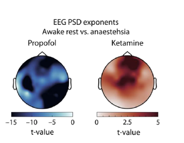
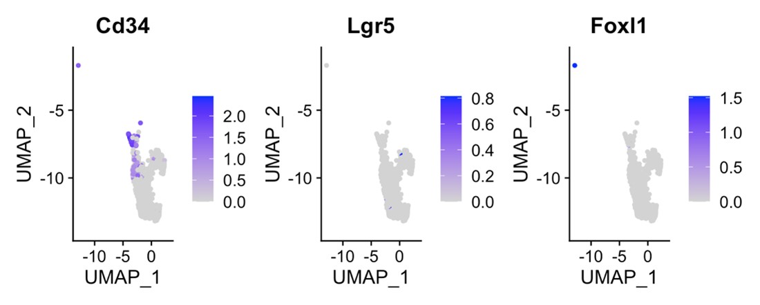
Batch effects in each of the pools preventing comparison across different residues. We think this is a serious design flaw and not standard for how these deep mutational scans are done. The standard would be to combine all 156 pools in a single experiment. Given the sequencing strategy of dividing up CDKN2A into 3 segments, the 156 pools could easily have been collapsed into 3 (1 to 53, 54 to 110, 111 to 156). This would significantly minimize variation in handling between variants at each residue and would be more manageable for performance of further replicates of the screen for reproducibility purposes. The huge variation in confluency time 16-40 days for each pool suggest that this batch effect is a strong source of variation in the experiment
Lack of experimental/biological replication: The functional assay was only performed once on all 156 CDKN2A residues and was repeated for only 28 out of 156 residues, with only ~80% concordance in functional classification between the first and second screens. This is not sufficiently robust for variant interpretation. Why was the experiment not performed more than once for most aa sites?
For the screen, the methods section states that PANC-1 cells were infected at MOI=1 while the standard is an MOI of 0.3-0.5 to minimize multiple variants integrating into a single cell. At an MOI =1 under a Poisson process which captures viral integration, ~25% of cells would have more than 1 lentiviral integrant. So in 25% of the cells the effect of a variant would be confounded by one or more other variants adding noise to the assay.
While the authors provide more explanation of the gamma GLM, we strongly advise that the heatmap and replicate correlations be shown with the log2 fold changes rather than the fit output of the p-values.
In this study, the authors only classify variants into the categories "neutral", "indeterminate", or "deleterious" but they do not address CDKN2A gain-of-function variants that may lead to decreased proliferation. For example, there is no discussion on variants at residue 104, whose proliferation values mostly consist of higher magnitude negative log2fold change values. These variants are defined as neutral but from the one replicate of the experiment performed, they appear to be potential gain-of-function variants.
-
Author response:
The following is the authors’ response to the original reviews.
Public Reviews:
Reviewer #1 (Public Review):
Summary:
Kimura et al performed a saturation mutagenesis study of CDKN2A to assess the functionality of all possible missense variants and compare them to previously identified pathogenic variants. They also compared their assay result with those from in silico predictors.
Strengths:
CDKN2A is an important gene that modulates cell cycle and apoptosis, therefore it is critical to accurately assess the functionality of missense variants. Overall, the paper reads well and touches upon major discoveries in a logical manner.
Weaknesses:
The paper lacks proper details for experiments and basic data, leaving the results less convincing. Analyses are superficial and do not provide variant-level resolution.
We thank the reviewer for their comments. We have updated the manuscript to include additional detail of experimental methods and variant level resolution of data and analyses. We have also conducted additional analyses to compare variant classifications using a gamma generalized linear model and log2 normalized fold change, establish the effect of low variant coverage on variant functional classifications, determine the performance of combining multiple in silico predictions, and determine the prevalence of functionally deleterious variants in gnomAD and functionally deleterious variants of uncertain significance in ClinVar compared all CDKN2A missense variants.
Reviewer #2 (Public Review):
This study describes a deep mutational scan across CDKN2A using suppression of cell proliferation in pancreatic adenocarcinoma cells as a readout for CDKN2A function. The results are also compared to in silico variant predictors currently utilized by the current diagnostic frameworks to gauge these predictors' performance. The authors also functionally classify CDKN2A somatic mutations in cancers across different tissues.
This study is a potentially important contribution to the field of cancer variant interpretation for CDKN2A, but is almost impossible to review because of the severe lack of details regarding the methods and incompleteness of the data provided with the paper. We do believe that the cell proliferation suppression assay is robust and works, but when it comes to the screening of the library of CDKN2A variants the lack of primary data and experimental detail prevents assessment of the scientific merit and experimental rigor.
We are grateful for the opportunity to clarify our experimental methods and to provide additional data in the revised manuscript. The manuscript has been updated to include, among other changes, additional information on assay design, analysis of variant representation in the library, inclusion of primary data with variant level resolution, and a comparison of variant classifications using a gamma generalized linear model and log2 normalized fold change.
Recommendations for the authors:
Reviewer #1 (Recommendations For The Authors):
Major issues:
(1) Can the pathogenicity values of individual amino acid changes be opened to the public? It would serve as a valuable asset to the community.
Thank you for your suggestion. We are happy to provide this information. Individual variant data and functional classifications from the functional assay are given in Appendix 1-table 4.
(2) In the method section, it is not clear (at least to the reviewer) whether the protocol describing the construction of the CDKN2A missense library was provided.
Thank you for your comment. We have included additional information in the manuscript describing construction of the CDKN2A missense library.
“CDKN2A expression plasmid libraries
Codon-optimized CDKN2A cDNA using p16INK4A amino acid sequence (NP_000068.1), was designed (Appendix 1-table 12) and pLJM1 containing codon optimized CDKN2A (pLJM1-CDKN2A) generated by Twist Bioscience (South San Francisco, CA). 156 plasmid libraries were then synthesized by using pLJM1-CDKN2A, such that each library contained all possible 20 amino acids variants (19 missense and 1 synonymous) at a given position, generating 500 ng of each plasmid library (Twist Bioscience, South San Francisco, CA). The proportion of variant in each library was shown in Appendix 1-table 2. Variants with a representation of less than 1% in a plasmid library were individually generated using the Q5 Site-Directed Mutagenesis kit (New England Biolabs, Ipswich, MA; catalog no. E0552), and added to each library to a calculated proportion of 5%. Primers used for site-directed mutagenesis are given in Appendix 1-table 13. Each library was then amplified to generate at least 5 ug of plasmid DNA using QIAGEN Plasmid Midi Kit (QIAGEN, Germantown, MD; catalog no. 12143).”
(3) The paper lacks basic experimental results. The results cover almost all possible missense variants, but it would be clearer if actual coverage values used for calculating relative enrichment were shown. Are all variants well covered? Isn't there any spurious signal due to low coverage? How many times were the experiments performed? Also, how many cells were used, what was the expected MOI, and what proportion of harvested cells is thought to have a single variant? How can you distinguish the effect of a single variant from a multiple variants effect?
We thank the reviewer for their comment. We have provided additional information in the manuscript to address these issues. Briefly, in response to each issue:
(1) We have provided read count data for all variants, used to determine functional classifications based on either gamma generalized linear model or normalized fold change, in Appendix 1-table 4.
(2) To assess if low variant coverage resulted in spurious signals, we compared prevalence of functionally deleterious classifications among variants binned by coverage in the Day 9 cell pool. We did not identify any statistically significant differences based on variant coverage.
“We also determined whether underrepresentation in the cell pool at Day 9 affected variant functional classifications. Fifty-three of 2,964 missense variants (1.8%) were present in the cell pool at Day 9 of the first assay replicate (experiment 1) at < 2%, as determined by the number of sequence reads supporting the variant (Figure 2 -figure supplement 4A, Appendix 1-table 4). There was no statistically significant difference in the proportion of variants classified as functionally deleterious for variants present in less than 2% of the cell pool at Day 9 (12 of 53 variants; 22.6%), and variants present in more than 2% of the cell pool (496 of 2,911 variants; 17.0%) (P value = 0.28) (Figure 2 -figure supplement 4B). We also found no significant differences in the proportion of variants classified as functionally deleterious for variants present in more than 2% of the cell pool at Day 9 when variants were binned in 1% intervals (Figure 2 -figure supplement 4B).”
(3) The assay was repeated in duplicate for 28 CDKN2A residues. For the remaining 128 residues of CDKN2A, the assay was completed once. We found good agreement between variant classifications in assay repeats. We have added to the text as follows:
“To confirm the reproducibility of our variant classifications, 28 amino acid residues were assayed in duplicate, and variants classified using the gamma GLM. The majority of missense variants, 452 of 560 (80.7%), had the same functional classification in each of the two replicates (Figure 2 -figure supplement 3A and B, Appendix 1-table 4).”
We have also added discussion of this study limitation to the manuscript:
“We repeated our functional assay twice for 28 CDKN2A residues. For the remaining 128 residues of CDKN2A, the functional assay was completed once. While we found general agreement between functional classifications from each replicate for the 28 residues assayed in duplicate, additional repeats for each residue are necessary to determine variability in variant functional classifications.”
(4) We have added additional information about the number of cells used for transduction and MOI to the method section:
“Lentiviral transduction
PANC-1 cells were used for CDKN2A plasmid library and single variant CDKN2A expression plasmid transductions. PANC-1 cells previously transduced with pLJM1-CDKN2A (PANC-1CDKN2A) and selected with puromycin were used for CellTag library transductions. Briefly, 1 x 10⁵ cells were cultured in media supplemented with 10 ug/ml polybrene and transduced with 4 x 10⁷ transducing units per mL of lentivirus particles. Cells were then centrifuged at 1,200 x g for 1 hour. After 48 hours of culture at 37oC and 5% CO2, transduced cells were selected using 3 µg/ml puromycin (CDKN2A plasmid libraries and single variant CDKN2A expression plasmids) or 5 µg/ml blasticidin (CellTag plasmid library) for 7 days. Expected MOI was one. After selection, cells were trypsinized and 5 x 10⁵ cells were seeded into T150 flasks. DNA was collected from remaining cells and this sample was named as (Day 9). T150 flasks were cultured until confluent and then DNA was collected. The time for cells to become confluent varied for each amino acid residue (Day 16 – 40, Appendix 1-table 5).”
(5) Our assay was not designed to distinguish multiple variant effects. However, we do not anticipate multiple transductions to significantly impact variant classifications in our assay. We found that our functional classifications were consistent with previously reported classifications:
“In general, our results were consistent with previously reported classifications. Of variants identified in patients with cancer and previously reported to be functionally deleterious in published literature and/or reported in ClinVar as pathogenic or likely pathogenic (benchmark pathogenic variants), 27 of 32 (84.4%) were functionally deleterious in our assay (Figure 2B, Figure 2 -figure supplement 1B and 1C, Appendix 1-table 4) (Chaffee et al., 2018; Chang et al., 2016; Horn et al., 2021; Hu et al., 2018; Kimura et al., 2022; McWilliams et al., 2018; Roberts et al., 2016; Zhen et al., 2015). Five benchmark pathogenic variants were characterized as indeterminate function, with log2 P values from -19.3 to -33.2. Of 156 synonymous variants and six missense variants previously reported to be functionally neutral in published literature and/or reported in ClinVar as benign or likely benign (benchmark benign variants), all were characterized as functionally neutral in our assay (Figure 2B, Figure 2 -figure supplement 1B and 1C, Appendix 1-table 4) (Kimura et al., 2022; McWilliams et al., 2018; Roberts et al., 2016). Of 31 VUSs previously reported to be functionally deleterious, 28 (90.3%) were functionally deleterious and 3 (9.7%) were of indeterminate function in our assay. Similarly, of 18 VUSs previously reported to be functionally neutral, 16 (88.9%) were functionally neutral and 2 (11.1%) were of indeterminate function in our assay, (Figure 2B, Figure 2 -figure supplement 1B and 1C, Appendix 1-table 4).”
(4) Comparison of functional classifications (shown in Figure 3) from this study and other in silico tools is superficial. The analysis is based on the presumption that their result is gold-standard, thereby calculating the sensitivity, accuracy, and PPV of individual predictors. But apparently, this won't be true, so it would be more reasonable to check the "correlation" of the study results and other predictors: e.g. which variants show consistent results between this study and other predictors? Are there any indicators of consistent vs inconsistent results? How does the consistency change by protein sequences or domains? Etc
Thank you for your comment. We have added additional analysis to our manuscript comparing our functional classifications with in silico variant effect predictions. Specifically, we have included analysis combining multiple predictors:
“We also tested the effect of combining multiple in silico predictors. 904 missense variants had in silico predictions from all 7 algorithms. The remaining 2,060 missense variants had in silico predictions from 5 algorithms. Of variants with in silico predictions from all 7 algorithms, 378 (41.8%) had predictions of deleterious or pathogenic effect from a majority of algorithms (≥ 4), and of these, 137 (36.2%) were functionally deleterious in our assay. Similarly, of 2,060 missense variants that had in silico predictions from 5 algorithms, 1107 (53.7%) had predictions of deleterious or pathogenic effect from a majority of algorithms (≥ 3), of which, 361 (32.6%) were functionally deleterious in our assay (Appendix 1-table 7).”
(5) Similarly, Figure 4 does not deliver much information, either. Rather than delivering a simple summary, it would be more informative if deeper analyses were conducted. e.g., do pathogenic variants show higher frequency among patients, or higher variant frequency in tumors (if data were available).
We have included additional analysis of somatic alterations in the manuscript. We found pathogenic/likely pathogenic somatic mutations were enriched in patients. This was also the case for somatic mutations that were classified as functionally deleterious in our assay. We also found statistically significant depletion of functionally deleterious mutations in colorectal adenocarcinoma. Interestingly, no patients with a somatic mutation in a mismatch repair gene had a functionally deleterious CDKN2A missense somatic mutation. However, this observation was not statistically significant. Future studies will determine whether CDKN2A and MMR gene somatic mutations are mutually exclusive in colorectal adenocarcinoma.
“We found that 34.2% - 53.4% of unique missense somatic mutations classified as functionally deleterious, with 61.4% - 67.6% of patients having a functionally deleterious somatic mutation (Figure 4A, Appendix 1-table 9). As with functionally deleterious variants, functionally deleterious missense somatic mutations were also not distributed evenly across CDKN2A, being enriched within the ankyrin repeat 3 (Figure 4B, Appendix 1-table 9). We found that 32.4% - 50.0% of all functionally deleterious missense somatic mutations occurred within ankyrin repeat 3, with 48.0% - 58.0% of patients in each cohort having a functionally deleterious missense somatic mutation in this domain. Notably, 65.7% - 76.0% of functionally deleterious missense somatic mutations in this domain were in residues 80-89 (Appendix 1-table 9).”
“We were also able to determine the functional classification of CDKN2A missense somatic mutations in COSMIC, TCGA, JHU, and MSK-IMAPCT by cancer type. We found that 22.2% - 100% of CDKN2A missense somatic mutations were functionally deleterious depending on cancer type (Figure 4-figure supplement 2A-D). When considering missense somatic mutation reported in any database, there was a statistically significant depletion of functionally deleterious mutations in colorectal adenocarcinoma (20.4%; adjusted P value = 5.4 x 10⁻⁹) (Figure 4C). As the proportion of missense somatic mutations that were functionally deleterious was less in colorectal carcinoma compared to other types of cancer, we assessed whether somatic mutations in mismatch repair genes (MLH1, MLH3, MSH2, MSH6, PMS1, and PMS2) were associated with the functional status of CDKN2A missense somatic mutations. Thirty-five patients in COSMIC had a CDKN2A missense somatic mutation, of which 12 (34.3%) had a somatic mutation in a mismatch repair gene. We found that no patients with a somatic mutation in a mismatch repair gene had a functionally deleterious CDKN2A missense somatic mutation compared to 6 of 23 samples (26.1%) without a somatic mutation in a mismatch repair gene (P value = 0.062).”
(6) It would be helpful to validate the neutral variants set. Are variants of UK biobank or gnomAD enriched on neutral population? Are synonymous variants exclusively found in neutral populations?
Thank you for the suggestion. All synonymous variants were found to functionally neutral in our assay. We also assessed VUSs from gnomAD and found a lower prevalence of functionally deleterious variants compared to all CDKN2A variants and CDKN2A missense somatic mutations:
“The Genome Aggregation Database (gnomAD) v4.1.0 reports 287 missense variants in CDKN2A, including the 13 pathogenic, 4 likely pathogenic, 3 likely benign, 3 benign, and 264 VUSs classified using ACMG variant interpretation guidelines (Figure 5A, Figure 5B, and Appendix 1-table 10). Of the 264 missense VUSs, 177 were functionally neutral (67.0%), 56 (21.2%) were indeterminate function, and 31 (11.7%) were functionally deleterious in our assay using the gamma GLM for classification (Figure 5C).”
(7) They used a pancreatic cancer cell line and assayed for cell proliferation. The limitations of this method and the possibility of complementing the limitations should be discussed.
Thank you for the suggestion. We have added discussion of this limitation to our manuscript:
“We characterized variants based upon a broad cellular phenotype, cell proliferation, in a single PDAC cell line. It is possible that CDKN2A variant functional classifications are cell-specific and assay-specific. Our assay may not encompass all cellular functions of CDKN2A and an alternative assay of a specific CDKN2A function, such as CDK4 binding, may result in different variant functional classifications. Furthermore, CDKN2A variants may have different effects if alternative cell lines are used for the functional assay. However, cell-specific effects appear to be limited. In our previous study, we characterized 29 CDKN2A VUSs in three PDAC cell lines, using cell proliferation and cell cycle assays, and found agreement between all functional classifications (Kimura et al., 2022).”
Minor issues:
(1) Figures 2B, C: it would be more intuitive to plot significance by logging p-values than raw p-values.
We used log2 P value (or log2 normalized fold change) for figures in the manuscript as appropriate.
(2) Figure 2D: annotate protein domain information at the side. Supplementary Figure 2 shows the domains but it would be more informative to show it in Figure 2D heatmap.
Thank you for the suggestion, we have annotated protein domain information on the left side of the heatmap in (the now) Figure 2C.
Reviewer #2 (Recommendations For The Authors):
Major Concerns:
(1) How many replicates of the screen were performed? It seems like only one library infection/ proliferation assay was done. If so this is insufficient to obtain any idea of the uncertainty of measurement for each variant.
The assay was repeated in duplicate for 28 CDKN2A residues. For the remaining 128 residues of CDKN2A, the assay was completed once. We found good agreement between variant classifications in assay repeats. We have added to the text as follows:
“To confirm the reproducibility of our variant classifications, 28 amino acid residues were assayed in duplicate, and variants classified using the gamma GLM. The majority of missense variants, 452 of 560 (80.7%), had the same functional classification in each of the two replicates (Figure 2 -figure supplement 3A and B, Appendix 1-table 4).”
We have also added discussion of this study limitation to the manuscript:
“We repeated our functional assay twice for 28 CDKN2A residues. For the remaining 128 residues of CDKN2A, the functional assay was completed once. While we found general agreement between functional classifications from each replicate for the 28 residues assayed in duplicate, additional repeats for each residue are necessary to determine variability in variant functional classifications.”
(2) The count data from the experiment and NGS pipeline to call variants need to be provided for each replication (i.e. the counts that were fed into the gamma model)
Accompanying this should be information about the depth of sequencing of the cells, the number of cells infected with the library, and standard metrics for pooled screens.
Quality metrics regarding the representation and completeness of the TWIST library need to be provided. See Brenan et al. Cell Reports (2016) Supplemental Figure 1
Thank you for your suggestion. We are happy to provide this additional information. Sequence read counts for each variant are given in Appendix 1-table 4. We have provided addition detail in the methods section on functional assay, including number of cells infected with each library:
“Lentiviral transduction
PANC-1 cells were used for CDKN2A plasmid library and single variant CDKN2A expression plasmid transductions. PANC-1 cells previously transduced with pLJM1-CDKN2A (PANC-1CDKN2A) and selected with puromycin were used for CellTag library transductions. Briefly, 1 x 10⁵ cells were cultured in media supplemented with 10 ug/ml polybrene and transduced with 4 x 10⁷ transducing units per mL of lentivirus particles. Cells were then centrifuged at 1,200 x g for 1 hour. After 48 hours of culture at 37oC and 5% CO2, transduced cells were selected using 3 µg/ml puromycin (CDKN2A plasmid libraries and single variant CDKN2A expression plasmids) or 5 µg/ml blasticidin (CellTag plasmid library) for 7 days. Expected MOI was one. After selection, cells were trypsinized and 5 x 10⁵ cells were seeded into T150 flasks. DNA was collected from remaining cells and this sample was named as (Day 9). T150 flasks were cultured until confluent and then DNA was collected. The time for cells to become confluent varied for each amino acid residue (Day 16 – 40, Appendix 1-table 5). DNA was extracted from PANC-1 cells using the PureLink Genomic DNA Mini Kit (Invitrogen, Carlsbad, CA; catalog no. K1820-01). The assay for CellTag library was repeated in triplicate. We repeated our CDKN2A assay in duplicate for 28 residues. For the remaining 128 CDKN2A residues the assay was completed once.”
We have also provided additional information on the TWIST library:
“CDKN2A expression plasmid libraries
Codon-optimized CDKN2A cDNA using p16INK4A amino acid sequence (NP_000068.1), was designed (Appendix 1-table 12) and pLJM1 containing codon optimized CDKN2A (pLJM1-CDKN2A) generated by Twist Bioscience (South San Francisco, CA). 156 plasmid libraries were then synthesized by using pLJM1-CDKN2A, such that each library contained all possible 20 amino acids variants (19 missense and 1 synonymous) at a given position, generating 500 ng of each plasmid library (Twist Bioscience, South San Francisco, CA). The proportion of variant in each library was shown in Appendix 1-table 2. Variants with a representation of less than 1% in a plasmid library were individually generated using the Q5 Site-Directed Mutagenesis kit (New England Biolabs, Ipswich, MA; catalog no. E0552), and added to each library to a calculated proportion of 5%. Primers used for site-directed mutagenesis are given in Appendix 1-table 13. Each library was then amplified to generate at least 5 ug of plasmid DNA using QIAGEN Plasmid Midi Kit (QIAGEN, Germantown, MD; catalog no. 12143).”
(3) It is unclear when barcode abundance is assessed in the cell proliferation assay/in the screen. The exact timepoints of "before and after in vitro culture" (line 91) need to be clarified in the text.
We are happy to clarify. We collected DNA on Day 9 post transfection and at confluency. Day of confluency for each residue is detailed in Appendix 1-table 5. The text of the manuscript has been updated appropriately.
(4) Is "before" day 9, as detailed in Figure 1 source data 1? If so, it is misleading to state that the experiment is in culture for 14 days but call day 9 "before... in vitro culture."
The "before" sample should be obtained immediately after viral infection and selection with the library to provide a representation of library representation.
We apologize for your confusion. We have clarified in the text and figures that our baseline measurement was at Day 9 post transfection. We also determined whether the proportion of each variant is maintained in the Day 9 cell pool compared to the amplified plasmid library for three CDKN2A amino acid residues (p.R24, p.H66, and p.A127) and updated the manuscript text:
“To confirm that the representation of each variant was maintained after transduction, we transduced three lentiviral libraries (amino acid residues p.R24, p.H66, and p.A127) individually into PANC-1 cells and determined the proportion of each variant in the amplified plasmid library and in the cell pool at Day 9 post-transduction. The proportion of each variant in the amplified plasmid library and in the cell pool at Day 9 were highly correlated (Figure 1 -figure supplement 2C and D, Appendix 1-table 3).”
(5) There is no information regarding the function of each variant, aside from just a p-value resulting from the final analysis with the gamma model. Some variants may cause loss of function, others may be neutral while others may be gain of function. Simply providing a p-value is not sufficient. The standard in the field is to provide a function score/ test-statistic giving the sign and magnitude of the effect. For proliferation assays at least a ratio of fold-change of (mut/ synonymous)[day 14] vs (mut/synonymous)[baseline] should be provided.
Thank you for your comment. We have provided read counts, P values, and functional classifications for each variant using the gamma GLM in Appendix 1-table 4. We have also analyzed variants using log2 normalized fold change. This data is presented in the text and compared to our classifications with the gamma GLM. We have provided normalized fold change and resulting classification for each variant in Appendix 1-table 6.
(6) A plot of the distribution of function scores for all variants is needed. This will serve as an effective visual to distinguish the control variants from those that are functionally deleterious or benign/neutral (see Findlay et al. Nature (2018) Figure 3A for an example visual).
Thank you for your suggestion. We have provided additional figures to visualize distribution of assay outputs using the gamma GLM in Figure 2 -figure supplement 1.
(7) Synonymous variants are used as a proxy for WT per variant library, but do all the synonymous variants truly behave like WT CDKN2A in their ability to suppress cell proliferation? A plot of the distribution of synonymous variant function relative to WT CDKN2A function would be effective here.
All 156 synonymous variants suppressed cell proliferation and were classified as functionally neutral in our assay using the gamma GLM. The manuscript has been updated to reflect this:
“Of 156 synonymous variants and six missense variants previously reported to be functionally neutral in published literature and/or reported in ClinVar as benign or likely benign (benchmark benign variants), all were characterized as functionally neutral in our assay (Figure 2B, Figure 2 -figure supplement 1B and 1C, Appendix 1-table 4)”
(8) The gamma generalized linear model is not commonly used to analyze the results of saturation mutagenesis screens. Please provide a justification for the use of this analysis method vs using log fold change as other dms scan studies have done (PMID: 27760319, PMID: 30224644).
Thank you for this important suggestion. We are happy to provide additional information. We used a gamma GLM to functionally characterize CDKN2A variants as it does not rely on an annotated set of pathogenic and benign variants to determine classification thresholds. Instead, classification thresholds are determined using the change in representation of 20 non-functional barcodes in a pool of PANC-1 cells stably expressing CDKN2A after a period of in vitro growth. As a gamma GLM is not commonly used for saturation mutagenesis screens, as noted by the reviewer, we also classified variants using log2 normalized fold change. We compared variant functional classifications using the gamma GLM and log2 normalized fold change and in general we found agreement between both methods with 98.5% of missense variants classified as functionally deleterious using a gamma GLM, similarly classified using log2 normalized fold change. We have updated the text to reflect this reasoning and additional analysis.
(9) The statistical methods used to calculate enrichment of deleterious variants per region of CDKN2A (Figure 2 supplement 1B; lines 163-168) are not described anywhere in the paper. Additionally, the same statistical analysis is not applied to the variants in the subregions near the ankyrin repeats (lines 168-172).
We are happy to clarify and have added text to the methods section:
“Z-tests with multiple test correction performed with the Bonferroni method was used in the following comparisons: 1) proportion of functionally deleterious variants present in < 2% of the cell pool and ≥ 2% of the cell pool at Day 9 binned in 1% intervals, 2) proportion of variants in each domain predicted to have deleterious or pathogenic effect by the majority of algorithms, 3) proportion of functionally deleterious variants in each domain, and 4) proportion of functionally deleterious missense variants and somatic mutations.”
Minor:
(1) Please review the manuscript for spelling and grammatical errors.
Sure.
-
-
www.biorxiv.org www.biorxiv.org
-
eLife Assessment
This study presents an important application of high-content image-based morphological profiling to quantitatively and systematically characterize induced pluripotent stem cell-derived mixed neural cultures cell type compositions. Compelling evidence through rigorous experimental and computational validations support new potential applications of this cheap and simple assay.
-
Joint Public Review:
Automatically identifying single cell types in heterogeneous mixed cell populations holds great promise to characterize mixed cell populations and to discover new rules of spatial organization and cell-cell communication. Although the current manuscript focuses on the application of quality control of iPSC cultures, the same approach can be extended to a wealth of other applications including in depth study of the spatial context. The simple and high-content assay democratizes use and enables adoption by other labs.
The authors also propose a new nucleocentric phenotyping pipeline, where a convolutional neural network is trained on the nucleus and some margins around it. This nucleocentric approach improves classification performance at high densities because nuclear segmentation is less prone to errors in dense cultures.
The manuscript is supported by comprehensive experimental and computational validations that raises the bar beyond the current state of the art in the field of high-content phenotyping and makes this manuscript especially compelling. These include (i) Explicitly assessing replication biases (batch effects); (ii) Direct comparison of feature-based (a la cell profiling) versus deep-learning-based classification (which is not trivial/obvious for the application of cell profiling); (iii) Systematic assessment of the contribution of each fluorescent channel; (iv) Evaluation of cell-density dependency; (v) explicit examination of mistakes in classification; (vi) Evaluating the performance of different spatial contexts around the cell/nucleus; (vii) generalization of models trained on cultures containing a single cell type (mono-cultures) to mixed co-cultures; (viii) application to multiple classification tasks.
-
Author response:
The following is the authors’ response to the previous reviews.
Public Review:
Summary:
The authors present a new application of the high-content image-based morphological profiling Cell Painting (CP) to single cell type classification in mixed heterogeneous induced pluripotent stem cell-derived mixed neural cultures. Machine learning models were trained to classify single cell types according to either "engineered" features derived from the image or from the raw CP multiplexed image. The authors systematically evaluated experimental (e.g., cell density, cell types, fluorescent channels) and computational (e.g., different models, different cell regions) parameters and convincingly demonstrated that focusing on the nucleus and its surroundings contain sufficient information for robust and accurate cell type classification. Models that were trained on mono-cultures (i.e., containing a single cell type) could generalize for cell type prediction in mixed co-cultures, and to describe intermediate states of the maturation process of iPSC-derived neural progenitors to differentiation neurons.
Strengths:
Automatically identifying single cell types in heterogeneous mixed cell populations hold great promise to characterize mixed cell populations and to discover new rules of spatial organization and cell-cell communication. Although the current manuscript focuses on the application of quality control of iPSC cultures, the same approach can be extended to a wealth of other applications including in depth study of the spatial context. The simple and high-content assay democratizes use and enables adoption by other labs.
The manuscript is supported by comprehensive experimental and computational validations that raises the bar beyond the current state of the art in the field of highcontent phenotyping and makes this manuscript especially compelling. These include (i) Explicitly assessing replication biases (batch effects); (ii) Direct comparison of featurebased (a la cell profiling) versus deep-learning-based classification (which is not trivial/obvious for the application of cell profiling); (iii) Systematic assessment of the contribution of each fluorescent channel; (iv) Evaluation of cell-density dependency; (v) explicit examination of mistakes in classification; (vi) Evaluating the performance of different spatial contexts around the cell/nucleus; (vii) generalization of models trained on cultures containing a single cell type (mono-cultures) to mixed co-cultures; (viii) application to multiple classification tasks.
Comments on latest version:
I have consulted with Reviewer #3 and both of us were impressed by revised manuscript, especially by the clear and convincing evidence regarding the nucleocentric model use of the nuclear periphery and its benefit for the case of dense cultures. However, there are two issues that are incompletely addressed (see below). Until these are resolved, the "strength of evidence" was elevated to "compelling".
First, the analysis of the patch size is not clearly indicating that the 12-18um range is a critical factor (Fig. 4E). On the contrary, the performance seems to be not very sensitive to the patch size, which is actually a desired property for a method. Still, Fig. 4B convincingly shows that the nucleocentric model is not sensitive to the culture density, while the other models are. Thus, the authors can adjust their text saying that the nucleocentric approach is not sensitive to the patch size and that the patch size is selected to capture the nucleus and some margins around it, making it less prone to segmentation errors in dense cultures.
We agree that there is a significant tolerance to different patch sizes, and have therefore reformulated the conclusion as suggested in the results and the discussion sections (page 10 and 16). As very large patch sizes (>40µm) do increase the variability of the predictions and the imbalance between recall and precision, we have left this observation in the results section, as it also motivates for using smaller patch sizes.
Second, the GitHub does not contain sufficient information to reproduce the analysis. Its current state is sparse with documentation that would make reproducing the work difficult. What versions of the software were used? Where should data be downloaded? The README contains references to many different argparse CLI arguments, but sparse details on what these arguments actually are, and which parameters the authors used to perform their analyses. Links to images are broken. Ideally, all of these details would be present, and the authors would include a step-by-step tutorial on how to reproduce their work. Fixing this will lead to an "exceptional" strength of evidence.
We have added additional information to the GitHub to increase the reproducibility of the analysis.
• The README now contains additional documentation and more extensive explanations. A flowchart has been added, making the dataflow and order of analyses more clear.
• The accompanying dataset is 20GB in size and can be downloaded as a .zip-file from https://figshare.com/articles/dataset/Nucleocentric-Profiling/27141441?file=49522557. This file contains 2x480 raw images and a layout file.
• The used software versions are included in the manuscript in table 4. To increase the reproducibility, a Conda environment file (.yaml) has been added to the GitHub. This can be installed and contains the correct package versions.
• The README now contains for each script and its arguments a short description on its meaning, on whether it is required or optional and its default setting.
• A step-by-step tutorial on the use of the test dataset has been included. This tutorial includes the arguments used to run the code from the command line terminal.
Recommendations for the authors:
There are no reference from the text to Fig. 2D and to Fig. 3C.
This has been changed. The text has been added to the manuscript at page 6 (fig. 2D) and the reference to Fig. 3C has been included at page 8.
-
-
www.biorxiv.org www.biorxiv.org
-
eLife Assessment
This work presents a valuable exploration of AI-assisted protein engineering, particularly in designing a VHH antibody with enhanced resistance and stability to extreme environments. However, the approach is weakened by incomplete support, with computational methods and experimental design appearing somewhat arbitrary and lacking clear justification. Further justification of the chosen methods and clearer exposition would strengthen the study's support and conclusions.
-
Reviewer #1 (Public review):
Summary:
In this manuscript, the model's capacity to capture epistatic interactions through multi-point mutations and its success in finding the global optimum within the protein fitness landscape highlights the strength of deep learning methods over traditional approaches.
Strengths:
It is impressive that the authors used AI combined with limited experimental validation to achieve such significant enhancements in protein performance. Besides, the successful application of the designed antibody in industrial settings demonstrates the practical and economic relevance of the study. Overall, this work has broad implications for future AI-guided protein engineering efforts.
Weaknesses:
However, the authors should conduct a more thorough computational analysis to complement their manuscript. While the identification of improved multi-point mutants is commendable, the manuscript lacks a detailed investigation into the mechanisms by which these mutations enhance protein properties. The authors briefly mention that some physicochemical characteristics of the mutants are unusual, but they do not delve into why these mutations result in improved performance. Could computational techniques, such as molecular dynamics simulations, be employed to explore the effects of these mutations? Additionally, the authors claim that their method is efficient. However, the selected VHH is relatively short (<150 AA), resulting in lower computational costs. It remains unclear whether the computational cost of this approach would still be acceptable when designing larger proteins (>1000 AA). Besides, the design process involves a large number of prediction tasks, including the properties of both single-site saturation and multi-point mutants. The computational load is closely tied to the protein length and the number of mutation sites. Could the authors analyze the model's capability boundaries in this regard and discuss how scalable their approach is when dealing with larger proteins or more complex mutation tasks?
-
Reviewer #2 (Public review):
In this paper, the authors aim to explore whether an AI model trained on natural protein data can aid in designing proteins that are resistant to extreme environments. While this is an interesting attempt, the study's computational contributions are weak, and the design of the computational experiments appears arbitrary.
(1) The writing throughout the paper is poor. This leaves the reader confused.
(2) The main technical issue the authors address is whether AI can identify protein mutations that adapt to extreme environments based solely on natural protein data. However, the introduction could be more concise and focused on the key points to better clarify the significance of this question.
(3) The authors did not develop a new model but instead used their previously developed Pro-PRIME model. This significantly weakens the novelty and contribution of this work.
(4) The computational experiments are not well-justified. For instance, the authors used a zero-shot setting for single-point mutation experiments but opted for fine-tuning in multiple-point mutation experiments. There is no clear explanation for this discrepancy. How does the model perform in zero-shot settings for multiple-point mutations? How would fine-tuning affect single-point mutation results? The choice of these strategies seems arbitrary and lacks sufficient discussion.
-
Author response:
Reviewer #1:
Weaknesses:
However, the authors should conduct a more thorough computational analysis to complement their manuscript. While the identification of improved multi-point mutants is commendable, the manuscript lacks a detailed investigation into the mechanisms by which these mutations enhance protein properties. The authors briefly mention that some physicochemical characteristics of the mutants are unusual, but they do not delve into why these mutations result in improved performance. Could computational techniques, such as molecular dynamics simulations, be employed to explore the effects of these mutations? Additionally, the authors claim that their method is efficient. However, the selected VHH is relatively short (<150 AA), resulting in lower computational costs. It remains unclear whether the computational cost of this approach would still be acceptable when designing larger proteins (>1000 AA). Besides, the design process involves a large number of prediction tasks, including the properties of both single-site saturation and multi-point mutants. The computational load is closely tied to the protein length and the number of mutation sites. Could the authors analyze the model's capability boundaries in this regard and discuss how scalable their approach is when dealing with larger proteins or more complex mutation tasks?
We agree that further analysis of the mechanisms by which the identified mutations enhance protein performance would strengthen our study. In the revised manuscript, we plan to conduct molecular dynamics simulations to explore the physicochemical effects of these mutations in more details. This analysis will help elucidate how the observed structural and dynamic changes contribute to the improved resistance and stability of the designed VHH antibody.
We acknowledge the need to assess the scalability of our method to larger proteins. To address this, we will include an analysis of the method’s performance when applied to longer proteins, including an estimation of computational cost and potential bottlenecks.
Reviewer #2:
(1) The writing throughout the paper is poor. This leaves the reader confused.
(2) The main technical issue the authors address is whether AI can identify protein mutations that adapt to extreme environments based solely on natural protein data. However, the introduction could be more concise and focused on the key points to better clarify the significance of this question.
(3) The authors did not develop a new model but instead used their previously developed Pro-PRIME model. This significantly weakens the novelty and contribution of this work.
(4) The computational experiments are not well-justified. For instance, the authors used a zero-shot setting for single-point mutation experiments but opted for fine-tuning in multiple-point mutation experiments. There is no clear explanation for this discrepancy. How does the model perform in zero-shot settings for multiple-point mutations? How would fine-tuning affect single-point mutation results? The choice of these strategies seems arbitrary and lacks sufficient discussion.
(1&2) We will revise the manuscript to improve the overall clarity and readability. Specifically, we will restructure the introduction to focus more concisely on the key scientific questions and contributions of our study.
(3) While the Pro-PRIME model was previously developed, this work focuses on designing proteins with properties that do not naturally exist and are scarce in the natural world. To address the concern about novelty, we will expand the discussion to highlight this unique contribution and its implications for advancing protein design.
(4) We appreciate the comment regarding the discrepancy between the zero-shot and fine-tuning strategies. In the revised manuscript, we will provide a detailed explanation for the choice of these settings, including an analysis of the trade-offs between zero-shot and fine-tuning approaches in multi-point mutation tasks. We will also explore the model’s performance in zero-shot settings for multi-point mutations and report these results in the supplementary materials to ensure completeness.
-
-
www.biorxiv.org www.biorxiv.org
-
eLife Assessment
This study follows up on Arimura et al's powerful new method MagIC-Cryo-EM for imaging native complexes at high resolution. Using a clever design embedding protein spacers between the antibody and the nucleosomes purified, thereby minimizing interference from the beads, the authors concentrate linker histone variant H1.8 containing nucleosomes. From these samples, the authors obtain convincing atomic structures of the H1.8 bound chromatosome purified from interphase and metaphase cells, finding a NPM2 chaperone bound form exists as well. Caveats include the use of formaldehyde crosslinking and tagged H1.8 which might affect the structures obtained; and the NPM2 work could be better incorporated into the main findings. Overall this is an important new tool in the arsenal of single molecule biologists, permitting a deep dive into structure of native complexes. This work will be of high interest to a broad swathe of scientists studying native macromolecules present at low concentrations in cells.
-
Reviewer #1 (Public review):
Summary:
In this manuscript, Arimura et al describe MagIC-Cryo-EM, an innovative method for immune-selective concentrating of native molecules and macromolecular complexes for Cryo-EM imaging and single-particle analysis. Typically, Cryo-EM imaging requires much larger concentrations of biomolecules than that are feasible to achieve by conventional biochemical fractionation. Overall, this manuscript is meticulously and clearly written and may become a great asset to other electron microscopists and chromatin researchers.
Strengths:
Previously, Arimura et al. (Mol. Cell 2021) isolated from Xenopus extract and resolved by Cryo-EM a sub-class of native nucleosomes conjugated containing histone H1.8 at the on-dyad position, similar to that previously observed by other researchers with reconstituted nucleosomes. Here they sought to analyze immuno-selected nucleosomes aiming to observe specific modes of H1.8 positioning (e.g. on-dyad and off-dyad) and potentially reveal structural motifs responsible for the decreased affinity of H1.8 for the interphase chromatin compared to metaphase chromosomes. The main strength of this work is a clever and novel methodological design, in particular the engineered protein spacers to separate captured nucleosomes from streptavidin beads for a clear imaging. The authors provide a detailed step-by-step description of MagIC-Cryo-EM procedure including nucleosome isolation, preparation of GFP nanobody attached magnetic beads, optimization of the spacer length, concentration of the nucleosomes on graphene grids, data collection and analysis, including their new DUSTER method to filter-out low signal particles. This tour de force methodology should facilitate considering of MagIC-Cryo-EM by other electron microscopists especially for analysis of native nucleosome complexes.<br /> In pursue of biologically important new structures, the immune-selected H1.8-containing nucleosomes were solved at about 4A resolution; their structure appears to be very similar to the previously determined structure of H1.8-reconstituted nucleosomes. There were no apparent differences between the metaphase and interphase complexes suggesting that the on-dyad and off-dyad positioning does not explain the differences in H1.8 - nucleosome binding. However, they were able to identify and solve complexes of H1.8-GFP with histone chaperone NPM2 in a closed and open conformation providing mechanistic insights for H1-NPM2 binding and the reduced affinity of H1.8 to interphase chromatin as compared to metaphase chromosomes.
Weaknesses:
Still, I feel that there are certain limitations and potential artifacts resulting from formaldehyde fixation, use of bacterial-expressed recombinant H1.8-GFP, and potential effects of magnetic beads and/or spacer on protein structure, that should be more explicitly discussed. Also, the GFP-pulled down H1.8 nucleosomes should be better characterized biochemically to determine the actual linker DNA lengths (which are known to have a strong effect of linker histone affinity) and presence or absence of other factors such as HMG proteins that may compete with linker histones and cause the multiplicity of nucleosome structural classes (such as shown on Fig. 3F) for which the association with H1.8 is uncertain.
-
Reviewer #2 (Public review):
Summary:
The authors present a straightforward and convincing demonstration of a reagent and workflow that they collectively term "MagIC-cryo-EM", in which magnetic nanobeads combined with affinity linkers are used to specifically immobilize and locally concentrate complexes that contain a protein-of-interest. As a proof of concept, they localize, image, and reconstruct H1.8-bound nucleosomes reconstructed from frog egg extracts. The authors additionally devised an image-processing workflow termed "DuSTER", which increases the true positive detections of the partially ordered NPM2 complex. The analysis of the NPM2 complex {plus minus} H1.8 was challenging because only ~60 kDa of protein mass was ordered. Overall, single-particle cryo-EM practitioners should find this study useful.
Strengths:
The rationale is very logical and the data are convincing.
Weaknesses: I have seen an earlier version of this study at a conference. The conference presentation was much easier to follow than the current manuscript. It is as if this manuscript had undergone review at another journal and includes additional experiments to satisfy previous reviewers. Specifically, the NPM2 results don't seem to add much to the main story (MagIC-cryo-EM), and read more like an addendum. The authors could probably publish the NPM2 results separately, which would make the core MagIC results (sans DusTER) easier to read.
-
Reviewer #3 (Public review):
Summary:
In this paper, Arimura et al report a new method, termed MagIC-Cryo-EM, which refers to the method of using magnetic beads to capture specific proteins out of a lysate via, followed immunoprecipitation and deposition on EM grids. The so-enriched proteins can be analzyed structurally. Importantly, the nanoparticles are further functionalized with protein-based spacers, to avoid a distorted halo around the particles. This is a very elegant approach and allows the resolution of the stucture of small amounts of native proteins at atomistic resolution.<br /> Here, the authors apply this method to study the chromatosome formation from nucleosomes and the oocyte-specific linker histone H1.8. This allows them to resolve H1.8-containing chromatomosomes from oocyte extract in both interphase and metaphase conditions at 4.3 A resolution, which reveal a common structure with H1 placed right at the dyad and contacting both entry-and exit linker DNA.<br /> They then investigate the origin of H1.8 loss during interphase. They identify a non-nucleosomal H1.8-containing complex from interphase preparations. To resolve its structure, the authors develop a protocol (DuSTER) to exclude particles with ambiguous center, revealing particles with five-fold symmetry, that matches the chaperone NPM2. MS and WB confirms that the protein is present in interphase samples but not metaphase. The authors further separate two isoforms, an open and closed form that coexist. Additional densities in the open form suggest that this might be bound H1.8.
Strengths:
Together this is an important addition to the suite of cryoEM methods, with broad applications. The authors demonstrate the method using interesting applications, showing that the methods work and they can get high resolution structures from nucleosomes in complex with H1 from native environments.
Weaknesses:
The structures of the NPM2 chaperone is less well resolved, and some of the interpretation in this part seems only weakly justified.
-
-
www.biorxiv.org www.biorxiv.org
-
eLife Assessment
This interesting study presents valuable information on how human cytomegalovirus (HCMV) infection disrupts the activity of the TEAD1 transcription factor, leading to widespread chromatin alterations. However, the precise mechanisms underlying this disruption and the extent to which these chromatin changes influence HCMV replication remain unclear. The study is supported by solid evidence, which would be made stronger by including functional analyses. This work will be of interest to virology, chromosome biology and transcriptional co-regulation fields.
-
Reviewer #1 (Public review):
The manuscript by Sayeed et al. uses a comprehensive series of multi-omics approaches to demonstrate that late-stage human cytomegalovirus (HCMV) infection leads to a marked disruption of TEAD1 activity, a concomitant loss of TEAD1-DNA interactions, and extensive chromatin remodeling. The data are thoroughly presented and provide evidence for the role of TEAD1 in the cellular response to HCMV infection. However, a key question remains unresolved: is the observed disruption of TEAD1 activity a direct consequence of HCMV infection, or could it be secondary to the broader innate antiviral response? In this respect, the study would benefit from experiments that assess the effect of TEAD1 overexpression or knockdown/deletion on HCMV replication dynamics. Such functional assays could help delineate whether TEAD1 perturbation directly influences viral replication or is part of a downstream/indirect cellular response, providing deeper mechanistic insights.
-
Reviewer #2 (Public review):
Summary:
This work uses genomic and biochemical approaches for HCMV infection in human fibroblasts and retinal epithelial cell lines, followed by comparisons and some validations using strategies such as immunoblots. Based on these analyses, they propose several mechanisms that could contribute to the HCMV-induced diseases, including closing of TEAD1-occupying domains and reduced TEAD1 transcript and protein levels, decreased YAP1 and phospho-YAP1 levels, and exclusion of TEAD1 exon 6.
Strengths:
The genomics experiments were done in duplicates and data analyses show good technical reproducibility. Data analyses are performed to show changes at the transcript and chromatin level changes, followed by some Western blot validations.
Weaknesses:
This work, at the current stage, is quite correlative since no functional studies are done to show any causal links. For readers who are outside the field, some clarifications of the system and design need to be stated.
-
-
www.biorxiv.org www.biorxiv.org
-
eLife Assessment
This study examines the impact of DNA methylation on CTCF binding in two cancer cell lines. Increased CTCF binding sites are enriched in gene bodies, and associate with nuclear speckles, indicating a potential role in increased transcription. However, the association with nuclear speckles needs to be more diligently demonstrated. Thus the strength of the evidence is considered incomplete. This work would be made more valuable to the community if these claims were buttressed by additional evidence and a deeper discussion of new findings in the light of previous relevant literature. This work will be of interest to the chromosome biology/epigenetics field.
-
Reviewer #1 (Public review):
Summary<br /> Roseman et al. use a new inhibitor of the maintenance DNA methyltransferase DNMT1 to probe the role of methylation on binding of the CTCF protein, which is known to be involved chromatin loop formation. As previous reported, and as expected based on our knowledge that CTCF binding is methylation-sensitive, the authors find that loss of methylation leads to additional CTCF binding sites and increased loop formation. By comparing novel loops with the binding of the pre-mRNA splicing factor SON, which localizes to the nuclear speckle compartment, they propose that these reactivated loops localize to near speckles. This behavior is dependent on CTCF whereas degradation of two speckle proteins does not affect CTCF binding or loop formation. The authors propose a model in which DNA methylation controls the association of genome regions with speckles via CTCF-mediated insulation.
Strengths<br /> The strengths of the study are 1) the use of a new, specific DNMT1 inhibitor and 2) the observation that genes whose expression is sensitive to DNMT1 inhibition and dependent on CTCF (cluster 2) show higher association with SON than genes which are sensitive to DNMT1 inhibition but are CTCF insensitive, is in line with the authors' general model.
Weaknesses<br /> There are a number of significant weaknesses that as a whole undermine many of the key conclusions, including the overall mechanistic model of a direct regulatory role of DNA methylation on CTCF-mediated speckle association of chromatin loops.
(1) The authors frequently make quasi-quantitative statements but do not actually provide the quantitative data, which they actually all have in hand. To give a few examples: "reactivated CTCF sites were largely methylated (p. 4/5), "many CTCF binding motifs enriched..." (p.5), "a large subset of reactivated peaks..."(p.5), "increase in strength upon DNMT1 inhibition" (p.5); "a greater total number....." (p.7). These statements are all made based on actual numbers and the authors should mention the numbers in the text to give an impression of the extent of these changes (see below) and to clarify what the qualitative terms like "largely", "many", "large", and "increase" mean. This is an issue throughout the manuscript and not limited to the above examples.<br /> Related to this issue, many of the comparisons which the authors interpret to show differences in behavior seem quite minor. For example, visual inspection suggests that the difference in loop strength shown in figure 1E is something like from 0 to 0.1 for K562 cells and a little less for KCT116 cells. What is a positive control here to give a sense of whether these minor changes are relevant. Another example is on p. 7, where the authors claim that CTCF partners of reactivated peaks tend to engage in a "greater number" of looping partners, but inspection of Figure 2A shows a very minor difference from maybe 7 to 7.5 partners. While a Mann-Whitney test may call this difference significant and give a significant P value, likely due to high sample number, it is questionable that this is a biologically relevant difference.
(2) The data to support the central claim of localization of reactivated loops to speckles is not overly convincing. The overlap with SON Cut&Tag (figure 2F) is partial at best and although it is better with the publicly available TSA-seq data, the latter is less sensitive than Cut&Tag and more difficult to interpret. It would be helpful to validate these data with FISH experiments to directly demonstrate and measure the association of loops with speckles (see below).
(3) It is not clear that the authors have indeed disrupted speckles from cells by degrading SON and SRRM2. Speckles contain a large number of proteins and considering their phase separated nature stronger evidence for their complete removal is needed. Note that the data published in ref 58 suffers from the same caveat.
(4) The authors ascribe a direct regulatory role to DNA methylation in controlling the association of some CTCF-mediated loops to speckles (p. 20). However, an active regulatory role of speckle association has not been demonstrated and the observed data are equally explainable by a more parsimonious model in which DNA methylation regulates gene expression via looping and that the association with speckles is merely an indirect bystander effect of the activated genes because we know that active genes are generally associated with speckles. The proposed mechanism of a regulatory role of DNA methylation in controlling speckle association is not convincingly demonstrated by the data. As a consequence, the title of the paper is also misleading.
(5) As a minor point, the authors imply on p. 15 that ablation of speckles leads to misregulation of genes by altering transcription. This is not shown as the authors only measure RNA abundance, which may be affected by depletion of constitutive splicing factors, but not transcription. The authors would need to show direct effects on transcription.
-
Reviewer #2 (Public review):
Summary:<br /> CTCF is one of the most well-characterized regulators of chromatin architecture in mammals. Given that CTCF is an essential protein, understanding how its binding is regulated is a very active area of research. It has been known for decades that CTCF is sensitive to 5-cystosine DNA methylation (5meC) in certain contexts. Moreover, at genomic imprints and in certain oncogenes, 5meC-mediated CTCF antagonism has very important gene regulatory implications. A number of labs (eg, Schubeler and Stamatoyannopoulos) have assessed the impact of DNA methylation on CTCF binding, but it is important to also interrogate the effect on chromatin organization (ie, looping). Here, Roseman and colleagues used a DNMT1 inhibitor in two established human cancer lines (HCT116 [colon] and K562 [leukemia]), and performed CTCF ChIPseq and HiChIP. They showed that "reactivated" CTCF sites-that is, bound in the absence of 5meC-are enriched in gene bodies, participate in many looping events, and intriguingly, appear associated with nuclear speckles. This last aspect suggests that these reactivated loops might play an important role in increased gene transcription. They showed a number of genes that are upregulated in the DNA hypomethylated state actually require CTCF binding, which is an important result.
Strengths:<br /> Overall, I found the paper to be succinctly written and the data presented clearly. The relationship between CTCF binding in gene bodies and association with nuclear speckles is an interesting result. Another strong point of the paper was combining DNMT1 inhibition with CTCF degradation.
Weaknesses:<br /> The most problematic aspect of this paper in my view is the insufficient evidence for the association of "reactivated" CTCF binding sites with nuclear speckles needs to be more diligently demonstrated (see Major Comment). One unfortunate aspect was that this paper neglected to discuss findings from our recent paper, wherein we also performed CTCF HiChIP in a DNA methylation mutant (Monteagudo-Sanchez et al., 2024 PMID: 39180406). It is true, this is a relatively recent publication, although the BioRxiv version has been available since fall 2023. I do not wish to accuse the authors of actively disregarding our study, but I do insist that they refer to it in a revised version. Moreover, there are a number of differences between the studies such that I find them more complementary rather than overlapping. To wit, the species (mouse vs human), the cell type (pluripotent vs human cancer), the use of a CTCF degron, and the conclusions of the paper (we did not make a link with nuclear speckles). Furthermore, we used a constitutive DNMT knockout which is not viable in most cell types (HCT116 cells being an exception), and in the discussion mentioned the advantage of using degron technology:
"With high-resolution techniques, such as HiChIP or Micro-C (119-121), a degron system can be coupled with an assessment of the cis-regulatory interactome (118). Such techniques could be adapted for DNA methylation degrons (eg, DNMT1) in differentiated cell types in order to gauge the impact of 5meC on the 3D genome."
The authors here used a DNMT1 inhibitor, which for intents and purposes, is akin to a DNMT1 degron, thus I was happy to see a study employ such a technique. A comparison between the findings from the two studies would strengthen the current manuscript, in addition to being more ethically responsible.
-
-
www.biorxiv.org www.biorxiv.org
-
eLife Assessment
This is an important study that reports the mechanism by which Ankle2 (LEM4 in humans) interacts with and recruits PP2A and the ER protein Vap33 to promote BAF dephosphorylation and mediate nuclear membrane reformation, using Drosophila as their model. Using Ankle2 mutants, they find that the ER protein Vap33 is key for the normal interphase localisation of Ankle2/LEM4 and also impacts on the function of Ankle2/LEM4 during mitosis. The authors use a variety of complementary techniques and provide convincing evidence to support the claims. The conclusions about the subcellular localization of Ankle2 might be incomplete since they are drawn from overexpression experiments.
-
Reviewer #1 (Public review):
Summary:
In organisms with open mitosis, nuclear envelope breakdown at mitotic entry and re-assembly of the nuclear envelope at the end of mitosis are important, highly regulated processes. One key regulator of nuclear envelope re-assembly is the BAF (Barrier-to-Autointegration) protein, which contributes to cross-linking of chromosomes to the nuclear envelope. Crucially, BAF has to be in a dephosphorylated form to carry out this function, and PP2A has been shown to be the phosphatase that dephosphorylates BAF. The Ankle2/LEM4 protein has previously been identified as an important regulator of PP2A in the dephosphorylation of BAF but its precise function is not fully understood, and Li and colleagues set out to investigate the function of Ankle2/LEM4 in both Drosophila flies and Drosophila cell lines.
Strengths:
The authors use a combination of biochemical and imaging techniques to understand the biology of Ankle2/LEM4. On the whole, the experiments are well conducted and the results look convincing. A particular strength of this manuscript is that the authors are able to study both cellular phenotypes and organismal effects of their mutants by studying both Drosophila D-mel cells and whole flies.
The work presented in this manuscript significantly enhances our understanding of how Ankle2/LEM4 supports BAF dephosphorylation at the end of mitosis. Particularly interesting is the finding that Ankle2/LEM4 appears to be a bona fide PP2A regulatory protein in Drosophila, as well as the localisation of Ankle2/LEM4 and how this is influenced by the interaction between Ankle2 and the ER protein Vap33. It would be interesting to see, though, whether these insights are conserved in mammalian cells, e.g. does mammalian Vap33 also interact with LEM4? Is LEM4 also a part of the PP2A holoenzyme complex in mammalian cells?
Weaknesses:
This work is certainly impactful but more discussion and comparison of the Drosophila versus mammalian cell system would be helpful. Also, to attract the largest possible readership, the Ankle2 protein should be referred to as Ankle2/LEM4 throughout the paper to make it clear that this is the same molecule.
A schematic model at the end of the final figure would be very useful to summarise the findings.
-
Reviewer #2 (Public review):
The authors first identify Ankle2 as a regulatory subunit and direct interactor of PP2A, showing they interact both in vitro and in vivo to promote BAF dephosphorylation. The Ankyrin domain of Ankle2 is important for the interaction with PP2A. They then show Ankle2 also interacts with the ER protein Vap33 through FFAT motifs and they particularly co-localize during mitosis. The recruitment of Ankle2 to Vap33 is essential to ER and nuclear envelop membrane in telophase while earlier in mitosis, it relies on the C terminus but not the FFAT motifs for recruitments to the nuclear membrane and spindle envelop in early mitosis. The molecular determinants and receptors are currently not known. The authors check the function of the PP2A recruitment to Ankle2/Vap33 in the context of embryos and show this recruitment pathway is functionally important. While the Ankle2/Vap33 interaction is dispensable in adult flies -looking at wing development, the PP2A/Ankle2 interaction is essential for correct wing and fly development. Overall, this is a very complete paper that reveals the molecular mechanism of PP2A recruitment to Ankle2 and studies both the cellular and the physiological effect of this interaction in the context of fly development.
Strengths:
The paper is well written and the narrative is well-developed. The figures are of high quality, well-controlled, clearly labelled, and easy to understand. They support the claims made by the authors.
Weaknesses:
The study would benefit from being discussed in the context of what is already known on Ankle2 biology in C.elegans and human cells. It is important to highlight the structures shown in the paper are alphafold models, rather than validated structures.
-
Reviewer #3 (Public review):
Summary:
The authors were interested in how Ankle2 regulates nuclear envelope reformation after cell division. Other published manuscripts, including those from the authors, show without a doubt that Ankle2 plays a role in this critical process. However, the mechanism by which Ankle2 functions was unclear. Previous work using worms and humans (Asencio et al., 2012) established that human ANKLE2 could bind endogenous PP2A subunits. The binding was direct and was mediated through a region before and including the first ankyrin repeat in human ANKLE2. In addition to its interaction with PP2A, Asencio et al., 2012 also show that ANKLE2 regulates VRK1 kinase activity. Together PP2A and VRK1 regulate BAF phosphorylation for proper nuclear envelope reformation. Here, the authors provide more evidence for interaction with PP2A by also mapping the domain of interaction to the ankyrin repeat in Drosophila. In addition, the ankyrin repeat is essential for nuclear envelope reformation after division. They show that Ankle2 can bind in a PP2A complex without other known regulatory subunits of PP2A. The authors also identify a novel interaction with ER protein Vap33, but functional relevance for this interaction in nuclear envelope reformation is not provided in the manuscript, which the authors explicitly state. This manuscript does not comment on the activity of Ballchen/VRK1 in relation to Ankle2 loss and BAF phosphorylation or nuclear envelope reformation, even though links were previously shown by multiple studies (Asencio et al., Link et al., Apridita Sebastian et al.,). Nuclear envelope defects were rescued by the reduction of VRK1 in two of these manuscripts. It is possible that BAF phosphorylation phenotypes can be contributed by both PP2A inactivity and VRK1 overactivity due to the loss of Ankle2.
Strengths:
This manuscript is a useful finding linking Ankle2 function during nuclear envelope reformation to the PP2A complex. The authors present solid data showing that Ankle2 can form a complex with PP2A-29B and Mts and generate a phosphoproteomic resource that is fundamentally important to understanding Ankle2 biology.
Weaknesses:
However, the main findings/conclusions about subcellular localization might be incomplete since they are drawn from overexpression experiments. In addition, throughout the text, some conclusions are overstated or are not supported by data.
-
-
www.biorxiv.org www.biorxiv.org
-
eLife Assessment
This valuable study reports the first characterization of the CG14545 gene in Drosophila melanogaster, which the authors name "Sakura." Acting during germline stem cell fate and differentiation, Sakura is required for both oogenesis and female fertility. The evidence supporting the claims of the authors is solid, but the manuscript would be strengthened by a more in-depth investigation into the cause-and-effect relationships for the different defects observed.
-
Reviewer #1 (Public review):
Summary:
In this manuscript, Azlan et al. identified a novel maternal factor called Sakura that is required for proper oogenesis in Drosophila. They showed that Sakura is specifically expressed in the female germline cells. Consistent with its expression pattern, Sakura functioned autonomously in germline cells to ensure proper oogenesis. In Sakura KO flies, germline cells were lost during early oogenesis and often became tumorous before degenerating by apoptosis. In these tumorous germ cells, piRNA production was defective and many transposons were derepressed. Interestingly, Smad signaling, a critical signaling pathway for GSC maintenance, was abolished in sakura KO germline stem cells, resulting in ectopic expression of Bam in whole germline cells in the tumorous germline. A recent study reported that Bam acts together with the deubiquitinase Otu to stabilize Cyc A. In the absence of sakura, Cyc A was upregulated in tumorous germline cells in the germarium. Furthermore, the authors showed that Sakura co-immunoprecipitated Otu in ovarian extracts. A series of in vitro assays suggested that the Otu (1-339 aa) and Sakura (1-49 aa) are sufficient for their direct interaction. Finally, the authors demonstrated that the loss of otu phenocopies the loss of sakura, supporting their idea that Sakura plays a role in germ cell maintenance and differentiation through interaction with Otu during oogenesis.
Strengths:
To my knowledge, this is the first characterization of the role of CG14545 genes. Each experiment seems to be well-designed and adequately controlled.
Weaknesses:
However, the conclusions from each experiment are somewhat separate, and the functional relationships between Sakura's functions are not well established. In other words, although the loss of Sakura in the germline causes pleiotropic effects, the cause-and-effect relationships between the individual defects remain unclear.
-
Reviewer #2 (Public review):
In this study, the authors identified CG14545 (and named it Sakura), as a key gene essential for Drosophila oogenesis. Genetic analyses revealed that Sakura is vital for both oogenesis progression and ultimate female fertility, playing a central role in the renewal and differentiation of germ stem cells (GSC).
The absence of Sakura disrupts the Dpp/BMP signaling pathway, resulting in abnormal bam gene expression, which impairs GSC differentiation and leads to GSC loss. Additionally, Sakura is critical for maintaining normal levels of piRNAs. Also, the authors convincingly demonstrate that Sakura physically interacts with Otu, identifying the specific domains necessary for this interaction, suggesting a cooperative role in germline regulation. Importantly, the loss of otu produces similar defects to those observed in Sakura mutants, highlighting their functional collaboration.
The authors provide compelling evidence that Sakura is a critical regulator of germ cell fate, maintenance, and differentiation in Drosophila. This regulatory role is mediated through the modulation of pMad and Bam expression. However, the phenotypes observed in the germarium appear to stem from reduced pMad levels, which subsequently trigger premature and ectopic expression of Bam. This aberrant Bam expression could lead to increased CycA levels and altered transcriptional regulation, impacting piRNA expression. Given Sakura's role in pMad expression, it would be insightful to investigate whether overexpression of Mad or pMad could mitigate these phenotypic defects (UAS-Mad line is available at Bloomington Drosophila Stock Center).
A major concern is the overstated role of Sakura in regulating Orb. The data does not reveal mislocalized Orb; rather, a mislocalized oocyte and cytoskeletal breakdown, which may be secondary consequences of defects in oocyte polarity and structure rather than direct misregulation of Orb. The conclusion that Sakura is necessary for Orb localization is not supported by the data. Orb still localizes to the oocyte until about stage 6. In the later stage, it looks like the cytoskeleton is broken down and the oocyte is not positioned properly, however, there is still Orb localization in the ~8-stage egg chamber in the oocyte. This phenotype points towards a defect in the transport of Orb and possibly all other factors that need to localize to the oocyte due to cytoskeletal breakdown, not Orb regulation directly. While this result is very interesting it needs further evaluation on the underlying mechanism. For example, the decrease in E-cadherin levels leads to a similar phenotype and Bam is known to regulate E-cadherin expression. Is Bam expressed in these later knockdowns?
The manuscript would benefit from a more balanced interpretation of the data concerning Sakura's role in Orb regulation. Furthermore, a more expanded discussion on Sakura's potential role in pMad regulation is needed. For example, since Otu and Bam are involved in translational regulation, do the authors think that Mad is not translated and therefore it is the reason for less pMad? Currently the discussion presents just a summary of the results and not an extension of possible interpretation discussed in context of present literature.
-
Reviewer #3 (Public review):
In this very thorough study, the authors characterize the function of a novel Drosophila gene, which they name Sakura. They start with the observation that sakura expression is predicted to be highly enriched in the ovary and they generate an anti-sakura antibody, a line with a GFP-tagged sakura transgene, and a sakura null allele to investigate sakura localization and function directly. They confirm the prediction that it is primarily expressed in the ovary and, specifically, that it is expressed in germ cells, and find that about 2/3 of the mutants lack germ cells completely and the remaining have tumorous ovaries. Further investigation reveals that Sakura is required for piRNA-mediated repression of transposons in germ cells. They also find evidence that sakura is important for germ cell specification during development and germline stem cell maintenance during adulthood. However, despite the role of sakura in maintaining germline stem cells, they find that sakura mutant germ cells also fail to differentiate properly such that mutant germline stem cell clones have an increased number of "GSC-like" cells. They attribute this phenotype to a failure in the repression of Bam by dpp signaling. Lastly, they demonstrate that sakura physically interacts with otu and that sakura and otu mutants have similar germ cell phenotypes. Overall, this study helps to advance the field by providing a characterization of a novel gene that is required for oogenesis. The data are generally high-quality and the new lines and reagents they generated will be useful for the field. However, there are some weaknesses and I would recommend that they address the comments in the Recommendations for the authors section below.
-
-
www.biorxiv.org www.biorxiv.org
-
eLife Assessment
This useful study presents findings on how some antibiotics, which inhibit protein synthesis in bacteria, affect the translation in mitochondrial ribosomes. The authors provide solid evidence that most tested antibiotics act similarly on bacterial and mitochondrial translation. Additionally, this work shows that alternative translation initiation events might exist in two specific mt-mRNAs (MT-ND1 and MT-ND5). The conclusions of this manuscript are of broad interest to the antibiotic and the mitochondrial fields.
-
Reviewer #1 (Public review):
Summary:
This study aimed to determine whether bacterial translation inhibitors affect mitochondria through the same mechanisms. Using mitoribosome profiling, the authors found that most antibiotics, except telithromycin, act similarly in both systems. These insights could help in the development of antibiotics with reduced mitochondrial toxicity.<br /> They also identified potential novel mitochondrial translation events, proposing new initiation sites for MT-ND1 and MT-ND5. These insights not only challenge existing annotations but also open new avenues for research on mitochondrial function.
Strengths:
Ribosome profiling is a state-of-the-art method for monitoring the translatome at very high resolution. Using mitoribosome profiling, the authors convincingly demonstrate that most of the analyzed antibiotics act in the same way on both bacterial and mitochondrial ribosomes, except for telithromycin. Additionally, the authors report possible alternative translation events, raising new questions about the mechanisms behind mitochondrial initiation and start codon recognition in mammals.
Weaknesses:
The main weaknesses of this study are:<br /> - While the authors highlight an interesting difference in the inhibitory mechanism of telithromycin on bacterial and mitochondrial ribosomes, mechanistic explanations or hypotheses are lacking.<br /> - The assignment of alternative start codons in MT-ND1 and MT-ND5 is very interesting but does not seem to fully align with structural data.<br /> - The newly proposed translation events in the ncRNAs are preliminary and should be further substantiated with additional evidence or interpreted with more caution.
-
Reviewer #2 (Public review):
In this study, the authors set out to explore how antibiotics known to inhibit bacterial protein synthesis also affect mitoribosomes in HEK cells. They achieved this through mitoribosome profiling, where RNase I and Mnase were used to generate mitoribosome-protected fragments, followed by sequencing to map the regions where translation arrest occurs. This profiling identified the codon-specific impact of antibiotics on mitochondrial translation.
The study finds that most antibiotics tested inhibit mitochondrial translation similarly to their bacterial counterparts, except telithromycin, which exhibited distinct stalling patterns. Specifically, chloramphenicol and linezolid selectively inhibited translation when certain amino acids were in the penultimate position of the nascent peptide, which aligns with their known bacterial mechanism. Telithromycin stalls translation at an R/K-X-R/K motif in bacteria, and the study demonstrated a preference for arresting at an R/K/A-X-K motif in mitochondria. Additionally, alternative translation initiation sites were identified in MT-ND1 and MT-ND5, with non-canonical start codons. Overall, the paper presents a comprehensive analysis of antibiotics in the context of mitochondrial translation toxicity, and the identification of alternative translation initiation sites will provide valuable insights for researchers in the mitochondrial translation field.
From my perspective as a structural biologist working on the human mitoribosome, I appreciate the use of mitoribosome profiling to explore off-target antibiotic effects and the discovery of alternative mitochondrial translation initiation sites. However, the description is somewhat limited by a focus on this single methodology. The authors could strengthen their discussion by incorporating structural approaches, which have contributed significantly to the field. For example, antibiotics such as paromomycin and linezolid have been modeled in the human mitoribosome (PMID: 25838379), while streptomycin has been resolved (10.7554/eLife.77460), and erythromycin was previously discussed (PMID: 24675956). The reason we can now describe off-target effects more meaningfully is due to the availability of fully modified human mitoribosome structures, including mitochondria-specific modifications and their roles in stabilizing the decoding center and binding ligands, mRNA, and tRNAs (10.1038/s41467-024-48163-x).<br /> These and other relevant studies should be acknowledged throughout the paper to provide additional context.
-
Reviewer #3 (Public review):
Summary:
Recently, the off-target activity of antibiotics on human mitoribosome has been paid more attention in the mitochondrial field. Hafner et al applied mitoribosome profilling to study the effect of antibiotics on protein translation in mitochondria as there are similarities between bacterial ribosome and mitoribosome. The authors conclude that some antibiotics act on mitochondrial translation initiation by the same mechanism as in bacteria. On the other hand, the authors showed that chloramphenicol, linezolid and telithromycin trap mitochondrial translation in a context-dependent manner. More interesting, during deep analysis of 5' end of ORF, the authors reported the alternative start codon for ND1 and ND5 proteins instead of previously known one. This is a novel finding in the field and it also provides another application of the technique to further study on mitochondrial translation.
Strengths:
This is the first study which applied mitoribosome profiling method to analyze mutiple antibiotics treatment cells.<br /> The mitoribosome profiling method had been optimized carefully and has been suggested to be a novel method to study translation events in mitochondria. The manuscript is constructive and written well.
Weaknesses:
This is a novel and interesting study, however, most of the conclusion comes from mitoribosome profiling analysis, as a result, the manuscript lacks the cellular biochemical data to provide more evidence and support the findings.
-
-
www.researchsquare.com www.researchsquare.com
-
eLife Assessment
The authors studied the relationship between structural and functional lateralization in the planum temporale region of the brain, whilst also considering the morphological presentation of a single or duplicated Heschl's gyrus. The analyses are compelling due to a large sample size, inter-rater reliability, and corrections for multiple comparisons. The associations in this important work might serve as a reference for future targeted-studies on brain lateralization.
-
Reviewer #1 (Public review):
Summary:
Qin and colleagues analysed data from the Human Connectome Project on four right-handed subgroups with different gyrification patterns in Heschl's gyrus. Based on these groups, the authors highlight the structure-function relationship of planum temporale asymmetry in lateralised language processing at the group level and next at the individual level. In particular, the authors propose that especially microstructural asymmetries are related to functional auditory language asymmetries in the planum temporale.
Strengths:
The study is interesting because of an ongoing and long-standing debate about the relationship between structural and functional brain asymmetries, and in particular whether structural brain asymmetries can be seen as markers of functional language brain lateralisation.
In this debate, the relationship between Heschl's gyrus asymmetry and planum temporale asymmetry is rare and therefore valuable here. A large sample size and inter-rater reliability support the findings.
Weaknesses:
The authors highlight the microstructural results, but could also emphasise on their interesting macrostructural results.
-
Author response:
The following is the authors’ response to the original reviews.
Reviewer #1 (Public Review):
Summary:
Qin and colleagues analysed data from the Human Connectome Project on four right-handed subgroups with different gyrification patterns in Heschl's gyrus. Based on these groups, the authors highlight the structure-function relationship of planum temporale asymmetry in lateralised language processing at the group level and next at the individual level. In particular, the authors propose that especially microstructural asymmetries are related to functional auditory language asymmetries in the planum temporale.
Strengths:
The study is interesting because of an ongoing and long-standing debate about the relationship between structural and functional brain asymmetries, and in particular whether structural brain asymmetries can be seen as markers of functional language brain lateralisation.
In this debate, the relationship between Heschl's gyrus asymmetry and planum temporale asymmetry is rare and therefore valuable here. A large sample size and inter-rater reliability support the findings.
Weaknesses:
In this case of multiple brain measures, it would be important to provide the reader with some sort of effect size (e.g. Cohen's d) to help interpret the results.
Thank you for pointing this out. In the revised version, the effect size, i.e., Cohen's d, has been incorporated into the results (page 8, line 159-160; page 9, line 181-186, supplementary page 14, Table S14).
In addition, the authors highlight the microstructural results in spite of the macrostructural results. However, the macrostructural surface results are also strong. I would suggest either reducing the emphasis on micro vs macrostructural results or adding information to justify the microstructural importance.
In the original manuscript, we highlighted the results of microstructural measures because the correlations between PT microstructural and functional measures were more pronounced both within the hemispheres and in terms of asymmetry, compared with the significant results of surface area. Following your comments here, we now lowered the tone of microstructure results (page 2, line 40; page 14, line 267), and added relevant discussion regarding the macrostructural results in the revised version (page 18, line 363-370; as copied below):
“As for macrostructural measures, the asymmetric PT surface area was also associated with speech comprehension AI. Given that the within-hemispheric coupling tendency between surface and speech comprehension existed only in the left PT, it was possible that the larger surface area of the left PT led to a less recruitment of its right homologous, and therefore the lateralization of functional activity would be more pronounced. Additionally, an opposite tendency was found between the correlation of speech perception and comprehension with surface area, potentially implying the segregation of the different speech processing in the PT area.”
Recommendations for the authors:
I have only some comments that I wish to be addressed by the authors:
(1) Please always specify "structural" or "functional" asymmetry or lateralisation, as the reader may be confused.
This has been done in relevant places.
(2) Please state that the scale is not the same between the results in Figure 3.
This have been specified, as suggested (see below).
“Notably, we did not standardize these structural measures, so the scales differed between indicators.”
(3) It may be of interest to the reader to learn more about interpretations of how Heschl's gyrus and planum temporale asymmetries are related.
Thank you for this comment. Given that the asymmetry of Heschl's gyrus was not analyzed in the present study, we do not have direct data/results for such an interpretation. Also, we reviewed the literature but found no relevant results on how Heschl's gyrus and planum temporale asymmetries are related. To address this, specific investigation targeting on this topic is needed. This has now been added in the discussion (page 20, line 415-417).
(4) As this manuscript builds somewhat on the Science Advances article by Ocklenburg et al. (2018), it would be important to discuss how this more liberal planum temporale definition might (or might not) affect the results compared to the more conservative planum temporale definition described here.
Yes, the definition of planum temporale varies across studies. Our current manual one is relatively more conservative than the Ocklenburg et al. (2018), in which the planum temporale was automatically derived from the Destrieux atlas. We believe that the definition of the planum temporale likely have non-trivial impact on the results, and our current manual definition with the consideration of the HG duplication should be more reliable and accurate, therefore favored, relative to the other ones. This has been briefly discussed in the revision (page 15-16, line 300-304).
(5) I would like the authors to briefly but critically discuss what exactly the MRI NODDI model measures and how this is interpreted as measuring microstructural properties of tissue.
We now provided relevant information regarding the NODDI measures (page 26, line 552-558; as copied below).
“NODDI is a highly effective method for detecting key features of neurite morphology, which employs a tissue model that detects three microstructural environments: the intracellular, extracellular and cerebrospinal fluid compartments (Zhang et al., 2012). In the grey matter of the cerebral cortex, the neurite density index (NDI) is an estimated volume fraction of the intracellular microstructural environment, with higher NDIs indicating greater neurite density (Jespersen et al., 2010; Zhang et al., 2012). The orientation dispersion index (ODI) is a measure of the alignment or dispersion of neurite, with higher ODIs indicating more dispersed neurite and lower ODIs indicating more aligned neurite (Jespersen et al., 2012; Zhang et al., 2012).”
(6) While not mandatory, I would be interested to read the authors' thoughts on the evolution of such a functional/(micro)structural lateralisation link of the planum temporale, in light of the literature on planum temporale asymmetries in (newborn) non-human primate species.
Thank you for this inspiring suggestion. We have incorporated relevant discussion into the revised version (page 15, line 281-288; as copied below).
“Moreover, there exist evolutionary evidence supporting the role of the PT as an anatomical substrate for language lateralization. For example, the leftward structural asymmetry of the PT have been observed in multiple non-human primates, including chimpanzees, macaques, and baboons (Becker et al., 2024; Gannon et al., 1998; Xia et al., 2019). Particularly, recent studies on baboons further demonstrated that PT structural leftward asymmetry in newborn baboons could predict future development of communicative gestures, implying a key role of PT structural asymmetry in the lateralized communication system for human and non-human brain evolution (Becker et al., 2024, 2021).”
Reference
Becker Y, Phelipon R, Marie D, Bouziane S, Marchetti R, Sein J, Velly L, Renaud L, Cermolacce A, Anton J-L, Nazarian B, Coulon O, Meguerditchian A. 2024. Planum temporale asymmetry in newborn monkeys predicts the future development of gestural communication’s handedness. Nat Commun 15:4791. doi:10.1038/s41467-024-47277-6
Becker Y, Sein J, Velly L, Giacomino L, Renaud L, Lacoste R, Anton J-L, Nazarian B, Berne C, Meguerditchian A. 2021. Early Left-Planum Temporale Asymmetry in newborn monkeys (Papio anubis): A longitudinal structural MRI study at two stages of development. NeuroImage 227:117575. doi:10.1016/j.neuroimage.2020.117575
Gannon PJ, Holloway RL, Broadfield DC, Braun AR. 1998. Asymmetry of Chimpanzee Planum Temporale: Humanlike Pattern of Wernicke’s Brain Language Area Homolog. Science 279:220–222. doi:10.1126/science.279.5348.220
Jespersen SN, Bjarkam CR, Nyengaard JR, Chakravarty MM, Hansen B, Vosegaard T, Østergaard L, Yablonskiy D, Nielsen NChr, Vestergaard-Poulsen P. 2010. Neurite density from magnetic resonance diffusion measurements at ultrahigh field: Comparison with light microscopy and electron microscopy. NeuroImage 49:205–216. doi:10.1016/j.neuroimage.2009.08.053
Jespersen SN, Leigland LA, Cornea A, Kroenke CD. 2012. Determination of Axonal and Dendritic Orientation Distributions Within the Developing Cerebral Cortex by Diffusion Tensor Imaging. IEEE Trans Med Imaging 31:16–32. doi:10.1109/TMI.2011.2162099
Xia J, Wang F, Wu Z, Wang L, Zhang C, Shen D, Li G. 2019. Mapping hemispheric asymmetries of the macaque cerebral cortex during early brain development. Hum Brain Mapp. doi:10.1002/hbm.24789
Zhang H, Schneider T, Wheeler-Kingshott CA, Alexander DC. 2012. NODDI: Practical in vivo neurite orientation dispersion and density imaging of the human brain. NeuroImage 61:1000–1016. doi:10.1016/j.neuroimage.2012.03.072
Reviewer #2 (Public Review):
Summary:
The authors assessed the link between structural and functional lateralization in area PT, one of the brain areas that is known to exhibit strong structural lateralization, and which is known to be implicated in speech processing. Importantly, they included the sulcal configuration of Heschl's gyrus (HG), presenting either as a single or duplicated HG, in their analysis. They found several significant associations between microstructural indices and task-based functional lateralization, some of which depended on the sulcal configuration.
Strengths:
A clear strength is the large sample size (n=907), an openly available database, and the fact that HG morphology was manually classified in each individual. This allows for robust statistical testing of the effects across morphological categories, which is not often seen in the literature.
Weaknesses:
- Unfortunately, no left-handers were included in the study. It would have been a valuable addition to the literature, to study the effect of handedness on the observed associations, as many previous studies on this topic were not adequately powered. The fact that only right-handers were studied should be pointed out clearly in the introduction or even the abstract.
Thank for pointing this out. We have explicitly specified this in the Abstract and Introduction.
- The tasks to quantify functional lateralization were not specifically designed to pick up lateralization. In the interest of the sample size, it is understandable that the authors used the available HCP-task-battery results, however, it would have been feasible to access another dataset for validation. A targeted subset of results, concerning for example the relationship between sulcal morphology and task-based functional lateralization, could be re-assessed using other open-access fMRI datasets.
Yes, the fMRI task was not specifically designed to evaluate PT functional lateralization, which has been acknowledged in the discussion (page 17, line 330-342). Given the observed small effect size of our current structural-functional relationship, reproducing similar results with other datasets would require a cohort with a large sample size. This would induce a quite labor-intensive work given our current manual protocol for outlining PT and HG for everyone. The lack of validation with independent dataset has been discussed as a limitation in the revised version. We will try to conduct such a validation in future work, likely after developing an automatic pipeline for accurately extracting the PT and HG in the individual space (like the manual outlining protocol).
- The study is mainly descriptive and the general discussion of the findings in the larger context of brain lateralization comes a bit short. For example, are the observed effects in line with what we know from other 'language-relevant' areas? What could be the putative mechanisms that give rise to functional lateralization based on the microstructural markers observed? And which mechanisms might be underlying the formation of a duplicated HG?
Thank you for these insightful comments. As suggested, we strengthened the discussion as below:
“Another possible explanation could be that higher myelin content and larger surface area in left PT potentially indicated more white matter connection with other language-related regions such as Broca’s area, and therefore is more involved in language tasks than its right homolog (Allendorfer et al., 2016; Catani et al., 2005; Giampiccolo and Duffau, 2022).
The distinct roles of left and right PT in speech processing have been well-documented. A number of studies substantiated that PT of the left hemisphere responded more strongly to lexical-semantic and syntactic aspects of sentence processing, whereas the right hemisphere demonstrated a greater involvement in the speech melody (Albouy et al., 2020; Meyer et al., 2002).
These findings are consistent with those reported for the arcuate fasciculus (AF). The left AF has been identified as a crucial structure for language function (Giampiccolo and Duffau, 2022; Zhang et al., 2021). Disruption to this pathway has been linked to multimodal phonological and semantic deficits (Agosta et al., 2010), while injuries in the right AF did not affect language function (Zeineh et al., 2015).”
Regarding the mechanism underlying the formation of a duplicated HG, we did not come up with good thoughts after careful literature review. Also, we feel that this is kind of out of the scope of the present study and therefore did not add more discussion on this topic.
Recommendations for the authors:
(1) The data availability statement makes no explicit mention of the manual labels of HG configuration. Would the authors consider making available a list of HCP-subject-ID with a morphological group (L1/R1, L1/R2, etc.) for replicability and for re-use by other researchers?
The list of HCP-subject-ID with a morphological group (L1/R1, L1/R2, etc.) is now available in the supplementary material 2. We have specified this in the revised version.
(2) It would be helpful to state again the statistical tests associated with the p-value in the figure/table caption, e.g. Table 2.
As suggested, we now specified the statistical method in the figure/table caption.
(3) Sometimes, the y-axis labels are missing or not clear, for example in Figure S2.
Sorry about these. We double-checked all the figures, and corrected the missing or unclear labels for Figure S2 and S3 in the revised version.
(4) In a few instances the font sizes vary within a figure caption.
This has been corrected in the revision.
Reference
Agosta F, Henry RG, Migliaccio R, Neuhaus J, Miller BL, Dronkers NF, Brambati SM, Filippi M, Ogar JM, Wilson SM, Gorno-Tempini ML. 2010. Language networks in semantic dementia. Brain J Neurol 133:286–299. doi:10.1093/brain/awp233
Albouy P, Benjamin L, Morillon B, Zatorre RJ. 2020. Distinct sensitivity to spectrotemporal modulation supports brain asymmetry for speech and melody. Science 367:1043–1047. doi:10.1126/science.aaz3468
Allendorfer JB, Hernando KA, Hossain S, Nenert R, Holland SK, Szaflarski JP. 2016. Arcuate fasciculus asymmetry has a hand in language function but not handedness. Hum Brain Mapp 37:3297–3309. doi:10.1002/hbm.23241
Catani M, Jones DK, Ffytche DH. 2005. Perisylvian language networks of the human brain. Ann Neurol 57:8–16. doi:10.1002/ana.20319
Giampiccolo D, Duffau H. 2022. Controversy over the temporal cortical terminations of the left arcuate fasciculus: a reappraisal. Brain J Neurol 145:1242–1256. doi:10.1093/brain/awac057
Meyer M, Alter K, Friederici AD, Lohmann G, von Cramon DY. 2002. FMRI reveals brain regions mediating slow prosodic modulations in spoken sentences. Hum Brain Mapp 17:73–88. doi:10.1002/hbm.10042
Zeineh MM, Kang J, Atlas SW, Raman MM, Reiss AL, Norris JL, Valencia I, Montoya JG. 2015. Right arcuate fasciculus abnormality in chronic fatigue syndrome. Radiology 274:517–526. doi:10.1148/radiol.14141079
Zhang H, Schneider T, Wheeler-Kingshott CA, Alexander DC. 2012. NODDI: Practical in vivo neurite orientation dispersion and density imaging of the human brain. NeuroImage 61:1000–1016. doi:10.1016/j.neuroimage.2012.03.072
Zhang J, Zhong S, Zhou L, Yu Yamei, Tan X, Wu M, Sun P, Zhang W, Li J, Cheng R, Wu Y, Yu Yanmei, Ye X, Luo B. 2021. Correlations between Dual-Pathway White Matter Alterations and Language Impairment in Patients with Aphasia: A Systematic Review and Meta-analysis. Neuropsychol Rev 31:402–418. doi:10.1007/s11065-021-09482-8
Reviewing Editor:
I encourage the authors to incorporate the suggestions of the reviewers, such as:
(1) to provide more in-depth interpretations about how and why structural and functional lateralization relate,
Done.
(2) to provide statistical effect sizes,
Done.
(3) to make their sulcal-morphology classification openly available,
Done.
(4) to provide statistical effect sizes,
Done
(5) to discuss the possible impact of diverging PT definitions with regard to previous studies,
Done.
(6) to provide more in-depth interpretations about how and why structural and functional lateralization relate.
Done.
Detailed comments:
In an impressive cohort of 907 human participants, the present paper presents a very interesting set of data on PT asymmetries not only at the macro-structural but also at the microstructural levels in order to investigate their potential correlates with PT functional asymmetry in relation to perceptual acoustic language tasks.
I believe this is a key paper for the following reasons:
(1) it provides critical data and results for addressing a controversial but important question: the relevance of measures of anatomical asymmetry for inferring its language-related functional hemispheric specialization;
(2) to do so, the authors made a very impressive effort to manually trace the anatomical delineation of the planum temporale at different levels in every participant, the best (but crazy time-consuming) approach so far to document interindividual variability of the PT and to address such a question;
(3) the contribution is particularly relevant regarding the statistical power of the study, the study and measures having been done in 907 participants!
(4) I also found the study well designed and well written with great relevance of the findings for the field.
As the results, the authors reported asymmetric measures of microstructural asymmetry (including intracortical myelin content, neurite density, and neurite orientation) but also of macrostructural asymmetries in relation to functional lateralization for language.
Comments:
I have only 2 additional minor comments of my own:
(1) In agreement with reviewer 2, I don't understand why the authors seem to downplay the links they found between gross PT asymmetry and functional lateralization. I recommend the authors to highlight and discuss this important result, just as the microstructural PT asymmetries and their functional links.
This has been done (page 18, line 363-370).
(2) PT structural asymmetry (both micro & macro) has been well documented in nonhuman primates (and their functional link with manual lateralization for gestural communication). Without detailing this literature, I recommend the authors at least mention this literature as a comparative perspective in the introduction and/or discussion in order to make the question of PT asymmetry less anthropocentric.
This has been done (page 15, line 281-288).
-
-
www.biorxiv.org www.biorxiv.org
-
eLife Assessment
This study investigates the molecular mechanisms underlying chronic pain-related memory impairment by focusing on S1P/S1PR1 signaling in the dentate gyrus (DG) of the hippocampus. Through behavioral tests (Y-maze and Morris water maze) and RNA-seq analysis, the researchers discovered that S1P/S1PR1 signaling is crucial for determining susceptibility to memory impairment, with decreased S1PR1 expression linked to structural plasticity changes and memory deficits. This work has important significance and a convincing level of evidence, thus offering new insights into the mechanisms underlying chronic pain-related memory impairment.
-
Reviewer #1 (Public review):
This work from Cui, Pan, Fan et al explores memory impairment in chronic pain mouse models, a topic of great interest for the neurobiology field. In particular, the work starts from a very interesting observation, that WT mice can be divided in susceptible and unsusceptible to memory impairment upon modelling chronic pain with CCI. This observation represents the basis of the work where the authors identify the sphingosine receptor S1PR1 as down-regulated in the dentate gyrus of susceptible animals and demonstrate through an elegant range of experiments involving AAV mediated knockdown or overexpression of S1PR1 that this receptor is involved in the memory impairment observed with chronic pain. Importantly for translational purposes, they also show that activation of S1PR1 through a pharmacological paradigm is able to rescue the memory impairment phenotype.
The authors also link these defects to reduced dendritic branching and reduced number of mature excitatory synapses in the DG to the memory phenotype.
They then proceed to explore possible mechanisms downstream of S1PR1 that could explain this reduction in dendritic spines. They identify integrin α2 as an interactor of S1PR1 and show a reduction in several proteins involved in actin dynamic, which is crucial for dendritic spine formation and plasticity.
They thus hypothesize that the interaction between S1PR1 and Integrin α2 is fundamental for the activation of Rac1 and Cdc42 and consequently for the polymerisation of actin; a reduction in this pathway upon chronic pain would thus lead to impaired actin polymerisation, synapse formation and thus impaired memory.
The work is of great interest and the experiments are of very good quality with results of great importance.
Comments on revisions:
The authors have replied satisfactorily to my previous concerns.
-
Reviewer #2 (Public review):
Summary:
The study investigates the molecular mechanisms underlying chronic pain-related memory impairment by focusing on S1P/S1PR1 signaling in the dentate gyrus (DG) of the hippocampus. Through behavioural tests (Y-maze and Morris water maze) and RNA-seq analysis, the researchers segregated chronic pain mice into memory impairment-susceptible and -unsusceptible subpopulations. They discovered that S1P/S1PR1 signaling is crucial for determining susceptibility to memory impairment, with decreased S1PR1 expression linked to structural plasticity changes and memory deficits.
Knockdown of S1PR1 in the DG induced a susceptible phenotype, while overexpression or pharmacological activation of S1PR1 promoted resistance to memory impairment and restored normal synaptic structure. The study identifies actin cytoskeleton-related pathways, including ITGA2 and its downstream Rac1/Cdc42 signaling, as key mediators of S1PR1's effects, offering new insights and potential therapeutic targets for chronic pain-related cognitive dysfunction.
This manuscript consists of a comprehensive investigation and significant findings. The study provides novel insights into the molecular mechanisms of chronic pain-related memory impairment, highlighting the critical role of S1P/S1PR1 signaling in the hippocampal dentate gyrus. The clear identification of S1P/S1PR1 as a potential therapeutic target offers promising avenues for future research and treatment strategies. The manuscript is well-structured, methodologically sound, and presents valuable contributions to the field.
Strengths:
(1) The manuscript is well-structured and written in clear, concise language. The flow of information is logical and easy to follow.
(2) The segregation of mice into memory impairment-susceptible and -unsusceptible subpopulations is innovative and well-justified. The statistical analyses are robust and appropriate for the data.
(3) The detailed examination of S1PR1 expression and its impact on synaptic plasticity and actin cytoskeleton reorganization is impressive. The findings are significant and contribute to the understanding of chronic pain-related memory impairment.
Comments on revisions:
The authors have satisfactorily addressed all the issues raised.
-
Author response:
The following is the authors’ response to the original reviews.
Public Reviews:
Reviewer #1 (Public Review):
This work from Cui, Pan, Fan, et al explores memory impairment in chronic pain mouse models, a topic of great interest in the neurobiology field. In particular, the work starts from a very interesting observation, that WT mice can be divided into susceptible and unsusceptible to memory impairment upon modelling chronic pain with CCI. This observation represents the basis of the work where the authors identify the sphingosine receptor S1PR1 as down-regulated in the dentate gyrus of susceptible animals and demonstrate through an elegant range of experiments involving AAV-mediated knockdown or overexpression of S1PR1 that this receptor is involved in the memory impairment observed with chronic pain. Importantly for translational purposes, they also show that activation of S1PR1 through a pharmacological paradigm is able to rescue the memory impairment phenotype.
The authors also link these defects to reduced dendritic branching and a reduced number of mature excitatory synapses in the DG to the memory phenotype.
They then proceed to explore possible mechanisms downstream of S1PR1 that could explain this reduction in dendritic spines. They identify integrin α2 as an interactor of S1PR1 and show a reduction in several proteins involved in actin dynamic, which is crucial for dendritic spine formation and plasticity.
They thus hypothesize that the interaction between S1PR1 and Integrin α2 is fundamental for the activation of Rac1 and Cdc42 and consequently for the polymerisation of actin; a reduction in this pathway upon chronic pain would thus lead to impaired actin polymerisation, synapse formation, and thus impaired memory.
The work is of great interest and the experiments are of very good quality with results of great importance. I have however some concerns. The main concern I have relates to the last part of the work, namely Figures 8 and 9, which I feel are not at the same level as the results presented in the previous 7 Figures, which are instead outstanding.
In particular:
- In Figure 8, given the reduction in all the proteins tested, the authors need to check some additional proteins as controls. One good candidate could be RhoA, considering the authors say it is activated by S1PR2 and not by S1PR1;
Thanks for your suggestion. We tested the expression level of RhoA in mice 7 days and 21 days post CCI as negative controls (Supplemental Figure 9).
- In addition to the previous point, could the authors also show that the number of neurons is not grossly different between susceptible and unsusceptible mice? This could be done by simply staining for NeuN or performing a western blot for a neuronal-specific protein (e.g. Map2 or beta3-tubulin);
As suggested, we performed immunofluorescence using NeuN antibody to detect the number of neurons in susceptible and unsusceptible mice. The number is not significantly different between the two populations (Supplementary Figure 7).
- In Figure 8, the authors should also evaluate the levels of activated RAC1 and activated Cdc42, which are much more important than just basal levels of the proteins to infer an effect on actin dynamics. This is possible through kits that use specific adaptors to pulldown GTP-Rac1 and GTP-Cdc42;
Thanks for your constructive suggestion. An elevated level and hyperactivation of Rac1 protein are both associated with actin dynamics and dendritic development [1]. We agree that showing the levels of activated RAC1 is better to infer its effect on actin dynamics. Here in Figure 8, the purpose of this experiment is to prove the levels of actin organization related proteins are altered according to the expression level of S1PR1, thus drawing a conclusion that the actin organization was disrupted, but not to specifically emphasize that S1PR1 activated these proteins. We apologize for the confusion made but we think the current data is enough to support the conclusion.
Thanks again for your advice. Your understanding is greatly appreciated.
- In Figure 9C, the experiment is performed in an immortalised cell line. I feel this needs to be performed at least in primary hippocampal neurons;
Thanks for your suggestion. As suggested, we performed the experiment in primary hippocampal neurons. Knockdown of S1pr1 in primary hippocampal neurons induced reduction in the number of branches and filamentous actin. Please refer to the updated Figure 9C.
- In Figure 9D, the authors use a Yeast two-hybrid system to demonstrate the interaction between S1PR1 and Integrin α2. However, as the yeast two-hybrid system is based on the proximity of the GAL4 activating domain and the GAL4 binding domain, which are used to activate the transcription of reporter genes, the system is not often used when probing the interaction between transmembrane proteins. Could the authors use other transmembrane proteins as negative controls?;
Thanks for your question. We apologize for the unclear description in the method part. Traditional yeast two-hybrid system can only detect protein interactions that occur in the nucleus, but cannot detect ones between membrane proteins. Here, we utilized the split-ubiquitin membrane-based Yeast two-hybrid system. Briefly, in the ubiquitin system, ubiquitin, a protein composed of 76 amino acid residues that can mediate the ubiquitination degradation of target proteins by proteasomes, is split into two domains, namely Cub at the C-terminus and NbuG at the N-terminus, which are fused and expressed with the bait protein “Bait” and the prey protein “Prey”, respectively. At the same time, Cub is also fused with transcription factors. If Bait and Prey proteins could bind, Cub and NbuG would be brought together and a complete ubiquitin would be formed, which would be recognized by the proteasome and the fused transcription factor would be cut off and enter the cell nucleus to activate the expression of the reporter gene. We then determine whether the Bait and Prey proteins interact with each other through the growth of the yeast.
Thanks again for pointing this out. We reworded the method in M&M (Line 678-696).
- In Figure 9E, the immunoblot is very unconvincing. The bands in the inputs are very weak for both ITGA2 and S1PR1, the authors do not show the enrichment of S1PR1 upon its immunoprecipitation and the band for ITGA2 in the IP fraction has a weird appearance. Were these experiments performed on DG lysates only? If so, I suggest the authors repeat the experiment using the whole brain (or at least the whole hippocampus) so as to have more starting material. Alternatively, if this doesn't work, or in addition, they could also perform the immunoprecipitation in heterologous cells overexpressing the two proteins;
Thanks for the question and suggestion. We used DG lysates from both the dentate gyrus of a single mouse as the starting material. We updated the result which showed clearer bands (Figure 9E).
- About the point above, even if the results were convincing, the authors can't say that they demonstrate an interaction in vivo. In co-IP experiments, the interaction is much more likely to occur in the lysate during the incubation period rather than being conserved from the in vivo state. These co-IPs demonstrate the ability of proteins to interact, not necessarily that they do it in vivo. If the authors wanted to demonstrate this, they could perform a Proximity ligation assay in primary hippocampal neurons, using antibodies against S1PR1 and ITGA2.
Thanks for your concern. Co-immunoprecipitation (Co-IP) is the gold standard to identify protein-protein interactions [2], and it is one of the most efficient techniques to study these protein-protein interactions in vivo [3]. We repeated the experiment and followed the experimental procedure exactly to avoid the protein interaction due to over-incubation. Over-incubation, particularly at room temperature, may result in non-specific binding and therefore high background, thus we performed Co-IPs at 4°C to preserve protein interactions. We agree that Proximity ligation assay is better suited for studies of endogenously expressed proteins in primary cells [4]. Since we optimized the experiment procedure to avoid non-specific binding and particularly, Co-IP utilized proteins from DG lysates which could validate the specificity of the protein interaction in native tissue, we prefer to keep the Co-IP result in Figure 9E.
Thanks again for your suggestion. We appreciate your understanding on this matter.
- In Figure 9H, could the authors increase the N to see if shItga2 causes further KD in the CCI?
As suggested, we repeated the experiment and increased the N to 6. As shown in the following picture, shItga2 did not cause further KD in the CCI.
Author response image 1.
- To conclusively demonstrate that S1PR1 and ITGA2 participate in the same pathway, they could show that knocking down the two proteins at the same time does not have additive effects on behavioral tests compared to the knockdown of each one of them in isolation.
Thanks for your suggestion. As suggested, we knocked down the two proteins at the same and did not observe additive effects on behavioral tests compared to the knockdown of each one of them in isolation. Please refer to Figure 9L-O.
Other major concerns:
- Supplementary Figure 5: the image showing colocalisation between S1PR1 and CamKII is not very convincing. Is the S1PR1 antibody validated on Knockout or knockdown in immunostaining?;
S1PR1 is a membrane receptor and the S1P1 antibody (PA1-1040, Invitrogen) shows membranous staining with diffuse dot-like signals (Please refer to the image “A” provided by ThermoFisher Scientific). Here, we utilized the antibody to detect the expression of S1PR1 in DG granule cells. We can see the diffuse dot-like signals aggregated in each single granule cell. CaMKII shows intense staining around the border of the granule cell soma (Image “B”) [5]. According to the images shown in Supplementary Figure 5B, we concluded that S1PR1 is expressed in CaMKII+ cells.
Besides, as suggested, we validated the S1PR1 antibody on knockdown in immunostaining (Image “C” and “D”). The expression of S1PR1 is significantly decreased compared with the control.
Author response image 2.
- It would be interesting to check S1PR2 levels as a control in CCI-chronic animals;
As suggested, we quantified the S1PR2 levels in Sham and CCI animals, and there is no significant difference between groups (Supplementary Figure 9).
- Figure 1: I am a bit concerned about the Ns in these experiments. In the chronic pain experiments, the N for Sham is around 8 whereas is around 20 for CCI animals. Although I understand higher numbers are necessary to see the susceptible and unsusceptible populations, I feel that then the same number of Sham animals should be used;
Thanks for your concern. In the preliminary experiment, we noticed that the ratio of susceptible and unsusceptible populations is around 1:1. After the behavioral tests, we need to further take samples to investigate molecular and cellular changes of each group. Thus, we set sham around 8 and CCI around 20 to ensure that after characterization into susceptible and unsusceptible groups, each group has relatively equal numbers for further investigations.
- Figures 1E and 1G have much higher Ns than the other panels. Why is that? If they have performed this high number of animals why not show them in all panels?;
Thanks for your concern. For Figure 1B, C, D and F, we showed the data for each batch of experiment, while for Figure 1E and 1G, we used data collected from all batches of experiment. To show the data from a single batch, we would like to demonstrate the ratio of susceptible to unsusceptible is relatively stable, but not only based on a big sample size.
- In the experiments where viral injection is performed, the authors should show a zoomed-out image of the brain to show the precision of the injection and how spread the expression of the different viruses was;
As suggested, we showed the zoomed-out image in Supplementary Figure 6. The viruses are mainly expressed in the hippocampal DG.
- The authors should check if there is brain inflammation in CCI chronic animals. This would be interesting to explain if this could be the trigger for the effects seen in neurons. In particular, the authors should check astrocytes and microglia. This is of interest also because the pathways altered in Figure 8A are related to viral infection.
- If the previous point shows increased brain inflammation, it would be interesting for the authors to check whether a prolonged anti-inflammatory treatment in CCI animals administered before the insurgence of memory impairment could stop it from happening;
- In addition, the authors should speculate on what could be the signal that can induce these molecular changes starting from the site of injury;
- Also, as the animals are all WT, the authors should speculate on what could render some animals prone to have memory impairments and others resistant.<br />
Thanks for the above four suggestions. We have observed inflammation including T cell infiltration and microglia activation in the hippocampal DG in CCI chronic animals and also used S1PR1 modulator which has anti-lymphocyte mediated inflammatory effect to prevent the insurgence of memory impairment from happening. We also examined the alteration in the numbers of peripheral T-lymphocyte subsets and the serum levels of cytokines. Furthermore, we found a neuron-microglia dialogue in the DG which may promote the resilience to memory impairment in CCI animals. Since these are unpublished results, we apologize that we would not give much detailed information to the public at the current stage. We will publish these data as soon as possible. Thanks for your understanding.
Reviewer #2 (Public Review):
Summary:
The study investigates the molecular mechanisms underlying chronic pain-related memory impairment by focusing on S1P/S1PR1 signaling in the dentate gyrus (DG) of the hippocampus. Through behavioural tests (Y-maze and Morris water maze) and RNA-seq analysis, the researchers segregated chronic pain mice into memory impairment-susceptible and -unsusceptible subpopulations. They discovered that S1P/S1PR1 signaling is crucial for determining susceptibility to memory impairment, with decreased S1PR1 expression linked to structural plasticity changes and memory deficits.
Knockdown of S1PR1 in the DG induced a susceptible phenotype, while overexpression or pharmacological activation of S1PR1 promoted resistance to memory impairment and restored normal synaptic structure. The study identifies actin cytoskeleton-related pathways, including ITGA2 and its downstream Rac1/Cdc42 signaling, as key mediators of S1PR1's effects, offering new insights and potential therapeutic targets for chronic pain-related cognitive dysfunction.
This manuscript consists of a comprehensive investigation and significant findings. The study provides novel insights into the molecular mechanisms of chronic pain-related memory impairment, highlighting the critical role of S1P/S1PR1 signaling in the hippocampal dentate gyrus. The clear identification of S1P/S1PR1 as a potential therapeutic target offers promising avenues for future research and treatment strategies. The manuscript is well-structured, methodologically sound, and presents valuable contributions to the field.
Strengths:
(1) The manuscript is well-structured and written in clear, concise language. The flow of information is logical and easy to follow.
(2) The segregation of mice into memory impairment-susceptible and -unsusceptible subpopulations is innovative and well-justified. The statistical analyses are robust and appropriate for the data.
(3) The detailed examination of S1PR1 expression and its impact on synaptic plasticity and actin cytoskeleton reorganization is impressive. The findings are significant and contribute to the understanding of chronic pain-related memory impairment.
Weaknesses:
(1) Results: While the results are comprehensive, some sections are data-heavy and could be more reader-friendly with summarized key points before diving into detailed data.
Thanks for the suggestion. For the first sentence in each part/paragraph, we used statement that summarises what will be investigating in the following experiments to make it more reader-friendly. They are labeled as blue in the main text.
(2) Discussion: There is a need for a more balanced discussion regarding the limitations of the study. For example, addressing potential biases in the animal model or limitations in the generalizability of the findings to humans would strengthen the discussion. Also, providing specific suggestions for follow-up studies would be beneficial.
As suggested, we discussed more on the limitations of this study and outlined some directions for future research (Line 481-498).
(3) Conclusion: The conclusion, while concise, could better highlight the study's broader impact on the field and potential clinical implications.
Thanks. We reworded the conclusion to better highlight the impacts of this study (Line 501-505).
Reviewer #3 (Public Review):
Summary of the Authors' Objectives:
The authors aimed to delineate the role of S1P/S1PR1 signaling in the dentate gyrus in the context of memory impairment associated with chronic pain. They sought to understand the molecular mechanisms contributing to the variability in memory impairment susceptibility and to identify potential therapeutic targets.
Major Strengths and Weaknesses of the Study:
The study is methodologically robust, employing a combination of RNA-seq analysis, viral-mediated gene manipulation, and pharmacological interventions to investigate the S1P/S1PR1 pathway. The use of both knockdown and overexpression approaches to modulate S1PR1 levels provides compelling evidence for its role in memory impairment. The research also benefits from a comprehensive assessment of behavioral changes associated with chronic pain.
However, the study has some weaknesses. The categorization of mice into 'susceptible' and 'unsusceptible' groups based on memory performance requires further validation. Additionally, the reliance on a single animal model may limit the generalizability of the findings. The study could also benefit from a more detailed exploration of the impact of different types of pain on memory impairment.
Assessment of the Authors' Achievements:
The authors successfully identified S1P/S1PR1 signaling as a key factor in chronic pain-related memory impairment and demonstrated its potential as a therapeutic target. The findings are supported by rigorous experimental evidence, including biochemical, histological, and behavioral data. However, the study's impact could be enhanced by further exploration of the molecular pathways downstream of S1PR1 and by assessing the long-term effects of S1PR1 manipulation.
Impact on the Field and Utility to the Community:
This study is likely to have a significant impact on pain research by providing a novel perspective on the mechanisms underlying memory impairment in chronic pain conditions. The identification of the S1P/S1PR1 pathway as a potential therapeutic target could guide the development of new treatments.
Additional Context for Readers:
The study's approach to categorizing susceptibility to memory impairment could inspire new methods for stratifying patient populations in clinical settings.
Recommendations:
(1) A more detailed explanation of the k-means clustering algorithm and its application in categorizing mice should be provided.
As suggested, we explained the k-means clustering algorithm in details (Line 697-711).
(2) The discussion on the potential influence of different pain types or sensitivities on memory impairment should be expanded.
Thanks for your suggestion. We discussed this point in the limitations of this study (Line 484-491).
(3) The protocol for behavioral testing should be clarified and the potential for learning or stress effects should be addressed.
Thanks for your suggestion. We clarified the order of the battery of behavioral tests in this study (Line 537-542). We start with the least stressful test (Y-maze) and leave the most stressful of all for last (Morris Water maze) [6]. Besides, we also conducted behavioral assays to prove that a one-day rest is enough to decrease carryover effects from prior test (Y-maze). We examined the stress related behaviors one day after Y-maze (23d post CCI) using open field test (OFT) and elevated plus maze (EPM). As shown in Author response image 3, the tests did not reflect the mice were under stressful circumstances. Thus, the order in which the tests were performed are appropriate in this study.
Author response image 3.
(4) Conduct additional behavioral assays for other molecular targets implicated in the study.
We agree that other molecular targets on susceptibility to memory impairment would be interesting to know. Our study was designed to focus specifically on ITGA2 this time and we'd like to keep the focus intact, but we have included your point as a consideration for future study (Lines 496-498). Thank you for the suggestion.
(5) The effective drug thresholds and potential non-specific effects of pharmacological interventions should be discussed in more detail.
As suggested, we emphasized this point of drug SEW2871 in Line 242-245.
Recommendations for the authors:
Reviewer #1 (Recommendations For The Authors):
Minor concerns:
- In Figure 6E the lines of the different groups are not visible. Showing the errors as error bars for each point would probably be better;
We apologize for the mistake of using mean±SD here instead of mean±SEM. After changing to mean±SEM, the lines of Figure 6E, Figure 7E and 7L become much clearer. It looks a little bit messy to show the error bars since there are numerous points, so we prefer to keep the line style.
- Do the authors have any speculation on why the % time in the quadrant is not further affected in the KD Itga2 in CCI animals (Figure 9K)?;
In CCI animals, the level of S1PR1 expression is decreased. ITGA2 may participate in the same pathway with S1PR1. Thus, knocking down ITGA2 in CCI animals will not further affect the animal behaviors. This has been proved by knocking down the two proteins at the same time and no additive effects were observed on behavioral tests compared to the knockdown of each one of them in isolation (Figure 9L-O).
- In the methods, it's unclear if in the multiple infusion, the animals were anaesthetised or kept awake;
We have clarified this point in the method. mice were deeply anesthetized by 1% pentobarbital sodium (40 mg/kg, i.p.). (Line 649-650)
- As the DG is quite small, could the authors clarify if, when performing western blots, they used the two DGs from one animal for each sample or if they pulled together the DGs of several animals?;
We used the two DGs from one animal for each sample. The amount of protein extracted from each sample is enough for 20-30 times of Western Blot assays. We have now added this to the method for clarity (Line 612).
- Is it possible to check the correlation between performance in the YM and MWM with S1PR1 levels?;
We would also be interested in this point. The data that we have cannot reveal this for it is difficult to manipulate the S1PR1 levels by using KD and overexpression viruses.
- EM images have a poor resolution in the figures, could the authors show higher-resolution images?;
We have inserted 300 DPI images for high resolution output.
- In line 268 there is a mention of an "ShLamb1"?
We apologize for the mistake and it was revised.
Reviewer #3 (Recommendations For The Authors):
This study explored the role of S1P/S1PR1 signaling within the dentate gyrus (DG) in chronic pain-related memory impairment using a murine model. The authors identified decreased expression of S1PR1 in the DG of mice susceptible to memory deficits. They demonstrated that S1PR1 knockdown increased susceptibility to memory deficits, whereas its overexpression or pharmacological activation mitigated these effects. Further biochemical and immunofluorescence analyses indicated that disruptions in S1P/S1PR1 signaling were related to disruptions in actin cytoskeleton dynamics, influenced by molecular pathways involving ITGA2, Rac1/Cdc42 signaling, and the Arp2/3 complex. These findings offer intriguing insights and suggest a potential therapeutic target for treating memory impairment in chronic pain.
Major Concerns:
The following five major concerns are the same with the five recommendations from Reviewer 3 on Page 9-10. Please refer to the answers above.
(1) The division of subjects into 'susceptible' and 'unsusceptible' categories requires further clarification regarding the methodologies and rationale employed, particularly concerning the use of the k-means clustering algorithm in data analysis. This explanation will strengthen the scientific grounding of the categorization process.
(2) The categorization of 'susceptible' and 'unsusceptible' groups might also benefit from a more detailed analysis or discussion concerning the influence of different pain sensitivities or types of pain assessments. Although the study mentions that memory impairment stands independent of pain thresholds, a more nuanced exploration could provide deeper insights.
(3) The article could benefit from more clarity on the protocol of behavioral testing, especially regarding the potential effects of repeated testing on performance outcomes due to learning or stress.
(4) While the connection between S1P/S1PR1 signaling and the molecular pathways highlighted (ITGA2, Rac1/Cdc42, Arp2/3) is intriguing, only ITGA2 underwent further behavioral validation in vivo. Conducting additional behavioral assays for one or more of the molecular targets could substantially strengthen these findings.
(5) Discussions regarding effective drug thresholds and the potential for non-specific effects are essential to fully evaluate the implications of pharmacological interventions utilized in the study.
Minor Concerns:
(1) Clarification of evidence of the specific infusion sites in pharmacological experiments would enhance the transparency and replicability of these methods.
For the infusion of S1PR1 agonist, guide cannula (internal diameter 0.34 mm, RWD) was unilaterally implanted into DG of hippocampus (-1.3 A/P, -1.95 M/L, and -2.02 D/V) as evidenced by Figure 5B.
(2) It would be beneficial if the manuscript provided details regarding the efficiency and reach of viral transfection within the neuronal population. This information would help in assessing the impact of genetic manipulations.
S1PR1 immunostaining showed that the efficiency is quite high and the reach of viral transfection is sufficient.
Author response image 4.
(3) The manuscript should make explicit the normalization techniques used in quantitative assessments such as Western blotting, including the housekeeping genes or proteins used for this purpose.
Here, we used housekeeping protein normalization for normalizing Western blot data. GAPDH was used as the internal control. First, the stained blot is imaged, a rectangle is drawn around the target protein in each lane, and the signal intensity inside the rectangle is measured by using ImageJ. The signal intensity obtained can then be normalized by being divided by the signal intensity of the loading internal control (GAPDH) detected on the same blot. The average of the ratios from the control group is calculated, and all individual ratios are divided by this average to obtain a new set of values, which represent the normalized values (Line 619-625).
(4) Details about the control groups in behavioral assessments were subjected to comparable handling and experimental conditions as the chronic pain groups are crucial, barring nerve injury, for maintaining the integrity of the comparative analysis.
We agree that a control group and an experimental group is identical in all respects except for one difference-nerve injury. We have added this point in the method (Line 520-522).
Minor Recommendations:
The following four minor recommendations are the same with the four minor concerns from Reviewer 3 on Page 12-13. Please refer to the answers above.
(1) Clarify the specifics of infusion site verification in pharmacological experiments.
(2) Provide details on the efficiency and neuronal reach of viral transfections.
(3) Explicitly describe the normalization techniques used in quantitative assessments.
(4) Ensure that control groups in behavioral assessments undergo comparable handling to maintain analysis integrity.
References
(1) Gualdoni, S., et al., Normal levels of Rac1 are important for dendritic but not axonal development in hippocampal neurons. Biology of the Cell, 2007. 99(8): p. 455-464.
(2) Alam, M.S., Proximity Ligation Assay (PLA). Curr Protoc Immunol, 2018. 123(1): p. e58.
(3) Song, P., S. Zhang, and J. Li, Co-immunoprecipitation Assays to Detect In Vivo Association of Phytochromes with Their Interacting Partners. Methods Mol Biol, 2021. 2297: p. 75-82.
(4) Krieger, C.C., et al., Proximity ligation assay to study TSH receptor homodimerization and crosstalk with IGF-1 receptors in human thyroid cells. Frontiers in Endocrinology, 2022. 13.
(5) Arruda-Carvalho, M., et al., Conditional Deletion of α-CaMKII Impairs Integration of Adult-Generated Granule Cells into Dentate Gyrus Circuits and Hippocampus-Dependent Learning. The Journal of Neuroscience, 2014. 34(36): p. 11919-11928.
(6) Wolf, A., et al., A Comprehensive Behavioral Test Battery to Assess Learning and Memory in 129S6/Tg2576 Mice. PLoS One, 2016. 11(1): p. e0147733.
-
-
www.biorxiv.org www.biorxiv.org
-
eLife Assessment
This important work investigates how orientation signals detected in higher brain areas may be transformed into motor responses in behaving animals. The authors characterize two types of descending neurons (DNs) that connect the brain to motor units and are involved in different aspects of turning control. They further show that orientation signals act by preferentially increasing relative stimulation onto left- or right-turn-inducing DNs. These convincing results, together with the independent work that they have inspired, represent significant progress in our understanding of mechanisms of animal navigation.
-
Reviewer #1 (Public review):
Summary:
The paper addresses the knowledge gap between the representation of goal direction in the central complex and how motor systems stabilize movement toward that goal. The authors focused on two descending neurons, DNa01 and 02, and showed that they play different roles in steering the fly toward a goal. They also explored the connectome data to propose a model to explain how these DNs could mediate response to lateralized sensory inputs. They finally used lateralized optogenetic activation/inactivation experiments to test the roles of these neurons in mediating turnings in freely walking flies.
Strengths:
The experiments are well-designed and controlled. The experiment in Figure 4 is elegant, and the authors put a lot of effort into ensuring that ATP puffs do not accidentally activate the DNs. They also have explained complex experiments well. I only have minor comments for the authors.
Weaknesses:
(1) I do not fully understand how the authors extracted the correlation functions from the population data in Figure 1. Since the ipsilateral DNs are anti-correlated with the contralateral ones, I expected that the average will drop to zero when they are pooled together (e.g., 1E-G). Of course, this will not be the case if all the data in Figure 1 are collected from the same brain hemisphere. It would be helpful if the authors could explain this.
(2) What constitutes the goal directions in Figures 1-3 and 8, as the authors could not use EPG activity as a proxy for goal directions? If these experiments were done in the dark, without landmarks, one would expect the fly's heading to drift randomly at times, and they would not engage the DNa01/02 for turning. Do the walking trajectories in these experiments qualify as menotactic bouts?
(3) In Figure 2B, the authors mentioned that DNa02 overpredicts and 01 underpredicts rapid turning and provided single examples. It would be nice to see more population-level quantification to support this claim.
-
Reviewer #2 (Public review):
The data is largely electrophysiological recordings coupled with behavioral measurements (technically impressive) and some gain-of-function experiments in freely walking flies. Loss-of-function was tested but had minimal effect, which is not surprising in a system with partially redundant control mechanisms. The data is also consistent with/complementary to subsequent manuscripts (Yang 2023, Feng 2024, and Ros 2024) showing additional descending neurons with contributions to steering in walking and flying.
The experiments are well executed, the results interesting, and the description clear. Some hypotheses based on connectome anatomy are tested: the insights on the pre-synaptic side - how sensory and central complex heading circuits converge onto these DNs are stronger than the suggestions about biomechanical mechanisms for how turning happens on the motor side.
Of particular interest is the idea that different sensory cues can converge on a common motor program. The turn-toward or turn-away mechanism is initiated by valence rather than whether the stimulus was odor or temperature or memory of heading. The idea that animals choose a direction based on external sensory information and then maintain that direction as a heading through a more internal, goal-based memory mechanism, is interesting but it is hard to separate conclusively.
The "see-saw", where left-right symmetry is broken to allow a turn, presumably by excitation on one side and inhibition of the other leg motor modules, is interesting but not well explained here. How hyperpolarization affects motor outputs is not clear.
The statement near Figure 5B that "DNa02 activity was higher on the side ipsilateral to the attractive stimulus, but contralateral to the aversive stimulus" is really important - and only possible to see because of the dual recordings.
-
Reviewer #3 (Public review):
Summary:
Rayshubskiy et al. performed whole-cell recordings from descending neurons (DNs) of fruit flies to characterize their role in steering. Two DNs implicated in "walking control" and "steering control" by previous studies (Namiki et al., 2018, Cande et al., 2018, Chen et al., 2018) were chosen by the authors for further characterization. In-vivo whole-cell recordings from DNa01 and DNa02 showed that their activity predicts spontaneous ipsilateral turning events. The recordings also showed that while DNa02 predicts transient turns DNa01 predicts slow sustained turns. However, optogenetic activation or inactivation showed relatively subtle phenotypes for both neurons (consistent with data in other recent preprints, Yang et al 2023 and Feng et al 2024). The authors also further characterized DNa02 with respect to its inputs and showed a functional connection with olfactory and thermosensory inputs as well as with the head-direction system. DNa01 is not characterized to this extent.
Strengths:
(1) In-vivo recordings and especially dual recordings are extremely challenging in Drosophila and provide a much higher resolution DN characterization than other recent studies that have relied on behavior or calcium imaging. Especially impressive are the simultaneous recordings from bilateral DNs (Figure 3). These bilateral recordings show clearly that DNa02 cells not only fire more during ipsilateral turning events but that they get inhibited during contralateral turns. In line with this observation, the difference between left and right DNa02 neuronal activity is a much better predictor of turning events compared to individual DNa02 activity.
(2) Another technical feat in this work is driving local excitation in the head-direction neuronal ensemble (PEN-1 neurons), while simultaneously imaging its activity and performing whole-cell recordings from DNa02 (Figure 4). This impressive approach provided a way to causally relate changes in the head-direction system to DNa02 activity. Indeed, DNa02 activity could predict the rate at which an artificially triggered bump in the PEN-1 ring attractor returns to its previous stable point.
(3) The authors also support the above observations with connectomics analysis and provide circuit motifs that can explain how the head direction system (as well as external olfactory/thermal stimuli) communicated with DNa02. All these results unequivocally put DNa02 as an essential DN in steering control, both during exploratory navigation as well as stimulus-directed turns.
Weaknesses:
(1) I understand that the first version of this preprint was already on biorxiv in 2020, and some of the "weaknesses" I list are likely a reflection of the fact that I'm tasked to review this manuscript in late 2024 (more than 4 years later). But given this is a 2024 updated version it suffers from laying out the results in contemporary terms. For instance, the manuscript lacks any reference to the DNp09 circuit implicated in object-directed turning and upstream to DNa02 even though the authors cite one of the papers where this was analyzed (Braun et al, 2024). More importantly, these studies (both Braun et al 2024 and Sapkal et al 2024) along with recent work from the authors' lab (Yang et al 2023) and other labs (Feng et al 2024) provide a view that the entire suite of leg kinematics changes required for turning are orchestrated by populations of heterogeneous interconnected DNs. Moreover, these studies also show that this DN-DN network has some degree of hierarchy with some DNs being upstream to other DNs. In this contemporary view of steering control, DNa02 (like DNg13 from Yang et al 2023) is a downstream DN that is recruited by hierarchically upstream DNs like DNa03, DNp09, etc. In this view, DNa02 is likely to be involved in most turning events, but by itself unable to drive all the motor outputs required for the said events. This reasoning could be used while discussing the lack of major phenotypes with DNa02 activation or inactivation observed in the current study, which is in stark contrast to strong phenotypes observed in the case of hierarchically upstream DNs like DNp09 or DNa03. In the section, "Contributions of single descending neuron types to steering behavior": the authors start off by asking if individual DNs can make measurable contributions to steering behavior. Once more, any citations to DNp09 or DNa03 - two DNs that are clearly shown to drive strong turning-on activation (Bidaye et al, 2020, Feng et al 2024) - are lacking. Besides misleading the reader, such statements also digress the results away from contemporary knowledge in the field. I appreciate that the brief discussion in the section titled "Ensemble codes for steering" tries to cover these recent updates. However, I think this would serve a better purpose in the introduction and help guide the results.
(2) The second major weakness is the lack of any immunohistochemistry (IHC) images quantifying the expression of the genetic tools used in these studies. Even though the main split-Gal4 tools for DNa01 and DNa02 were previously reported by Namiki et al, 2018, it is important to document the expression with the effectors used in this work and explicitly mention the expression in any ectopic neurons. Similarly, for any experiments where drivers were combined together (double recordings, functional connectivity) or modified for stochastic expression (Figure 8), IHC images are absolutely necessary. Without this evidence, it is difficult to trust many of the results (especially in the case of behavioral experiments in Figure 8). For example, the DNa01 genetic driver used by the authors is also expressed in some neurons in the nerve cord (as shown on the Flylight webpage of Janelia Research Campus). One wonders if all or part of the results described in Figure 8 are due to DNa01 manipulation or manipulation of the nerve cord neurons. The same applies for optic lobe neurons in the DNa02 driver.
(3) The paper starts off with a comparative analysis of the roles of DNa01 and DNa02 during steering. Unfortunately, after this initial analysis, DNa01 is largely ignored for further characterization (e.g. with respect to inputs, connectomics, etc.), only to return in the final figure for behavioral characterization where DNa01 seems to have a stronger silencing phenotype compared to DNa02. I couldn't find an explanation for this imbalance in the characterization of DNa01 versus DNa02. Is this due to technical reasons? Or was it an informed decision due to some results? In addition to being a biased characterization, this also results in the manuscript lacking a coherent thread, which in turn makes it a bit inaccessible to the non-specialist.
(4) There seems to be a discrepancy with regard to what is emphasized in the main text and what is shown in Figures S3/S4 in relation to the role of these DNs in backward walking. There are only two sentences in the main text where these figures are cited.<br /> a) "DNa01 and DNa02 firing rate increases were not consistently followed by large changes in forward velocity (Figs. 1G and S3)."<br /> b) "We found that rotational velocity was consistently related to the difference in right-left firing rates (Fig. 3B). This relationship was essentially linear through its entire dynamic range, and was consistent across paired recordings (Fig. 3C). It was also consistent during backward walking, as well as forward walking (Fig. S4)."<br /> These main text sentences imply the role of the difference between left and right DNa02 in turning. However, the actual plots in the Figures S3 and S4 and their respective legends seem to imply a role in "backward walking". For instance, see this sentence from the legend of Figure S3 "When (ΔvoltageDNa02>>ΔvoltageDNa01), the fly is typically moving backward. When (firing rateDNa02>>firing rateDNa01), the fly is also often moving backward, but forward movement is still more common overall, and so the net effect is that forward velocity is small but still positive when (firing rateDNa02>>firing rateDNa01). Note that when we condition our analysis on behavior rather than neural activity, we do see that backward walking is associated with a large firing rate differential (Fig. S4)." This sort of discrepancy in what is emphasized in the text, versus what is emphasized in the figures, ends up confusing the reader. More importantly, I do not agree with any of these conclusions regarding the implication of backward walking. Both Figures S3 and S4 are riddled with caveats, misinterpretations, and small sample sizes. As a result, I actually support the authors' decision to not infer too much from these figures in the "main text". In fact, I would recommend going one step further and removing/modifying these figures to focus on the role of "rotational velocity". Please find my concerns about these two figures below:<br /> a) In Figures S3 and S4, every heat map has a different scale for the same parameter: forward velocity. S3A is -10 to +10mm/s. S3B is -6 to +6 S4B (left) is -12 to +12 and S4B (right) is -4 to +4. Since the authors are trying to depict results based on the color-coding this is highly problematic.<br /> b) Figure S3A legend "When (ΔvoltageDNa02>>ΔvoltageDNa01), the fly is typically moving backward." There are also several instances when ΔvoltageDNa02= ΔvoltageDNa01 and both are low (lower left quadrant) when the fly is typically moving backwards. So in my opinion, this figure in fact suggests DNa02 has no role in backward velocity control.<br /> c) Based on the example traces in S4A, every time the fly walks backwards it is also turning. Based on this it is important to show absolute rotational velocity in Figure S4C. It could be that the fly is turning around the backward peak which would change the interpretation from Figure S4C. Also, it is important to note that the backward velocities in S4A are unprecedentedly high. No previous reports show flies walking backwards at such high velocities (for example see Chen et al 2018, Nat Comm. for backward walking velocities on a similar setup).<br /> d) In my opinion, Figure S4D showing that right-left DNa02 correlates with rotational velocity, regardless of whether the fly is in a forward or backward walking state, is the only important and conclusive result in Figures S3/S4. These figures should be rearranged to only emphasize this panel.
(5) Figure 3 shows a really nice analysis of the bilateral DNa02 recordings data. While Figure S5 shows that authors have a similar dataset for DNa01, a similar level analysis (Figures 3D, E) is not done for DNa01 data. Is there a reason why this is not done?
(6) In Figure 4 since the authors have trials where bump-jump led to turning in the opposite direction to the DNa02 being recorded, I wonder if the authors could quantify hyperpolarization in DNa02 as is predicted from connectomics data in Figure 7.
(7) Figure 6 suggests that DNa02 contains information about latent steering drives. This is really interesting. However, in order to unequivocally claim this, a higher-resolution postural analysis might be needed. Especially given that DNa02 activation does not reliably evoke ipsilateral turning, these "latent" steering events could actually contain significant postural changes driven by DNa02 (making them "not latent"). Without this information, at least the authors need to explicitly mention this caveat.
(8) Figure 7 would really benefit from connectome data with synapse numbers (or weighted arrows) and a corresponding analysis of DNa01.
(9) In Figure 8E, the most obvious neuronal silencing phenotype is decreased sideways velocity in the case of DNa01 optogenetic silencing. In Figure S2, the inverse filter for sideways velocity for DNa01 had a higher amplitude than the rotational velocity filter. Taken together, does this point at some role for DNa01 in sideways velocity specifically?
(10) In Figure 8G, the effect on inner hind leg stance prolongation is very weak, and given the huge sample size, hard to interpret. Also, it is not clear how this fits with the role of DNa01 in slow sustained turning based on recordings.
-
Author response:
We thank the reviewers for their feedback. We are currently revising the manuscript to address their questions and concerns. Here we briefly summarize our planned revisions.
Reviewer 1 requested clarification on three points. We will clarify all these points with text edits. One point is brief enough to be addressed here: in cases when we pooled data from the left and right hemispheres, the reviewer wants to know how this was done. Simply put, we defined the “ipsi” side of the body as the side where the recorded DN resided, and we defined “contra” as the other side.
Reviewer 2 requested clarification on two minor points. We will clarify these points with text edits and with an additional analysis.
Reviewer 3 had a number of substantive concerns. Briefly:
(1) The reviewer asks us to improve its discussion of some relevant literature. We will provide updated information on the DN steering network, and in particular, we will cite Bidaye et al. 2020 and Sapkal et al. 2024. We apologize for the oversight.
(2) The reviewer asks us for immunofluorescent images documenting the expression patterns of our effector transgenes. With regard to GtACR1::eYPF expression, we will include these images in our resubmission. With regard to ReachR expression, we expressed this reagent stochastically under hs-FLP control, and so different brains had different expression patterns; however, we carefully documented the number of DNa02 cells that expressed ReachR in each brain. With regard to GFP expression, these expression patterns are available online from the FlyLight documentation associated with Namiki et al. eLife 2018 (https://splitgal4.janelia.org/precomputed/Descending%20Neurons%202018.html). The UAS-GFP transgene used by Namiki et al. 2018 (pJFRC200-10XUASIVS-myr::smGFP-HA in attP18) is different from the UAS-GFP transgene we used (10XUAS-IVS-mCD8::GFP(su(Hw)attP8), and so there may be minor differences in expression pattern. However, it should be noted that we only used GFP expression to target somata for patch clamp recording, and DNa01 and DNa02 somata have a distinctive location and a distinctive size; when we performed these recordings, we only targeted a soma in this location, and we verified that there were no “distractor” somata in this vicinity with similar size and appearance. The same applies to patch clamp recordings targeted via Halo7 expression (SiR110-HaloTag fluorescence). In paired recordings from both DNa02 and DN01, we verified the identity of each cell as described in Fig. S1.
(3) The reviewer asks why we focused on DNa02 in the latter part of the manuscript, rather than DNa01. We made this decision because DNa02 is more highly predictive of steering behavior, as compared to DNa01 (Fig. 1H). Also, an impulse of DNa02 activity is followed by a relatively large turning maneuver, on average, whereas an impulse of DNa01 activity is followed by a relatively small turning maneuver (Fig. 1E-F). Moreover, DNa02 has many more synaptic inputs in the brain (Fig. 7A), and it has many more direct synaptic connections onto motor neurons (Fig. 1B).
(4) The reviewer highlights difficulties in interpreting DN activity during backward movement (Figs. S3/S4). We included this material in the spirit of completeness, but we agree with the reviewer that it is difficult to interpret. In our revision, we will omit Fig. S3C and Fig. S4A-B, and we will revise these legends to improve clarity.
(5) The reviewer asks why do a systematic analysis of paired DNa01 recordings, as we did for DNa02. It is difficult to get paired right/left recordings from two DNs of the same type in the same fly, while the fly is walking vigorously, and we were only able to get two such paired recordings from DNa01. We did not feel this was a sufficiently large sample size to support a systematic analysis. We chose not to invest more time in getting more paired DNa01 recordings because we thought that DNa02 was more important, for the reasons noted above.
(6) The reviewer asks for an analysis of trials where bump-jump led to turning in the opposite direction to the DNa02 being recorded. We will provide this analysis in the revision.
(7) The reviewer points out that “latent” steering drives might not be latent, as they might produce small postural changes we are not capturing. This is a fair point, and we will note this in our revision.
(8) The reviewer asks for a systematic analysis of DNa01 inputs in Figure 7, similar to our analysis of DNa02 inputs. Here we would prefer to focus on DNa02, for three reasons. First, we think DNa02 is likely more important, for the reasons noted above. Second, there has been some uncertainty as to the identity of DNa01 in connectome data; indeed, in the hemibrain data set, the cell recently identified as DNa01 was annotated as VES006 (Schlegel et al. Nature 634: 139-152). Third, the cell now identified as DNa01 does not receive direct input from either the central complex or the mushroom body, and for this reason, we felt that the inputs to DNa01 might be less interesting to a general audience.
(9) The reviewer wonders whether DNa01 is more involved in sideways movement, rather than rotational movement. Our data do not support this conclusion: rather, our data show that DNa01 is only weakly correlated with sideways movement. Thus, the forward filter (Fig. 1F) shows that an impulse of DNa01 activity is (on average) followed by a relatively small amount of sideways movement. Conversely, the reverse filter (in Fig. S2I) shows that an impulse of sideways movement is (on average) preceded by a relatively large amount of DNa01 activity.
(10) The reviewer points out that the phenotype associated with optogenetic suppression in Fig. 8G is weak. We will highlight this point and discuss potential reasons for this weak phenotype in the revision.
-
-
www.biorxiv.org www.biorxiv.org
-
eLife Assessment
This study presents an important finding on sperm flagellum and HTCA stabilization. The evidence supporting the authors' claims is convincing. The work will be of broad interest to cell and reproductive biologists working on cilium and sperm biology.
-
Reviewer #1 (Public review):
In this paper, Wu et al. investigated the physiological roles of CCDC113 in sperm flagellum and HTCA stabilization by using CRISPR/Cas knockouts mouse models, co-IP and single sperm imaging. They find that CCDC113 localizes in the linker region among radial spokes, the nexin-dynein regulatory complex (N-DRC), and doublet microtubules (DMTs) RS, N-DRC and DMTs and interacts with axoneme-associated proteins CFAP57 and CFAP91, acting as an adaptor protein that facilitates the linkage between RS, N-DRC and DMTs within the sperm axoneme. They show the disruption of CCDC113 produced spermatozoa with disorganized sperm flagella and CFAP91, DRC2 could not colocalize with DMTs in Ccdc113-/- spermatozoa. Interestingly, the data also indicate that CCDC113 could localize on the HTCA region, and interact with HTCA-associated proteins. The knockout of Ccdc113 could also produce acephalic spermatozoa. By using Sun5 and Centlein knockout mouse models, the authors further find SUN5 and CENTLEIN are indispensable for the docking of CCDC113 to the implantation site on the sperm head. Overall, the experiments were designed properly and performed well to support the authors' observation in each part. Furthermore, the study's findings offer valuable insights into the physiological and developmental roles of CCDC113 in the male germ line, which can provide insight into impaired sperm development and male infertility. The conclusions of this paper are mostly well supported by data, but some points need to be clarified and discussed.
(1) In Fig. 1, a sperm flagellum protein, which is far way from CCDC113, should be selected as a negative control to exclude artificial effects in co-IP experiments.<br /> (2) Whether the detachment of sperm head and tail in Ccdc113-/- mice is a secondary effect of the sperm flagellum defects? The author should discuss this point.<br /> (3) Given that some cytoplasm materials could be observed in Ccdc113-/- spermatozoa (Fig. 5A), whether CCDC113 is also essential for cytoplasmic removal?<br /> (4) Although CCDC113 could not bind to PMFBP1, the localization of CCDC113 in Pmfbp1-/- spermatozoa should be also detected to clarify the relationship between CCDC113 and SUN5-CENTLEIN-PMFBP1.
Comments on revisions:
The authors addressed all my concerns. The manuscript was greatly improved.
-
Reviewer #2 (Public review):
Summary:
In the present study, the authors select the coiled-coil protein CCDC113 and revealed its expression in the stages of spermatogenesis in the testis as well as in the different steps of spermiogenesis with expression also mapped in the different parts of the epididymis. Gene deletion led to male infertility in CRISPR-Cas9 KO mice and PAS staining showed defects mapped in the different stages of the seminiferous cycle and through the different steps of spermiogenesis. EM and IF with several markers of testis germ cells and spermatozoa in the epididymis indicated defects in flagella and head-to-tail coupling for flagella as well as acephaly. The authors' co-IP experiments of expressed CCDC113 in HEK293T cells indicated an association with CFAP91 and DRC2 as well as SUN5 and CENTLEIN.
The authors propose that CCDC113 connects CFAP91 and DRC2 to doublet microtubules of the axoneme and CCDC113's association with SUN5 and CENTLEIN to stabilize the sperm flagellum head-to-tail coupling apparatus. Extensive experiments mapping CCDC13 during postnatal development are reported as well as negative co-IP experiments and studies with SUN5 KO mice as well as CENTLEIN KO mice.
Strengths:
The authors provide compelling observations to indicate the relevance of CCDC113 to flagellum formation with potential protein partners. The data are relevant to sperm flagella formation and its coupling to the sperm head.
Weaknesses:
The authors' observations are consistent with the model proposed but the authors' conclusions for the mechanism may require direct demonstration in sperm flagella. The Walton et al paper shows human CCDC96/113 in cilia of human respiratory epithelia. An application of such methodology to the proteins indicated by Wu et al for the sperm axoneme and head-tail coupling apparatus is eagerly awaited as a follow-up study.
-
Author response:
The following is the authors’ response to the original reviews.
This study presents a valuable finding on sperm flagellum and HTCA stabilization. The evidence supporting the authors' claims is incomplete. The work will be of broad interest to cell and reproductive biologists working on cilium and sperm biology.
We thank the Editor and the two reviewers for their time and thorough evaluation of our manuscript. We greatly appreciate their valuable guidance on improving our study. In the revised manuscript, we have conducted additional experiments and provided quantitative data in response to the reviewers' comments. Furthermore, we have refined the manuscript and added further context to elucidate the significance of our findings for the readers.
Public Reviews:
Reviewer #1 (Public Review):
In this paper, Wu et al. investigated the physiological roles of CCDC113 in sperm flagellum and HTCA stabilization by using CRISPR/Cas knockouts mouse models, co-IP, and single sperm imaging. They find that CCDC113 localizes in the linker region among radial spokes, the nexin-dynein regulatory complex (N-DRC), and doublet microtubules (DMTs) RS, N-DRC, and DMTs and interacts with axoneme-associated proteins CFAP57 and CFAP91, acting as an adaptor protein that facilitates the linkage between RS, N-DRC, and DMTs within the sperm axoneme. They show the disruption of CCDC113 produced spermatozoa with disorganized sperm flagella and CFAP91, DRC2 could not colocalize with DMTs in Ccdc113-/- spermatozoa. Interestingly, the data also indicate that CCDC113 could localize on the HTCA region, and interact with HTCA-associated proteins. The knockout of Ccdc113 could also produce acephalic spermatozoa. By using Sun5 and Centlein knockout mouse models, the authors further find SUN5 and CENTLEIN are indispensable for the docking of CCDC113 to the implantation site on the sperm head. Overall, the experiments were designed properly and performed well to support the authors' observation in each part. Furthermore, the study's findings offer valuable insights into the physiological and developmental roles of CCDC113 in the male germ line, which can provide insight into impaired sperm development and male infertility. The conclusions of this paper are mostly well supported by data, but some points need to be clarified and discussed.
We thank Reviewer #1 for his or her critical reading and the positive assessment.
(1) In Figure 1, a sperm flagellum protein, which is far away from CCDC113, should be selected as a negative control to exclude artificial effects in co-IP experiments.
We greatly appreciate Reviewer #1’s insightful suggestion. In response, we selected two sperm outer dense fiber proteins, ODF1 and ODF2, which are located distant from the sperm axoneme, as negative controls in the co-IP experiments. As shown in Figure 1- figure supplement 1A and B, neither ODF1 nor ODF2 bound to CCDC113, indicating the interaction observed in Figure 1 is not an artifact.
(2) Whether the detachment of sperm head and tail in Ccdc113-/- mice is a secondary effect of the sperm flagellum defects? The author should discuss this point.
Good question. Considering that CCDC113 is localized in the sperm neck region and interacts with SUN5 and CENTLEIN, it may play a direct role in connecting the sperm head and tail. Indeed, PAS staining revealed that Ccdc113–/– sperm heads exhibit abnormal orientation in stages V–VIII of the seminiferous epithelia (Figure 6C-D). Furthermore, transmission electron microscopy (TEM) analysis indicated that the absence of CCDC113 caused detachment of the damaged coupling apparatus from the sperm head in step 9–11 spermatids (Figure 6E). These results suggest that the detachment of the sperm head and tail in Ccdc113–/– mice may not be a secondary effect of sperm flagellum defects. We have discussed this point further below:
“CCDC113 can interact with SUN5 and CENTLEIN, but not PMFBP1 (Figure 7A-C), and left on the tip of the decapitated tail in Sun5–/– and Centlein–/– spermatozoa (Figure 7K and L). Furthermore, CCDC113 colocalizes with SUN5 in the HTCA region, and immunofluorescence staining in spermatozoa shows that SUN5 is positioned closer to the sperm nucleus than CCDC113 (Figure 7G and H). Therefore, SUN5 and CENTLEIN may be closer to the sperm nucleus than CCDC113. PAS staining revealed that Ccdc113–/– sperm heads are abnormally oriented in stages V–VIII seminiferous epithelia (Figure6 C and D), and TEM analysis further demonstrated that the disruption of CCDC113 causes the detachment of the destroyed coupling apparatus from the sperm head in step 9–11 spermatids (Figure 6E). All these results suggest that the detachment of sperm head and tail in Ccdc113–/– mice may not be a secondary effect of sperm flagellum defects.”
(3) Given that some cytoplasm materials could be observed in Ccdc113-/- spermatozoa (Fig. 5A), whether CCDC113 is also essential for cytoplasmic removal?
Good question. Unremoved cytoplasm could be detected in spermatozoa by using transmission electron microscopy (TEM) analysis, including disrupted mitochondria, damaged axonemes, and large vacuoles. These observations indicate defects in cytoplasmic removal in Ccdc113–/– mice. We have discussed this point as below:
“Moreover, TEM analysis detected excess residual cytoplasm in spermatozoa, including disrupted mitochondria, damaged axonemes, and large vacuoles, indicating defects in cytoplasmic removal in Ccdc113–/– mice (Figure 5A).”
(4) Although CCDC113 could not bind to PMFBP1, the localization of CCDC113 in Pmfbp1-/- spermatozoa should be also detected to clarify the relationship between CCDC113 and SUN5-CENTLEIN-PMFBP1.
We appreciate Reviewer #1’s suggestion. We have analyzed the localization of CCDC113 in Pmfbp1-/- spermatozoa and found that CCDC113 was located at the tip of the decapitated tail in Pmfbp1-/- spermatozoa (Figure 7K and L). This finding has been incorporated into the revised manuscript as below:
“To further elucidate the functional relationships among CCDC113, SUN5, CENTLEIN, and PMFBP1 at the sperm HTCA, we examined the localization of CCDC113 in Sun5-/-, Centlein–/–, and Pmfbp1–/– spermatozoa. Compared to the control group, CCDC113 was predominantly localized on the decapitated flagellum in Sun5-/-, Centlein–/–, and Pmfnp1–/– spermatozoa (Figure 7K and L), indicating SUN5, CENTLEIN, and PMFBP1 are crucial for the proper docking of CCDC113 to the implantation site on the sperm head. Taken together, these data demonstrate that CCDC113 cooperates with SUN5 and CENTLEIN to stabilize the sperm HTCA and anchor the sperm head to the tail.”
Reviewer #2 (Public Review):
Summary:
In the present study, the authors select the coiled-coil protein CCDC113 and revealed its expression in the stages of spermatogenesis in the testis as well as in the different steps of spermiogenesis with expression also mapped in the different parts of the epididymis. Gene deletion led to male infertility in CRISPR-Cas9 KO mice and PAS staining showed defects mapped in the different stages of the seminiferous cycle and through the different steps of spermiogenesis. EM and IF with several markers of testis germ cells and spermatozoa in the epididymis indicated defects in flagella and head-to-tail coupling for flagella as well as acephaly. The authors' co-IP experiments of expressed CCDC113 in HEK293T cells indicated an association with CFAP91 and DRC2 as well as SUN5 and CENTLEIN.
The authors propose that CCDC113 connects CFAP91 and DRC2 to doublet microtubules of the axoneme and CCDC113's association with SUN5 and CENTLEIN to stabilize the sperm flagellum head-to-tail coupling apparatus. Extensive experiments mapping CCDC13 during postnatal development are reported as well as negative co-IP experiments and studies with SUN5 KO mice as well as CENTLEIN KO mice.
Strengths:
The authors provide compelling observations to indicate the relevance of CCDC113 to flagellum formation with potential protein partners. The data are relevant to sperm flagella formation and its coupling to the sperm head.
We are grateful to Reviewer #2 for his or her recognition of the strength of this study.
Weaknesses:
The authors' observations are consistent with the model proposed but the authors' conclusions for the mechanism may require direct demonstration in sperm flagella. The Walton et al paper shows human CCDC96/113 in cilia of human respiratory epithelia. An application of such methodology to the proteins indicated by Wu et al for the sperm axoneme and head-tail coupling apparatus is eagerly awaited as a follow-up study.
We thank Reviewer 2 for his/her kindly help in improving the manuscript. We now understand that directly detection of CCDC113 precise localization in sperm axoneme and head-tail coupling apparatus (HTCA) using cryo-electron microscopy (cryo-EM) could powerfully strengthen our model. Recent advances in cryo-EM have indeed advanced our understanding of axonemal structures analysis of axonemal structures and determined the structures of native axonemal DMTs from mouse, bovine, and human sperm (Leung et al., 2023; Zhou et al., 2023). However, high-resolution structures of sperm axoneme and HTCA regions, including those involving CCDC113, have yet to be fully characterized. Thus, we would like to discuss this point and consider it a valuable direction for future research.
“Given that the cryo-EM of sperm axoneme and HTCA could powerfully strengthen the role of CCDC113 in stabilizing sperm axoneme and head-tail coupling apparatus, it a valuable direction for future research.”
References:
Bazan, R., Schröfel, A., Joachimiak, E., Poprzeczko, M., Pigino, G., & Wloga, D. (2021). Ccdc113/Ccdc96 complex, a novel regulator of ciliary beating that connects radial spoke 3 to dynein g and the nexin link. PLoS Genet, 17(3), e1009388.
Ghanaeian, A., Majhi, S., McCafferty, C. L., Nami, B., Black, C. S., Yang, S. K., Legal, T., Papoulas, O., Janowska, M., Valente-Paterno, M., Marcotte, E. M., Wloga, D., & Bui, K. H. (2023). Integrated modeling of the Nexin-dynein regulatory complex reveals its regulatory mechanism. Nat Commun, 14(1), 5741.
Leung, M. R., Zeng, J., Wang, X., Roelofs, M. C., Huang, W., Zenezini Chiozzi, R., Hevler, J. F., Heck, A. J. R., Dutcher, S. K., Brown, A., Zhang, R., & Zeev-Ben-Mordehai, T. (2023). Structural specializations of the sperm tail. Cell, 186(13), 2880-2896.e2817
Walton, T., Gui, M., Velkova, S., Fassad, M. R., Hirst, R. A., Haarman, E., O'Callaghan, C., Bottier, M., Burgoyne, T., Mitchison, H. M., & Brown, A. (2023). Axonemal structures reveal mechanoregulatory and disease mechanisms. Nature, 618(7965), 625-633.
Zhou, L., Liu, H., Liu, S., Yang, X., Dong, Y., Pan, Y., Xiao, Z., Zheng, B., Sun, Y., Huang, P., Zhang, X., Hu, J., Sun, R., Feng, S., Zhu, Y., Liu, M., Gui, M., & Wu, J. (2023). Structures of sperm flagellar doublet microtubules expand the genetic spectrum of male infertility. Cell, 186(13), 2897-2910.e2819.
Recommendations for the authors:
Reviewer #1 (Recommendations For The Authors):
(1) Please provide full gel for the Figure 2C experiment (could be as a supplementary file).
Thanks for your insightful suggestions. We have replaced Figure 2C and provided the full gel in Figure 2-figure supplement 1A.
(2) The authors write on Line 163 "In contrast, the flagellum staining appeared reduced in Ccdc113-/- seminiferous tubules (Fig. 2J, red asterisk)." However, the magnification of the pictures is not sufficient to distinguish anything in the panel mentioned, please provide others.
Many thanks for pointing this out. We have provided the iconic figure to show the flagella defect in seminiferous tubules.
(3) Please add statistical p-values for figures.
Thanks for your valuable advice. We have added statistical p-values to the figures in the revised manuscript.
(4) Line 128: Should "speculate" be "speculated"?
Thank you for pointing out this problem. We have corrected it in the revised manuscript, as shown below:
“Given that CFAP91 has been reported to stabilize RS on the DMTs (Bicka et al., 2022; Dymek et al., 2011; Gui et al., 2021) and cryo-EM analysis shows that CCDC113 is closed to DMTs, we speculated that CCDC113 may connect RS to DMTs by binding to CFAP91 and microtubules.”
(5) In lines 384-385, more "-" is typed.
Thank you for pointing out this problem. We have corrected it in the revised manuscript, as shown below:
“Furthermore, CCDC113 colocalizes with SUN5 in the HTCA region, and immunofluorescence staining in spermatozoa shows that SUN5 is closer to the sperm nucleus than CCDC113 (Figure 7G and H). Therefore, SUN5 and CENTLEIN may be closer to the sperm nucleus than CCDC113.”
(6) In general, the article has many typos and should be professionally proofread.
Many thanks for pointing this out. We have thoroughly revised the manuscript with the assistance professional proofreading.
Reviewer #2 (Recommendations For The Authors):
Can the authors indicate in the Materials and Methods if n=3 biological replicates were done for all co-IP, EM, LM, and IF studies? The statistical analysis section indicates this but quantification is missing for most figures including co-IP, most IF, PAS staining, EM, etc.
We thank Reviewer 2 for the insightful comments and guidance to improve our data quality. All the experiments in this study were repeated at least three times to ensure reproducibility. We have quantified the co-IP experiments in Figures 1C-H and 7A-F, the IF data in Figures 2K, 5C, and 5D, as well as the PAS staining in Figure 6C. Since electron microscopy samples require very little testicular tissue and the sections obtained are very thin, the likelihood of capturing sections specifically at the sperm head-tail junction is considerably low. This challenge makes it difficult to perform quantitative analysis and statistical evaluation in the TEM experiment. To address this limitation, we have quantified the percentage of _Ccdc113-/-_sperm heads with abnormal orientation in stages V–VIII of the seminiferous epithelium to indicate impaired head-to-tail anchorage.
Figure S2 is compelling and might be indicated as a major figure instead of a supplementary figure.
We appreciate the positive comment. We have included it as a major figure in Figure 3F.
Figure 4A may be incomplete. Data sets for RNA expression suggest high expression in the ovary and other organs in males and females including the brain and are not indicated by the authors. Figure 4A may be considered for removal with a more complete study for another paper.
Thank you for pointing out this issue. We reviewed RNA expression data from various tissues using RNA-Seq data from Mouse ENCODE (https://www.ncbi.nlm.nih.gov/gene/244608) and found that CCDC113 is highly expressed in the testis, but not significantly in the ovary and brain (Figure 4- figure supplement 1A). Additionally, we re-evaluated CCDC113 protein levels in the spleen, lung, kidney, testis, intestine, stomach, brain, and ovary, confirming that it is highly expressed in the testes, with negligible expression in the ovary and brain (Figure 4- figure supplement 1B). In line with Reviewer 2's suggestion, we have removed Figure 4A in the revised manuscript.
There are grammatical errors throughout the manuscript and Figure 7 is truncated.
Thank you for pointing out this problem. We have thoroughly revised the manuscript with the assistance professional proofreading.
The Introduction and Discussion parts of the paper may need some clarification for the general reader. The material in the "Additional Context " section of the critique below may be a helpful place to introduce what a stage is, and the steps in germ cell development in the testis with the latter of course where and when the flagellum develops.
We appreciate your valuable suggestions. We have referred to the material in the “Additional Context” section to introduce the stages of spermatogenesis and the steps in germ cell development in the testis in the introduction and results.
“Male fertility relies on the continuous production of spermatozoa through a complex developmental process known as spermatogenesis. Spermatogenesis involves three primary stages: spermatogonia mitosis, spermatocyte meiosis, and spermiogenesis. During spermiogenesis, spermatids undergo complex differentiation processes to develop into spermatozoa, which includes nuclear elongation, chromatin remodeling, acrosome formation, cytoplasm elimination, and flagellum development (Hermo et al., 2010).”
Hermo, L., Pelletier, R. M., Cyr, D. G., & Smith, C. E. (2010). Surfing the wave, cycle, life history, and genes/proteins expressed by testicular germ cells. Part 1: background to spermatogenesis, spermatogonia, and spermatocytes. Microscopy research and technique, 73(4), 241–278. https://doi.org/10.1002/jemt.20783
“Pioneering work in the mid-1950s used the PAS stain in histologic sections of mouse testis to visualize glycoproteins of the acrosome and Golgi in seminiferous tubules (Oakberg, 1956). The pioneers discovered in cross-sectioned seminiferous tubules the association of differentiating germ cells with successive layers to define different stages that in mice are twelve, indicated as Roman numerals (XII). For each stage, different associations of maturing germ cells were always the same with early cells in differentiation at the periphery and more mature cells near the lumen. In this way, progressive differentiation from stem cells to mitotic, meiotic, acrosome-forming, and post-acrosome maturing spermatocytes was mapped to define spermatogenesis with the XII stages in mice representing the seminiferous cycle. The maturation process from acrosome-forming cells to mature spermatocytes is defined as spermiogenesis with 16 different steps that are morphologically distinct spermatids (O'Donnell L, 2015).”
Oakberg, E. F. (1956). A description of spermiogenesis in the mouse and its use in analysis of the cycle of the seminiferous epithelium and germ cell renewal. The American journal of anatomy, 99(3), 391-413. https://doi.org/10.1002/aja.1000990303
O'Donnell L. (2015). Mechanisms of spermiogenesis and spermiation and how they are disturbed. Spermatogenesis, 4(2), e979623. https://doi.org/10.4161/21565562.2014.979623
For the Discussion, the authors indicate that the function of CCDC113 in mammals is unknown yet the authors point to the work of Walton et al on human respiratory epithelia that points to a function for CCDC96/113. The work in the manuscript here does indicate a role in sperm flagella and the head-to-tail coupling apparatus but remains descriptive until the methodology of Walton et al is applied. Hopefully, the authors will consider it for a follow-up study.
Thank you for pointing out this problem. We have revised this part and highlighted the Walton et al’s work in the Discussion.
“CCDC113 is a highly evolutionarily conserved component of motile cilia/flagella. Studies in the model organism, Tetrahymena thermophila, have revealed that CCDC113 connects RS3 to dynein g and the N-DRC, which plays essential role in cilia motility (Bazan et al., 2021; Ghanaeian et al., 2023). Recent studies have also identified the localization of CCDC113 within the 96-nm repeat structure of the human respiratory epithelial axoneme, and localizes to the linker region among RS, N-DRC and DMTs (Walton et al., 2023). In this study, we reveal that CCDC113 is indispensable for male fertility, as Ccdc113 knockout mice produce spermatozoa with flagellar defects and head-tail linkage detachment (Figure 3D).”
“Overall, we identified CCDC113 as a structural component of both the flagellar axoneme and the HTCA, where it performs dual roles in stabilizing the sperm axonemal structure and maintaining the structural integrity of HTCA. Given that the cryo-EM of sperm axoneme and HTCA could powerfully strengthen the role of CCDC113 in stabilizing sperm axoneme and head-tail coupling apparatus, it a valuable direction for future research.”
The Discussion may be focused on the key aspects of CCDC113 related to sperm flagella and the head-to-tail coupling apparatus that represent a genuine advance. The more speculative parts of the Discussion that have not been addressed by experimentation in the Results section may be considered for removal in the Discussion section.
Thank you for pointing out this. We have removed the speculative parts of the Discussion that have not been addressed by experimentation in the Results section.
Additional Context to help readers understand the significance of the work:
Pioneering work in the mid-1950s used the periodic acid Schiff (PAS) stain in histologic sections of rodent testis to visualize glycoproteins of the acrosome and Golgi in seminiferous tubules. The pioneers discovered in cross-sectioned seminiferous tubules the association of differentiating germ cells with successive layers to define different stages that in mice are twelve, indicated as Roman numerals (XII). For each stage, different associations of maturing germ cells were always the same with early cells in differentiation at the periphery and more mature cells near the lumen. In this way, progressive differentiation from stem cells to mitotic, meiotic, acrosome-forming, and post-acrosome maturing spermatocytes was mapped to define spermatogenesis with the XII stages in mice representing the seminiferous cycle. The maturation process from acrosome-forming cells to mature spermatocytes is defined as spermiogenesis with 19 different steps that are morphologically distinct spermatids. It is from steps 8-19 of spermiogenesis that the formation of the flagellum takes place. Final maturation occurs in the epididymis as sperm move through the caput, corpus, and cauda of the organ with motile spermatozoa generated.
Thank you very much!
-
-
www.biorxiv.org www.biorxiv.org
-
eLife Assessment
This valuable study investigates the oscillatory activity of gonadotropin-releasing hormone (GnRH) neurones in mice using GCaMP fiber photometry. It demonstrates three distinct patterns of oscillatory activity that occur in GnRH neurons comprising low-level rapid baseline activity, abrupt short-duration oscillations that drive pulsatile gonadotropin secretion, and, in females, a gradual and prolonged oscillating increase in activity responsible for the relatively short-lived preovulatory LH surge. The evidence presented in the study is solid, offering theoretical implications for understanding the behaviour of GnRH neurones in the context of reproductive physiology, and will be of interest to researchers in neuroendocrinology and reproductive biology.
-
Reviewer #1 (Public review):
Summary:
The authors aimed to investigate the oscillatory activity of GnRH neurones in freely behaving mice. By utilising GCaMP fiber photometry, they sought to record real-time neuronal activity to understand the patterns and dynamics of GnRH neuron firing and their implications for reproductive physiology.
Strengths:
- The use of GCaMP fiber photometry allows for high temporal resolution recordings of neuronal activity, providing real-time data on the dynamics of GnRH neurones.<br /> - Recording in freely behaving animals ensures that the findings are physiologically relevant and not artifacts of a controlled laboratory environment.<br /> - The authors used statistical methods to characterise the oscillatory patterns, ensuring the reliability of their findings.
Weaknesses:
- While the study identifies distinct oscillatory patterns in GnRH neurones' calcium dynamics, it falls short in exploring the functional implications of these patterns for GnRH pulsatility and overall reproductive physiology.<br /> - The study lacks broader discussion to include comparisons with existing studies on GnRH neurone activity and pulsatility and highlight how the findings of this study align with or differ from previous research and what novel contributions are made.<br /> - The authors aimed to characterise the oscillatory activity of GnRH neurons and successfully identified distinct oscillatory patterns. The results support the conclusion that GnRH neurons exhibit complex oscillatory behaviours, which are critical for understanding their role in reproductive physiology. However, it has not been made clear what exactly do the authors mean by "multi-dimensional oscillatory patterns" and how has this been shown.
-
Reviewer #2 (Public review):
Summary:
In this manuscript, the authors report GCaMP fiber-photometry recordings from the GnRH neuron distal projections in the ventral arcuate nucleus. The recording are taken from intact, male and female, freely behaving mice. The report three patterns of neuronal activity:
1) abrupt increases in the Ca2+ signals that are perfectly correlated with LH pulses.
2) a gradual, yet fluctuating (with a slow ultradian frequency), increase in activity, which is associated with the onset of the LH surge in female animals.
3) clustered (high frequency) baseline activity in both female and male animals.
Strengths:
The GCaMP fiber-photometry recordings reported here are the first direct recordings from GnRH neurones in free behaving mice. These recordings suggest a rich repertoire of activity, including the integration of distinct "surge" and "pulse" generation signals, and an ultradian rhythm during the onset of the surge.
Weaknesses:
The data analysis methods used for the characterisation of the oscillatory behaviour could be complemented with more advanced wavelet methods to quantify and analyse how the frequency content of the observed Ca2+ signal changes over the cycle.
-
Author response:
The following is the authors’ response to the original reviews.
Public Reviews:
Reviewer #1 (Public Review):
Summary:
The authors aimed to investigate the oscillatory activity of GnRH neurones in freely behaving mice. By utilising GCaMP fiber photometry, they sought to record real-time neuronal activity to understand the patterns and dynamics of GnRH neuron firing and their implications for reproductive physiology.
Strengths:
(1) The use of GCaMP fiber photometry allows for high temporal resolution recordings of neuronal activity, providing real-time data on the dynamics of GnRH neurones.
(2) Recording in freely behaving animals ensures that the findings are physiologically relevant and not artifacts of a controlled laboratory environment.
(3) The authors used statistical methods to characterise the oscillatory patterns, ensuring the reliability of their findings.
Weaknesses:
(1) While the study identifies distinct oscillatory patterns in GnRH neurones' calcium dynamics, it falls short in exploring the functional implications of these patterns for GnRH pulsatility and overall reproductive physiology.
The functional roles of pulsatile and surge patterns of GnRH release are extremely well established. We have found perfect correlations between GnRH neuron dendron GCaMP activity and LH pulses as well as the LH surge clearly indicating the function of these activity patterns. We do not know the functional role of the clustered high-frequency basal activity that we have discovered and, as noted in the Discussion, are unsure of its physiological importance. Although it may be minor, it will require future investigation.
(2) The study lacks a broader discussion to include comparisons with existing studies on GnRH neurone activity and pulsatility and highlight how the findings of this study align with or differ from previous research and what novel contributions are made.
The Reviewer fails to recognise that these are first recordings of GnRH neurons in vivo. There are no prior studies for comparison. We have noted the only other in vivo study (undertaken by ourselves) many years ago in anaesthetized mice. It was never expected that electrophysiological recordings of GnRH neurons in acute brain slices (by ourselves and others) would reflect their activity in vivo. Now that we know this to be the case, it would be churlish to point this out explicitly. We have made some modifications to the Discussion by comparing the present data more thoroughly with other in vivo GnRH secretion and kisspeptin neuron activity studies.
(3) The authors aimed to characterise the oscillatory activity of GnRH neurons and successfully identified distinct oscillatory patterns. The results support the conclusion that GnRH neurons exhibit complex oscillatory behaviours, which are critical for understanding their role in reproductive physiology. However, it has not been made clear what exactly the authors mean by "multi-dimensional oscillatory patterns" and how has this been shown.
The study shows three types of GnRH neuron activity; two of which would be classified as oscillatory in nature and these show different temporal dimensions.
Reviewer #2 (Public Review):
Summary:
In this manuscript, the authors report GCaMP fiber-photometry recordings from the GnRH neuron distal projections in the ventral arcuate nucleus. The recordings are taken from intact, male and female, freely behaving mice. The report three patterns of neuronal activity:
(1) Abrupt increases in the Ca2+ signals that are perfectly correlated with LH pulses.
(2) A gradual, yet fluctuating (with a slow ultradian frequency), increase in activity, which is associated with the onset of the LH surge in female animals.
(3) Clustered (high frequency) baseline activity in both female and male animals.
Strengths:
The GCaMP fiber-photometry recordings reported here are the first direct recordings from GnRH neurones in vivo. These recordings have uncovered a rich repertoire of activity suggesting the integration of distinct "surge" and "pulse" generation signals, and an ultradian rhythm during the onset of the surge.
Weaknesses:
The data analysis method used for the characterisation of the ultradian rhythm observed during the onset of the surge is not detailed enough. Hence, I'm left wondering whether this rhythm is in any way correlated with the clusters of activity observed during the rest of the cycle and which have similar duration.
We have provided further information on the characterisation of the ultradian rhythm observed at the time of the surge. Whether this is related to the clustered basal activity is an interesting point but very difficult to resolve. We note that the “basal” and “surge” ultradian oscillations have very different durations of ~30 and ~80 min suggesting that they may be independent phenomenon. However, the only way to really exclude a similar genesis will be to establish the origin of each type of oscillatory activity. Preliminary data in the lab show that the RP3V kisspeptin neurons exhibit an identical pattern of ultradian oscillation at the time of the surge leading us to suspect that the surge oscillation is driven by this input. As noted in the Discussion it is presently difficult to determine where the high basal activity originates.
Recommendations for the authors:
Reviewer #1 (Recommendations For The Authors):
(1) Evidence of Multi-Dimensional Oscillatory Patterns: The manuscript presents data showing the oscillatory activity of GnRH neurones with distinct frequency and amplitude characteristics. The analysis includes statistical tests that illustrate the variability in neuronal firing patterns. However, the multi-dimensional nature of this activity has not been demonstrated. It is not clear what is meant by "dimension" with regard to the calcium recordings (oscillatory activity). If the authors refer to the frequency content of the calcium signal then a proper Fourier or Wavelet analysis should be carried out to characterise the multiple frequencies present in the calcium dynamics in male mice and during various stages of the cycle in female mice
The study shows three types of GnRH neuron activity; two of which would be classified as oscillatory in nature. One occurs for ~10 min every hour or so and the other occurs for ~ 12 hours once every 4-5 days. This does not require any analysis to distinguish between the two or claim that they are different i.e. multidimensional.
(2) Data Interpretation: Expand the discussion on the physiological relevance of the identified oscillatory patterns. Specifically, explore how these patterns might influence GnRH pulsatility, hormone secretion dynamics, and reproductive cycles.
The functional roles of pulsatile and surge patterns of GnRH release are extremely well established. We have found perfect correlations between GnRH neuron dendron GCaMP activity and LH pulses as well as the LH surge clearly indicating the function of these activity patterns. We do not know the functional role of the clustered high-frequency basal activity that we have discovered and, as noted in the Discussion, are unsure of its physiological importance. Although it may be minor, it will require future investigation.
(3) Literature Contextualisation: Broaden the discussion to include comparisons with existing studies on GnRH neuron activity and pulsatility. Highlight how the findings of this study align with or differ from previous research and what novel contributions are made.
The Reviewer fails to recognise that these are first recordings of GnRH neurons in vivo. There are no prior studies for comparison. We have noted the only other in vivo study (undertaken by ourselves) many years ago in anaesthetized mice. It would be naive to expect that electrophysiological recordings of GnRH neurons in acute brain slices (by ourselves and others) would reflect their activity in vivo. Now that we know this to be the case, it would be churlish to point this out explicitly. We have made some modifications to the Discussion by comparing the present data more thoroughly with other in vivo GnRH secretion and kisspeptin neuron activity studies.
(4) Future Directions: Suggest potential follow-up experiments to explore the regulatory mechanisms underlying the observed oscillatory patterns. This could include investigating the role of neurotransmitters, hormonal feedback mechanisms, and other factors that might influence GnRH neuron activity.
By addressing these recommendations, the authors can further strengthen their manuscript and enhance its impact on the field.
Reviewer #2 (Recommendations For The Authors):
Suggestions:
(1) The authors might want to analyse their inter-peak interval data by fitting them to a simple parametric statistical model (the gamma distribution would be a good choice to capture the skewness of these data). This way they would be able to describe the observed variability, and if the fits are not good back up to their claims "The dSEs occurred on average ... and showed no clear modal distribution pattern (Fig. 2D)".
Thank you for the suggestion. We have carried out Shapiro-Wilk tests for male inter-peak interval distribution and found a W value of 0.87 and P value <0.0001****, providing strong evidence that the data is not normally distributed. Skewness and Kurtosis values are 1.39 and 1.81 respectively, indicating that the distribution is right-skewed with a platykurtic distribution, indicating that the data is less peaked and more spread out than the normal distribution (with a kurtosis of 3). This has now been added to the manuscript.
(2) If I understand correctly, in Figure 3D, inter-peak intervals from all 4 stages of the estrus cycle are pooled together. It would also be interesting if the authors gave the interval histograms for the different stages of the cycle separately.
We have now plotted the inter-peak interval distribution histograms for each individual cycle next to the example traces in Figure 3. The descriptions of the distribution pattern are also updated in the figure legends.
(3) In Figure 3C, one can see the mean interval for different animals (as open circles), is that right? Is the statistical test run on these animals mean, or is the entire dSEs dataset used? In any case, it's not clear to the reader how variable intervals are in individual recordings from each animal. Could the authors add this information (could be easily added in the figure caption)?
The reviewer is correct, that each open circle is the mean interval for each animal. The statistical test was run on the animals mean. Now this information is added to the figure legend.
(4) The authors should explain how they identify the regions (clusters) of high-frequency baseline activity, which they present in Figure 4.
The relevant information is now added to the methods section under the heading ‘GCaMP6 fiber photometry and blood sampling’.
(5) The authors should detail how to identify and characterise the ultradian rhythm they observe at the onset of the surge.
The relevant information is now added to the methods section under the heading ‘GCaMP6 fiber photometry and blood sampling’.
(6) The author could perform some kind of wavelet-type analysis to quantify and analyse how the frequency content of the observed Ca2+ signal changes over the cycle. From their current analysis, I am not sure whether the ultradian oscillations they observe during the surge are related to the low-activity cluster events they observe during the other stages of the cycle.
This is an interesting point but very difficult to resolve. We note that the “basal” and “surge” ultradian oscillations have very different durations of ~30 and ~80 min suggesting that they may be independent phenomenon. However, the only way to really exclude a similar genesis will be to establish the origin of each type of oscillatory activity. Preliminary data in the lab show that the RP3V kisspeptin neurons exhibit an identical pattern of ultradian oscillation at the time of the surge leading us to suspect that the surge oscillation is driven by this input. As noted in the Discussion it is presently difficult to determine where the high basal activity originates.
-
-
www.medrxiv.org www.medrxiv.org
-
Author response:
The following is the authors’ response to the original reviews.
Response to Reviewer’s comments
We are most grateful for the opportunity to address the reviewer comments. Point-by-point responses are presented below.
Overall, the paper has several strengths, including leveraging large-scale, multi-modal datasets, using computational reasonable tools, and having an in-depth discussion of the significant results.
We thank the reviewer for the very supportive comments.
Based on the comments and questions, we have grouped the concerns and corresponding responses into three categories.
(1) The scope and data selection
The results are somewhat inconclusive or not validated.
The overall results are carefully designed, but most of the results are descriptive. While the authors are able to find additional evidence either from the literature or explain the results with their existing knowledge, none of the results have been biologically validated. Especially, the last three result sections (signaling pathways, eQTLs, and TF binding) further extended their findings, but the authors did not put the major results into any of the figures in the main text.”
The goal of this manuscript is to provide a list of putative childhood obesity target genes to yield new insights and help drive further experimentation. Moreover, the outputs from signaling pathways, eQTLs, and TF binding, although noteworthy and supportive of our method, were not particularly novel. In our manuscript we placed our focus on the novel findings from the analyses. We did, however, report the part of the eQTLs analysis concerning ADCY3, which brought new insight to the pathology of obesity, in Figure 4C.
The manuscript would benefit from an explanation regarding the rationale behind the selection of the 57 human cell types analyzed. it is essential to clarify whether these cell types have unique functions or relevance to childhood development and obesity.
We elected to comprehensively investigate the GWAS-informed cellular underpinnings of childhood development and obesity. By including a diverse range of cell types from different tissues and organs, we sought to capture the multifaceted nature of cellular contributions to obesity-related mechanisms, and open new avenues for targeted therapeutic interventions.
There are clearly cell types that are already established as being key to the pathogenesis of obesity when dysregulated: adipocytes for energy storage, immune cell types regulating inflammation and metabolic homeostasis, hepatocytes regulating lipid metabolism, pancreatic cell types intricately involved in glucose and lipid metabolism, skeletal muscle for glucose uptake and metabolism, and brain cell types in the regulation of appetite, energy expenditure, and metabolic homeostasis.
While it is practical to focus on cell types already proven to be associated with or relevant to obesity, this approach has its limitations. It confines our understanding to established knowledge and rules out the potential for discovering novel insights from new cellular mechanisms or pathways that could play significant roles in the pathogenesis if obesity. Therefore, it was essential to reflect known biology against the unexplored cell types to expand our overall understanding and potentially identify innovative targets for treatment or prevention.
I wonder whether the used epigenome datasets are all from children. Although the authors use literature to support that body weight and obesity remain stable from infancy to adulthood, it remains uncertain whether epigenomic data from other life stages might overlook significant genetic variants that uniquely contribute to childhood obesity.
The datasets utilized in our study were derived from a combination of sources, both pediatric and adult. We recognize that epigenetic profiles can vary across different life stages but our principal effort was to characterize susceptibility BEFORE disease onset.
Given that the GTEx tissue samples are derived from adult donors, there appears to be a mismatch with the study's focus on childhood obesity. If possible, identifying alternative validation strategies or datasets more closely related to the pediatric population could strengthen the study's findings.
We thank the reviewer for raising this important point. We acknowledge that the GTEx tissue samples are derived from adult donors, which might not perfectly align with the study's focus on childhood obesity. The ideal strategy would be a longitudinal design that follows individuals from childhood into adulthood to bridge the gap between pediatric and adult data, offering systematic insights into how early-life epigenetic markers influencing obesity later in life. In future work, we aim to carry out such efforts, which will represent substantial time and financial commitment.
Along the same lines, the Developmental Genotype-Tissue Expression (dGTEx) Project is a new effort to study development-specific genetic effects on gene expression at 4 developmental windows spanning from infant to post-puberty (0-18 years). Donor recruitment began in August 2023 and remains ongoing. Tissue characterization and data production are underway. We hope that with the establishment of this resource, our future research in the field of pediatric health will be further enhanced.
Figure 1B: in subplots c and d, the results are either from Hi-C or capture-C. Although the authors use different colors to denote them, I cannot help wondering how much difference between Hi-C and capture-C brings in. Did the authors explore the difference between the Hi-C and capture-C?
Thank you for your comment. It is not within the scope of our paper to explore the differences between the Hi-C and Capture-C methods. In the context of our study, both methods serve the same purpose of detecting chromatin loops that bring putative enhancers to sometimes genomically distant gene promoters. Consequently, our focus was on utilizing these methods to identify relevant chromatin interactions rather than comparing their technical differences.
(2) Details on defining different categories of the regions of interest
Some technical details are missing.
While the authors described all of their analysis steps, a lot of the time, they did not mention the motivation. Sometimes, the details were also omitted.”
We have added a section to the revision to address the rationale behind different OCRs categories.
Line 129: should "-1,500/+500bp" be "-500/+500bp"?
A gene promoter was defined as a region 1,500 bases upstream to 500 bases downstream of the TSS. Most transcription factor binding sites are distributes upstream (5’) from TSS, and the assembly of transcription machinery occurs up to 1000 bases 5’ from TSS. Given our interest in SNPs that can potentially disrupt transcription factor binding, this defined promoter length allowed us to capture such SNPs in our analyses.
How did the authors define a contact region?
Chromatin contact regions identified by Hi-C or Capture-C assays are always reported as pairs of chromatin regions. The Supplementary eMethods provide details on the method of processing and interaction calling from the Hi-C and Capture-C data.
The manuscript would benefit from a detailed explanation of the methods used to define cREs, particularly the process of intersecting OCRs with chromatin conformation data. The current description does not fully clarify how the cREs are defined.
In the result section titled "Consistency and diversity of childhood obesity proxy variants mapped to cREs", the authors introduced the different types of cREs in the context of open chromatin regions and chromatin contact regions, and TSS. Figure 2A is helpful in some way, but more explanation is definitely needed. For example, it seems that the authors introduced three chromatin contacts on purpose, but I did not quite get the overall motivation.
We apologize for the confusion. Our definition of cREs is consistent throughout the study. Figure 2A will be the first Figure 1A in the revision in order to aid the reader.
The 3 representative chromatin loops illustrate different ways the chromatin contact regions (pairs of blue regions under blue arcs) can overlap with OCRs (yellow regions under yellow triangles – ATAC peaks) and gene promoters.
(1) The first chromatin loop has one contact region that overlaps with OCRs at one end and with the gene promoter at the other. This satisfies the formation of cREs; thus, the area under the yellow ATAC-peak triangle is green.
(2) The second loop only overlapped with OCR at one end, and there was no gene promoter nearby, so it is unqualified as cREs formation.
(3) The third chromatin loop has OCR and promoter overlapping at one end. We defined this as a special cRE formation; thus, the area under the yellow ATAC-peak triangle is green.
To avoid further confusion for the reader, we have eliminated this variation in the new illustration for the revised manuscript.
Figure 2A: The authors used triangles filled differently to denote different types of cREs but I wonder what the height of the triangles implies. Please specify.
The triangles are illustrations for ATAC-seq peaks, and the yellow chromatin regions under them are OCRs. The different heights of ATAC-seq peaks are usually quantified as intensity values for OCRs. However, in our study, when an ATAC-seq peak passed the significance threshold from the data pipeline, we only considered their locations, regardless of their intensities. To avoid further confusion for the reader, we have eliminated this variation in the new illustration for the revised manuscript.
Figure 1B-c. the title should be "OCRs at putative cREs". Similarly in Figure 1B-d.
cREs are a subset of OCRs.
- In the section "Cell type specific partitioned heritability", the authors used "4 defined sets of input genomic regions". Are you corresponding to the four types of regions in Figure 2A?
Figure 2A is the first Figure 1A in the revision and is modified to showcase how we define OCRs and cREs.
It seems that the authors described the 771 proxies in "Genetic loci included in variant-to-genes mapping" (ln 154), and then somehow narrowed down from 771 to 94 (according to ln 199) because they are cREs. It would be great if the authors could describe the selection procedure together, rather than isolated, which made it quite difficult to understand.
In the Methods section entitled “Genetic loci included in variant-to-genes mapping," we described the process of LD expansion to include 771 proxies from 19 sentinel obesity-significantly associated signals. Not all of these proxies are located within our defined cREs. Figure 2B, now Figure 2A in the revision, illustrates different proportions of these proxies located within different types of regions, reducing the proxy list to 94 located within our defined cREs.
Figure 2. What's the difference between the 771 and 758 proxies?
13 out of 771 proxies did not fall within any defined regions. The remaining 758 were located within contact regions of at least one cell type regardless of chromatin state.
(3) Typos
In the paragraph "Childhood obesity GWAS summary statistics", the authors may want to describe the case/control numbers in two stages differently. "in stage 1" and "921 cases" together made me think "1,921" is one number.
This has been amended in the revision.
Hi-C technology should be spelled as Hi-C. There are many places, it is miss-spelled as "hi-C". In Figure 1, the author used "hiC" in the legend. Similarly, Capture-C sometime was spelled as "capture-C" in the manuscript.
At the end of the fifth row in the second paragraph of the Introduction section: "exisit" should be "exist".
In Figure 2A: "Within open chromatin contract region" should be "Within open chromatin contact region”
These typos and terminology inconsistencies have been amended in the revision.
-
eLife Assessment
This important study presents genome-wide high-resolution chromatin-based 3D genomic interaction maps for over 50 diverse human cell types and integrates these data with pediatric obesity GWAS. The work provides convincing evidence that multiple pancreatic islet cell types are key effector cell types. The authors also perform variant-to-gene mapping to nominate genes underlying several GWAS hits. Overall, the results will be of interest to both the fields of 3D genome architecture and pediatric obesity.
-
Joint Public Reviews:
Summary:
This paper studies the genetic factors contributing to childhood obesity. Through a comprehensive analysis integrating genome-wide association study (GWAS) data with 3D genomic datasets across 57 human cell types, consisting of Capture-C/Hi-C, ATAC-seq, and RNA-seq, the study identifies significant genetic contributions to obesity using stratified LD score regression, emphasizing the enrichment of genetic signals in pancreatic alpha cells and identification of significant effector genes at obesity-associated loci such as BDNF, ADCY3, TMEM18, and FTO. Additionally, the study implicated ALKAL2, a gene responsive to inflammation in nerve nociceptors, as a novel effector gene at the TMEM18 locus, suggesting a role for inflammatory and neurological pathways in obesity's pathogenesis which was supported through colocalization analysis using eQTL derived from the GTEx dataset. This comprehensive genomic analysis sheds light on the complex genetic architecture of childhood obesity, highlighting the importance of cellular context for future research and the development of more effective strategies.
Strengths:
Overall, the paper has several strengths, including leveraging large-scale, multi-modal datasets, using appropriate computational tools, and in-depth discussion of their significant results.
-
-
www.biorxiv.org www.biorxiv.org
-
Author response:
The following is the authors’ response to the original reviews.
Public Reviews:
Reviewer #1 (Public Review):
Summary:
In this manuscript, Zhang et al. report a genetic screen to identify novel transcriptional regulators that could coordinate mitochondrial biogenesis. They performed an RNAi-based modifier screen wherein they systematically knocked down all known transcription factors in the developing Drosophila eye, which was already sensitised and had decreased mitochondrial DNA content. Through this screen, they identify CG1603 as a potential regulator of mitochondrial content. They show that protein levels of mitochondrial proteins like TFAM, SDHA, and other mitochondrial proteins and mtDNA content are downregulated in CG1603 mutants. RNA-Seq and ChIP-Seq further show that CG1603 binds to the promoter regions of several known nuclear-encoded mitochondrial genes and regulates their expression. Finally, they also identified YL-1 as an upstream regulator of CG1603. Overall, it is a very important study as our understanding of the regulation of mitochondrial biogenesis remains limited across metazoans. Most studies have focused on PGC-1α as a master regulator of mitochondrial biogeneis, which seems a context-dependent regulator. Also, PGC-1α mediated regulation could not explain the regulation of 1100 genes that are required for mitochondrial biogenesis. Therefore, identifying a new regulator is crucial for understanding the overall regulation of mitochondrial biogenesis.
Reviewer #2 (Public Review):
Summary:
In this study, the authors aim to identify the nuclear genome-encoded transcription factors that regulate mtDNA maintenance and mitochondrial biogenesis. They started with an RNAi screening in developing Drosophila eyes with reduced mtDNA content and identified a number of putative candidate genes. Subsequently, using ChIP-seq data, they built a potential regulatory network that could govern mitochondrial biogenesis. Next, they focused on a candidate gene, CG1603, for further characterization. Based on the expression of different markers, such as TFAM and SDHA, in the RNAi and OE clones in the midgut cells, they argue that CG1603 promotes mitochondrial biogenesis and the expression of ETC complex genes. Then, they used a mutant of CG1603 and showed that both mtDNA levels and mitochondrial protein levels were reduced. Using clonal analyses, they further show a reduction in mitochondrial biogenesis and membrane potential upon loss of CG1603. They made a reporter line of CG1603, showed that the protein is localized to the mitochondria, and binds to polytene chromosomes in the salivary gland. Based on the RNA-seq results from the mutants and the ChIP data, the authors argue that the nucleus-encoded mitochondrial genes that are downregulated >2 folds in the CG1603 mutants and that are bound by CG1603 are related to ETC biogenesis. Finally, they show that YL-1, another candidate in the network, is an upstream regulator of CG1603.
Strengths:
This is a valuable study, which identifies a potential regulator and a network of nucleus-encoded transcription factors that regulate mitochondrial biogenesis. Through in-vivo and in-vitro experimental evidence, the authors identify the role of CG1603 in this process. The screening strategy was smart, and the follow-up experiments were nicely executed.
Weaknesses:
Some additional experiments showing the effects of CG1603 loss on ETC integrity and functionality would strengthen the work.
Recommendations for the authors:
Reviewer #1 (Recommendations For The Authors):
(1) Fig 3F: SDHA levels are severely downregulated in CG1603 RNAi clones. Therefore, estimating mitochondrial volume based on the SDHA reporter might be misleading. I suggest the authors perform this experiment with an independent marker of mitochondria, like mitoTracker Green or other dyes. I also suggest checking for mitochondrial number/quantity/size by electron microscopy.
Even though being downregulated, the SDHA-mNeon signal in EC clones clearly outlined mitochondria and the overall mitochondrial network, allowing us to quantify the total mitochondrial volume. Examining mitochondrial number/quantity/size by electron microscopy would further strengthen this statement, and we will consider it in future studies.
(2) The authors might comment on whether there was any decrease in the volume of CG1603i clone cells. And whether this was taken into account while normalising the mitochondrial volume.
The size/volume of CG1603i clone cells were indeed decreased, which was considered while normalizing the mitochondrial volume. We clarified this point in methods section (page 18, line 511-512 (revised version page 18, line 515-517)).
(3) Line 230-234: Collectively, these results demonstrate that CG1603 promotes the expression of both nuclear and mtDNA-encoded ETC genes and boosts mitochondrial biogenesis. CG1603 RNAi produced very few EC clones, consistent with the notion that mitochondrial respiration is necessary for ISCs differentiation.
(4) Quantifying the number of EC clone cells observed might help support this statement.
This is a great point. We quantified the number of EC clone cells, and the data was included in the revised Figure 3—figure supplement.
(5) Figure 5: The intensity of MTGreen in CH1603 clones seems comparable to that in control cells, at least visually. Since the authors claim a reduction in mitochondrial volume in CG1603 mutants, it is crucial to estimate mitochondrial volume based on MTGreen intensity in mutant and control cells.
There are two types of clones shown in Figure 5: germ cell clones including all 16 germ cells in the same egg chamber and follicle cell clones. We highlight these two types of clones in the revised Figure 5, to emphasize this point. The total MT Green intensity in both germ cell and follicle cell CG1603PBac clones were reduced, compared to germ cells in adjacent egg chambers and adjacent follicle cells in the same egg chamber, respectively. We included the quantification of MTGreen intensity in the revised Figure 5—figure supplement C. Examining mitochondrial number/quantity/size by electron microscopy would further strengthen this statement, and we will consider it in future studies.
(6) Figure 8: It would be interesting to know what happens to steady-state mtDNA levels during YL-1 knockdown. If decreased, could overexpressing CG1603 in YL-1 knockdown cells rescue the phenotype?
YL-1 knockdown reduced steady-state mtDNA levels in eyes, and overexpressing CG1603 restored mtDNA level in YL-1 knockdown cells. These results are included in the revised Figure 8-figure supplement C.
Minor comments:
(7) The paper is lucidly written, but there are minor typos in several places. The authors might proofread it to remove these errors.
We corrected typos and other minor errors in the manuscript.
(8) Quantification for Figure 8 - Supplementary needs to be included.
We performed the quantification, and the result is shown in Figure 8—figure supplement B.
Reviewer #2 (Recommendations For The Authors):
(1) In lines 275-276 and Figure 6E, the authors mention that more than 800 nuclear-encoded mitochondrial genes were reduced by >2-folds in CG1603 mutants. One gene related to mitochondrial replication and three genes related to mtDNA transcription were among them. Was TFAM one of these candidates? What were the reduction levels of TFAM mRNA in RNA seq results? Can the author confirm it via RT-PCR?
In RNAseq analyses, TFAM was differentially expressed with a log2 Fold-Change of “ -0.74”, corresponding to ~1.6-fold decrease, and hence was not one of these candidates that were down-regulated more than two folds in CG1603 mutant. Per reviewer’s suggestion, we carried out RT-PCR and found TFAM was downregulated about 2-fold in CG1603 mutant. We included this result in the revised Figure 6F and listed all differentially expressed genes in Supplementary file 5a.
(2) In many places, the authors argued about the role of CG1603 in ETC biogenesis. Also, the RNA-seq data shows that 64 genes related to the ETC complex were reduced by > 2-fold in CG1603 mutant. Therefore, it would be critical to expand a little on this aspect. For example, what are these genes and related to which of the ETC complex? Can the authors show the reduced levels of some of the candidate genes from each complex via RT-PCR?
We listed all ETC genes that were down-regulated more than two folds in CG1603 mutant in a separate sheet in Supplementary file 5b. We further validated the reduced expression of ETC genes by RT-PCR on three randomly selected candidate genes from each complex. The result is included in the revised Figure 6F.
(3) To make their argument solid on the role of CG1603 on ETC biogenesis, it is important to show the assembly/integrity of ETC complexes as well as the functionality/activity of the ETC complexes in CG1603 mutants.
We purified mitochondria, and assayed assembly/integrity of three ETC complexes (Complex I, II and IV) and their activities, using blue native PAGE analysis and in gel activity analysis, respectively. The amount of these three complexes, and accordingly, their activities were all markedly reduced in CG1603 mutant compared to wt. The result is included as Figure 4—figure supplement A.
(4) CG1603 has already been named as cliff. Why do the authors not use this name, or alternatively propose one?
We thank the reviewer for the note. The CG1603 has not been named as cliff when we were preparing this manuscript.
(5) In lines 230-231, based on the TFAM-GFP and SDHA-mNG levels, the authors claim that "these results demonstrate that CG1603 promotes the expression of both nuclear and mtDNA-encoded ETC genes..." The authors may tone down this statement since it sounds overstating. It would be prudent to claim that a subset of genes are regulated by CG1603.
We appreciate the reviewer’s suggestion. We revised the text to tone down this statement (page 8, line 201; page 9, line 229-230).
-
eLife Assessment
This study's findings substantially advance our understanding of an important aspect of mitochondrial metabolism. The data are compelling and the study is well executed. The work is relevant to all who are interested in the biogenesis of mitochondria.
-
Reviewer #1 (Public review):
In this manuscript, Zhang et al. report a genetic screen to identify novel transcriptional regulators that coordinate mitochondrial biogenesis. They performed an RNAi-based modifier screen wherein they systematically knocked down all known transcription factors in the developing Drosophila eye, which was sensitized and had decreased mitochondrial DNA content. Through this screen, they identify CG1603 as a potential regulator of mitochondrial volume. They show that protein levels of mitochondrial proteins like TFAM, SDHA, and other mitochondrial proteins and mtDNA content are downregulated in CG1603 mutants. RNA-Seq and ChIP-Seq further show that CG1603 binds to the promoter regions of several known nuclear-encoded mitochondrial genes and regulates their expression. Finally, they also identified YL-1 as an upstream regulator of CG1603. Most studies have focused on PGC-1α as a master regulator of mitochondrial biogenesis. which seems to be a context-dependent regulator. Also, PGC-1α mediated regulation does not explain the regulation of 1100 genes that are required for mitochondrial biogenesis. Therefore, identifying new regulators in this work is crucial for the advancement of our understanding of mitochondrial biogenesis.
-
Reviewer #2 (Public review):
Summary:
In this study, the authors identified nuclear genome-encoded transcription factors that regulate mtDNA maintenance and mitochondrial biogenesis. They started with an RNAi screening in developing Drosophila eyes with reduced mtDNA content and identified several putative candidate genes. Subsequently, using ChIP-seq data, they built a potential regulatory network that seems to govern mitochondrial biogenesis. Next, they focused on a candidate gene, CG1603 /clifford, for further characterization. Based on the expression of different markers, such as TFAM and SDHA, in RNAi and overexpression clones in the midgut, they argued that CG1603 promotes mitochondrial biogenesis and the expression of ETC complex genes. They used a CG1603 mutant to show reduced mtDNA and mitochondrial protein levels. Clonal analyses showed a reduction in mitochondrial biogenesis and membrane potential upon loss of CG1603. They further showed that the protein is localized to the mitochondria, and binds to polytene chromosomes in the salivary gland. Based on the RNA-seq results from the mutants and the ChIP data, the authors argued that the nucleus-encoded mitochondrial genes are downregulated >2 folds in the CG1603 mutants and that the regulatory elements bound by CG1603 are related to ETC biogenesis. Finally, they showed that YL-1, another candidate in the network, is an upstream regulator of CG1603. The screening strategy was well-designed, and the follow-up experiments were nicely executed.
Comments on revisions:
The authors have addressed my previous comments satisfactorily.
-
-
www.biorxiv.org www.biorxiv.org
-
Author response:
Reviewer #1:
Weaknesses:
However, given that S1P is upstream NF-κB signaling, it is unclear if it offers conceptual innovations as compared to previous studies from the same team (Palazzo et al. 2020; 2022, 2023)
We find distinct differences between the impacts of S1P- and NFkB-signaling on glial activation, neuronal differentiation of the progeny of MGPCs and neuronal survival in damaged retinas. In the current study we demonstrate that 2 consecutive daily intravitreal injections of S1P selectively activated mTor (pS6) and Jak/Stat3 (pStat3), but not MAPK (pERK1/2) signaling in Müller glia. Further, inhibition of S1P synthesis (SPHK1 inhibitor) decreased ATF3, mTor (pS6) and pSmad1/5/9 levels in activated Müller glia in damaged retinas. Inhibition of NFkB-signaling in damaged chick retinas did not impact the above-mentioned cell signaling pathways (Palazzo et al., 2020). Thus, S1P-signaling impacts cell signaling pathways in MG that are distinct from NFκB, but we cannot exclude the possibility of cross-talk between NFkB and these pathways. Further, inhibition of NFκB-signaling potently decreases numbers of dying cells and increases numbers of surviving ganglion cells (Palazzo et al 2020). Consistent with these findings, a TNF orthologue, which presumably activates NFκB-signaling, exacerbates cell death in damage retinas (Palazzo et al., 2020). By contrast, 5 different drugs targeting S1P-signaling had no effect on numbers of dying cells and only one S1PR1 inhibitor modestly decreased numbers of dying cells (current study). In addition, inhibition of NFκB does not influence the neurogenic potential of MGPCs in damaged chick retinas (Palazzo et al., 2020), whereas inhibition of S1P receptors (S1PR1 and S1PR3) and inhibition of S1P synthesis (SPHK1) significantly increased the differentiation of amacrine-like neurons in damaged retinas (current study). Collectively, in comparison to the effects of pro-inflammatory cytokines and NFκB-signaling, our current findings indicate that S1P-signaling through S1PR1 and S1PR3 in Müller glia has distinct effects upon cell signaling pathways, neuronal regeneration and cell survival in damaged retinas. We will revise text in the Discussion to better highlight these important distinctions between NFκB- and S1P-signaling.
Reviewer #2:
Weaknesses:
The methodology is not very clean. A number of drugs (inhibitors/ antagonists/agonists signal modulators) are used to modulate S1P expression or signaling in the retina without evidence that these drugs are reaching the target cells. No alternative evaluation if the drugs, in fact, are effective. The drug solubility in the vehicle and in the vitreous is not provided, and how did they decide on using a single dose of each drug to have the optimal expected effect on the S1P pathway?
Müller glia are the predominant retinal cell type that expresses S1P receptors. Consistent with these patterns of expression, we report Müller glia-specific effects of different agonists and antagonists that increase or decrease S1P-signaling. Since we compare cell-level changes within contralateral eyes wherein one retina is exposed to vehicle and the other is exposed to vehicle plus drug, it seems highly probable that the drugs are eliciting effects upon the Müller glia. It is possible, but very unlikely, that the responses we observed could have resulted from drugs acting on extra-retinal tissues, which might secondarily release factors that elicit cellular responses in Müller glia. However, this seems unlikely given the distinct patterns of expression for different S1P receptors in Müller glia, and the outcomes of inhibiting Sphk1 or S1P lyase on retinal levels of S1P.
For example, we provide evidence that S1PR1 and S1PR3 expression is predominant in Müller glia in the chick retina using single cell-RNA sequencing and fluorescence in situ hybridization (FISH). Thus, we expect that S1PR1/3-targeting small molecule inhibitors to directly act on Müller glia, which is consistent with our read-outs of cell signaling with injections of S1P in undamaged retinas. We show that SPHK1 and SGPL1, which encode the enzymes that synthesize or degrade S1P, are expressed by different retinal cell types, including the Müller glia. The efficacy of the drugs that target SPHK1 and SGPL1 was assessed by measuring levels of S1P in the retina. By using liquid chromatography and tandem mass spectroscopy (LC-MS/MS), we provide data that inhibition of S1P synthesis (inhibition of SPHK1) significantly decreased levels of S1P in normal retinas, whereas inhibition of S1P degradation (inhibition of SGPL1) increased levels of S1P in damaged retinas (Fig. 5). These data suggest that the SPHK1 inhibitor and the SGPL1 inhibitor specifically act at the intended target to influence retinal levels of S1P. Further, inhibition of SPHK1 (to decrease levels S1P) results in decreased levels of ATF3, pS6 (mTor) and pSMAD1/5/9 in Müller glia, consistent with the notion that reduced levels of S1P in the retina impacts signaling at Müller glia. Finally, we find similar cellular responses to chemically different agonists or antagonists, and we find opposite cellular responses to agonists and antagonists, which are expected to be complimentary if the drugs are specifically acting at the intended targets in the retina. We will revise the Discussion to better address caveats and concerns regarding the actions and specificity of different drugs within the retina following intravitreal delivery.
We will provide the drug solubility specifications and estimates of the initial maximum dose per eye for each drug. For chick eyes between P7 and P14, these estimates will assume a volume of about 100 µl of liquid vitreous, 800 µl gel vitreous and an average eye weight of 0.9 grams. We will revise Table 1 (pharmacological compounds) with ranges of reported in vivo ED50’s (mg/kg) for drugs and we will list the calculated initial maximum dose (mg/kg equivalent per eye). Doses were chosen based on estimates of the initial maximum ocular dose that were within the range of reported ED50’s. However, as is the case for any in vivo model system, it is difficult to predict rates of drug diffusion out of the vitreous, how quickly the drugs are cleared from the entire eye, how much of the compound enters the retina, and how quickly the drug is cleared from the retina. Accordingly, we assessed drug specificity and sites of activation by relying upon readouts of cell signaling pathways, parsed with S1P receptor expression patterns, together with measurements of retinal levels of S1P following exposure to drugs targeting enzymes that catalyze synthesis or degradation of S1P, as described above.
-
eLife Assessment
This valuable study investigates the signaling pathways regulating retina regeneration. Solid evidence shows that the sphingosine-1-phosphate (S1P) signaling pathway is inhibited following retinal injury. Small-molecule activators and inhibitors support a model in which S1P signaling must be inhibited to generate Müller glia progenitor cells-a key step in retinal regeneration. The presented results support the major conclusions. However, the methodology concerning drug treatments is unclear, and the conceptual innovation is, to some extent, incremental.
-
Reviewer #1 (Public review):
Summary:
This study shows that the pro-inflammatory S1P signaling regulates the responses of muller glial cells to damage. The authors describe the expression of S1P signaling components. Using agonist and antagonist of the pathways they also investigate their effect on the de-differentiation and proliferation of Muller glial cells in damaged retina of postnatal chicks. They show that S1PR1 is highly expressed in resting MG and non-neurogenic MGPCs. This receptor suppresses the proliferation and neuronal activity promotes MGPC cell cycle re-entry and enhanced the number of regenerated amacrine-like cells after retinal damage. The formation of MGPCs in damaged retinas is impaired in the absence of microglial cells. This study further shows that ablation of microglial cells from the retina increases the expression of S1P-related genes in MG, whereas inhibition of S1PR1 and SPHK1 partially rescues the formation of MGPCs in damaged retinas depleted of microglia. The studies also show that expression of S1P-related genes is conserved in fish and human retinas.
Strengths:
This is well-conducted study, with convincing images and statistically relevant data
Weaknesses:
However, given that S1P is upstream N NF-κB signaling, it is unclear if it offers conceptual innovations as compared to previous studies from the same team (Palazzo et al. 2020; 2022, 2023)
-