ola mundo
prueba seleccion
ola mundo
prueba seleccion
eLife Assessment
This useful study presents an improved protocol for long-term in vitro culture of Schistosoma mansoni that enables progression toward sexually dimorphic stages, representing a meaningful advance for studying parasite development and reducing reliance on animal models. The findings show that host-specific culture conditions support essential developmental and metabolic functions required for parasite maturation, although development remains delayed compared to in vivo conditions. The evidence is solid overall, but limited pairing efficiency and the absence of egg production indicate that the system does not yet fully recapitulate complete reproductive development.
Reviewer #1 (Public review):
Pichon, Rémi et al. describe an in vitro method for transforming Schistosoma cercariae into mature adult worms. The authors show that human serum (HS) supports parasite growth and differentiation more effectively than fetal bovine serum (FBS). They also observed differences in parasite growth and activity, with worms cultured in HS efficiently digesting human red blood cells (hRBC). Cultured worms were able to pair with ex vivo adult worms and produce eggs, indicating functional maturation suitable for downstream applications such as drug screening. While the experimental approach is comprehensive and supports the advantages of HS culture conditions, the pairing efficiency was low (≈7%) and required long culture periods (70-80 days), highlighting limitations that may affect reproducibility.
A major strength of the study, in particular, is that the authors clearly differentiate the effects of FBS versus HS on developmental progression. The conversion rate observed in HS cultures is significant and consistent with previously published data.
While the study has several strengths, some aspects of the work are not fully explored. In particular, the role of hRBC supplementation requires further clarification. Although HS-cultured worms were shown to digest hRBC more readily, the implications of this observation remain unclear. Specifically, it would be useful to understand whether hRBC supplementation influences (1) long-term culture stability, (2) molecular pathways associated with development and differentiation, or (3) the pairing capacity of the worms. While addressing these questions may not be the main objective of the study, further discussion of these points would strengthen the manuscript.
The manuscript is clearly written and represents a valuable contribution to the field. Overall, the experimental approach is sound, and the results support a useful methodological framework for the in vitro culture of Schistosoma worms and the attainment of sexual maturity, particularly for adult male worms.
Reviewer #2 (Public review):
Summary:
The authors perform confirmation studies of Paul Basch's seminal schistosome work from 1981, demonstrating the development of transformed schistosomules into sexually dimorphic adult parasites, albeit without successful egg production. In addition to the findings from Basch's earlier work, the authors add some new molecular data in the form of an analysis of proliferative cells in in-vitro-derived animals.
Strengths:
The authors successfully confirm experimental results from earlier schistosome researchers, providing a potential new tool for studying schistosome biology without the need for vertebrate hosts.
Weaknesses:
The display of data from the authors is sometimes difficult to follow/understand where it comes from. For example:
(1) Line 136: The authors claim that parasites in HS and FBS conditions have substantially different mortality rates (11.3 +/- 2.7 vs 5 +/- 2.3) but a quite high p-value (0.8). Analyzing the raw data myself, I obtained a mean of 8.2 +/- 1.7% vs 4.8% +/- 4.3% with a p-value of 0.15. Either the data are not clearly presented, and I did not follow them, or the data presented in the text do not match the raw data in the supplemental files.
(2) Line 187/Figure 4: Though it is not clearly stated, it appears that the authors treat their EdU counts as an ordinal data set of 61 steps (from 0 to >60) rather than a continuous measure of EdU+ cells per animal. In this author's opinion, the graph strongly suggests a continuous data set, and the fact that this reviewer had to dig through poorly-labeled raw data to discover the nature of the data is problematic. The authors should either switch to a continuous data set or make it explicit that the data shown are ordinal. If counting EdU+ cells is too arduous, the authors could consider comparing the amount of EdU+ area to the amount of DAPI+ area in maximum intensity projections of their confocal images, as this would roughly approximate the amount of proliferative cells in the animals.
There are some minor issues as well:
(1) Line 122: It is perhaps incorrect to refer to humans as "the" definitive host of schistosomes, as S. japonicum is primarily considered a zoonotic infection with water buffalo/cows being the primary definitive host.
(2) Line 185/298: The authors refer to EdU pulse-chase experiments, but the experiments described here are EdU pulse experiments.
Reviewer #3 (Public review):
Summary:
This study is significant as it established a protocol for the long-term culture of Schistosoma mansoni newly transformed cercariae, which developed in vitro into sexually dimorphic forms. The impact of two different sera, Fetal Bovine Serum (FBS) and Human Serum (HS), added to the culture medium supplemented with human red blood cells was evaluated. The authors demonstrated that HS-cultured parasites were able to digest red blood cells, a critical step for long-term parasite development. Furthermore, while most FBS-cultured parasites did not progress beyond an early liver stage, sexual dimorphism was clearly evident in the HS-cultured worms, albeit delayed compared to in vivo development.
Strengths:
This study could contribute to further in vitro studies for a better understanding of the unique sexual biology of Schistosoma mansoni and for screening novel schistosomicidal compounds. By increasing parasite development in in vitro studies, this protocol could have a positive impact on the principles of the 3Rs (Replacement, Reduction and Refinement) for animal research.
Weaknesses:
As the authors mentioned, "pairing between male and female parasites was rare. Pairing was observed in approximately ~7% of the experiments, usually after day ~ 80 in culture. Egg production was also not achieved with this protocol.
Author response:
eLife Assessment
This useful study presents an improved protocol for long-term in vitro culture of Schistosoma mansoni that enables progression toward sexually dimorphic stages, representing a meaningful advance for studying parasite development and reducing reliance on animal models. The findings show that host-specific culture conditions support essential developmental and metabolic functions required for parasite maturation, although development remains delayed compared to in vivo conditions. The evidence is solid overall, but limited pairing efficiency and the absence of egg production indicate that the system does not yet fully recapitulate complete reproductive development.
On behalf of the co-authors, we thank the three reviewers and the editors for their complimentary remarks as well as the major and minor comments/ concerns. Addressing these concerns have led to revisions that improved the manuscript. In particular, further analyses have generated an updated Figures 3 and 4, and Supplementary Tables S1, and S4-S6.
Public Reviews:
Reviewer #1 (Public review):
Pichon, Rémi et al. describe an in vitro method for transforming Schistosoma cercariae into mature adult worms. The authors show that human serum (HS) supports parasite growth and differentiation more effectively than fetal bovine serum (FBS). They also observed differences in parasite growth and activity, with worms cultured in HS efficiently digesting human red blood cells (hRBC). Cultured worms were able to pair with ex vivo adult worms and produce eggs, indicating functional maturation suitable for downstream applications such as drug screening. While the experimental approach is comprehensive and supports the advantage of HS culture conditions, the pairing efficiency was low (≈7%) and required long culture periods (70-80 days), highlighting limitations that may affect reproducibility.
We acknowledge the reviewer for the positive highlights. Regarding the low in vitro pairing efficiency, we have now edited the manuscript to clarify a misleading statement related to 7%. We decided to remove the value of 7% — which corresponds to the percentage of experiments in which couples were observed, as it does not accurately represent the actual number of observed worm pairs and it is probably misleading. We have updated the text as follows:
Results, lines 230 ff.:
“While the establishment of sexual dimorphism was robust and reproducible across more than 15 independent experiments, pairing between male and female parasites was rare. Pairing was observed only in experiments lasting more than 80 days in which we were only able to observe a few couples. In addition, these pairings were temporary (Figures 6A, B; Supplementary Video S4).”
We also agree with the reviewer that the extended culture periods required to obtain fully sexually dimorphic parasites remain a limitation. As elaborated in Discussion (see below), key factors, probably derived from the host, are missing in the in vitro system explaining both the slow in vitro development and low rate of spontaneous pairing between in vitro developed, sexually dimorphic male and female worms. This was discussed as follows (lines 340-343): “That said, while our system was highly efficient in producing sexually dimorphic worms, spontaneous pairing between male and female parasites was extremely rare, mainly in aged in vitro cultures (from 80 to 100 days in culture) indicating that other factors, e.g., cholesterol, may be missing[35].”
A major strength of the study, in particular, is that the authors clearly differentiate the effects of FBS versus HS on developmental progression. The conversion rate observed in HS cultures is significant and consistent with previously published data.
While the study has several strengths, some aspects of the work are not fully explored. In particular, the role of hRBC supplementation requires further clarification. Although HS-cultured worms were shown to digest hRBC more readily, the implications of this observation remain unclear. Specifically, it would be useful to understand whether hRBC supplementation influences (1) long-term culture stability, (2) molecular pathways associated with development and differentiation, or (3) the pairing capacity of the worms. While addressing these questions may not be the main objective of the study, further discussion of these points would strengthen the manuscript.
We agree that deciphering the role of the human Red Blood Cells (hRBCs) supplementation is critical. Regarding the influence of hRBCs on the long-term culture stability in parasite development it has been well established for more than four decades that schistosomes do need red blood cells to grow in culture [Basch, P. F. Cultivation of Schistosoma mansoni in vitro. II. production of infertile eggs by worm pairs cultured from cercariae. J Parasitol 67, 186-190 (1981); Basch, P. F. Cultivation of Schistosoma mansoni in vitro. I. Establishment of cultures from cercariae and development until pairing. J. Parasitol. 67, 179-185 (1981)]. The molecular pathways underlying development, sexual differentiation and pairing and modulated by hRBCs in culture is currently being investigated by our team. We decided not to include these data and analyses in the current manuscript, as they fall outside its scope.
The manuscript is clearly written and represents a valuable contribution to the field. Overall, the experimental approach is sound, and the results support a useful methodological framework for the in vitro culture of Schistosoma worms and the attainment of sexual maturity, particularly for adult male worms.
We thank the reviewer for highlighting the manuscript’s strengths.
Reviewer #2 (Public review):
Summary:
The authors perform confirmation studies of Paul Basch's seminal schistosome work from 1981, demonstrating the development of transformed schistosomules into sexually dimorphic adult parasites, albeit without successful egg production. In addition to the findings from Basch's earlier work, the authors add some new molecular data in the form of an analysis of proliferative cells in in-vitro-derived animals.
Strengths:
The authors successfully confirm experimental results from earlier schistosome researchers, providing a potential new tool for studying schistosome biology without the need for vertebrate hosts.
We thank the reviewer for highlighting the manuscript’s strengths.
Weaknesses:
The display of data from the authors is sometimes difficult to follow/understand where it comes from. For example:
(1) Line 136: The authors claim that parasites in HS and FBS conditions have substantially different mortality rates (11.3 +/- 2.7 vs 5 +/- 2.3) but a quite high p-value (0.8). Analyzing the raw data myself, I obtained a mean of 8.2 +/- 1.7% vs 4.8% +/- 4.3% with a p-value of 0.15. Either the data are not clearly presented, and I did not follow them, or the data presented in the text do not match the raw data in the supplemental files.
We thank the reviewer for pointing this out; we have now edited Supplementary Tables S1 and S6 by turning them into a long format for the sake of clarity. Accordingly, Results, Methods sections, and indicated supplementary tables were edited as follows:
Results, lines 142 ff.:
“No morphological differences were observed between parasites cultured either in FBS or HS within the first week in culture; in both conditions most parasites were classified as early schistosomula [category 1: 76% ± 30 (average ± SD) in FBS and 73% ± 29 (average ± SD) in HS] with few lung (category 2) and early liver schistosomula (category 3) (Figure 1B, week 1; Supplementary Figure S1). The mean mortality (category 0) at week 1 was slightly higher, but not statistically significant (P= 0.42), in worms cultured in HS [9.75% ± 2.76 (average ± SD)] compared to the mortality registered in FBS-cultured parasites [5.52% ± 5.18 (average ± SD), Supplementary Table S6], consistent with previous findings[39].”
Methods, lines 463-465:
“To evaluate differences in mortality between HS- and FBS-cultured parasites, data from 5 experiments were combined and analysed using a Shapiro-Wilk normality test to test normality of the data and a non-parametric Wilcoxon rank sum exact test (Supplementary Tables S1 and S6).”
Supplementary Tables:
Supplementary Table S1. “Raw counts of parasites within each developmental stage category. Each row corresponds to a picture of parasites in culture medium containing FBS or HS. Each column corresponds to the raw parasite counts at indicated stage development (categories 0 to 5), time in culture (Time in days - D), and experimental condition.”
Supplementary Table S6. “Summary of all statistical tests employed in this study. 1. Statistical tests of parasite mortality and the raw data table used for this test. 2. Statistical tests for worm size comparisons (correspond to Figure 2). 3. Statistical tests for worm black gut comparisons (correspond to Figure 3). BG: Black gut. 4. Statistical tests for EdU positive cells comparisons (correspond to Figure 4). Replicate code: E, M and L correspond to day 2, 8 and 15 respectively; R and W correspond to the presence (R) or absence (W) of RBCs added 13 days after transformation.”
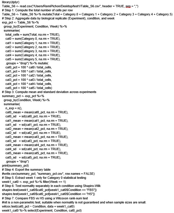
For clarity, in Author response image 1 we provide the R script used to perform the statistical tests on the data shown in Supplementary Table S6 (column Raw count of parasite developmental category per image and experiment)
Author response image 1.
(2) Line 187/Figure 4: Though it is not clearly stated, it appears that the authors treat their EdU counts as an ordinal data set of 61 steps (from 0 to >60) rather than a continuous measure of EdU+ cells per animal. In this author's opinion, the graph strongly suggests a continuous data set, and the fact that this reviewer had to dig through poorly-labeled raw data to discover the nature of the data is problematic. The authors should either switch to a continuous data set or make it explicit that the data shown are ordinal. If counting EdU+ cells is too arduous, the authors could consider comparing the amount of EdU+ area to the amount of DAPI+ area in maximum intensity projections of their confocal images, as this would roughly approximate the amount of proliferative cells in the animals.
As the reviewer correctly pointed out, the data were treated as ordinal because counting worms with more than 60 Edu+ cells became extremely difficult and highly inaccurate. Therefore, we decided to group in a single category, “60 EdU+ cells”, all worms showing more than 60 EdU+ cells. We have now updated Figure 4 where medians are shown instead of media values, Supplementary Table S5 to provide more comprehensive access to the raw counts, and Supplementary Table S6 to indicate the data for EdU+ cells per worm were considered ordinal. Accordingly, we have revised the corresponding sections as follows:
Results, lines 211 ff.:
“HS-cultured schistosomula showed higher numbers of proliferating stem cells, with a median of >48 and >60 EdU+ cells per worm at days 8 and 15, respectively (Figure 4). On the other hand, most FBS-cultured parasites displayed no more than an average of 20 EdU+ cells per worm (Figure 4).”
Methods, lines 520 ff.:
“EdU+ cells per parasite were counted for an average of 100 parasites across three independent experiments (Supplementary Table S5). Worms were grouped based on the number of cells per individual, but all those showing ⪰ 60 EdU+ cells were counted in the same group named ‘60 EdU+ cells'. Therefore, the data were considered ordinal data. Statistical analysis was performed by Kruskal-Wallis test with Dunn multiple comparison post-hoc test, with P≤0.05 considered significant (Supplementary Table S6).”
Figure 4 legend, lines 830 ff.:
“A. Violin plots showing the number of Edu+ cells per worm at indicated time points (2, 8, and 15 days post cercarial transformation) in parasites cultured either in Foetal Bovine Serum (FBS, blue) or Human Serum (HS, light brown). Human Red Blood Cells (hRBCs) were added in the culture at day 13 post cercarial transformation. The small black dots indicate individual worms, and the big black point indicates the median of EdU+ cells per worm. All worms showing ⪰ 60 EdU+ cells were counted and clustered together in the group named ‘60 EdU+ cells’. Hence, the data were treated as ordinal and statistical analysis performed by Kruskal-Wallis test with Dunn multiple comparison post-hoc test, with P≤0.05 (*) considered significant (Supplementary Tables S5 and S6).”
We thank the reviewer for the very interesting suggestion to quantify cell proliferation by calculating the ratio between EdU+ area to DAPI+ area in maximum intensity projections images. Measuring the fluorescence area for each worm in maximum projection is an excellent idea; however, due to the number of EdU+ cells present in some samples, we think this technique would not provide additional information or produce more detailed data compared with our analysis when the number of Edu+ cells exceeds 60 per worm. We will certainly consider this approximation for future studies.
There are some minor issues as well:
(1) Line 122: It is perhaps incorrect to refer to humans as "the" definitive host of schistosomes, as S. japonicum is primarily considered a zoonotic infection with water buffalo/cows being the primary definitive host.
We thank the reviewer for pointing this out; we have now replaced “schistosomes” with “Schistosoma mansoni” (current line 131)
(2) Line 185/298: The authors refer to EdU pulse-chase experiments, but the experiments described here are EdU pulse experiments.
This is a very good point, we thank the reviewer for bringing this up and have accordingly edited by replacing “EdU pulse-chase” with “EdU pulse” experiments in lines 37, 204, and 321.
Reviewer #3 (Public review):
Summary:
This study is significant as it established a protocol for the long-term culture of Schistosoma mansoni newly transformed cercariae, which developed in vitro into sexually dimorphic forms. The impact of two different sera, Fetal Bovine Serum (FBS) and Human Serum (HS), added to the culture medium supplemented with human red blood cells was evaluated. The authors demonstrated that HS-cultured parasites were able to digest red blood cells, a critical step for long-term parasite development. Furthermore, while most FBS-cultured parasites did not progress beyond an early liver stage, sexual dimorphism was clearly evident in the HS-cultured worms, albeit delayed compared to in vivo development.
Strengths:
This study could contribute to further in vitro studies for a better understanding of the unique sexual biology of Schistosoma mansoni and for screening novel schistosomicidal compounds. By increasing parasite development in in vitro studies, this protocol could have a positive impact on the principles of the 3Rs (Replacement, Reduction and Refinement) for animal research.
We thank the reviewer for highlighting the manuscript’s strengths.
Weaknesses:
As the authors mentioned, "pairing between male and female parasites was rare. Pairing was observed in approximately ~7% of the experiments, usually after day ~ 80 in culture. Egg production was also not achieved with this protocol.
Following the reviewer’s point and to clarify a misleading point, we have now decided to remove the value of 7% — which corresponds to the percentage of experiments in which couples were observed. However, this value does not accurately reflect the actual number of observed worm pairs, and it is probably misleading. We have updated the text as follows:
Results, lines 230 ff.:
“While the establishment of sexual dimorphism was robust and reproducible across more than 15 independent experiments, pairing between male and female parasites was rare. Pairing was observed only in experiments lasting more than 80 days in which we were only able to observe a few couples. In addition, these pairings were temporary (Figures 6A, B; Supplementary Video S4).”
desarrollada en la Sección 4.9
que se presentará (poner enlace a la sección 4.9)
es (Hillier y Lieberman 2015).
quitar la cita
un conjunto definido por intersección de semiespacios
a la región factible
remuestreo
Más detalles de esta distribución, consultar
r al
más detalles de esta técnica, consultar
bootstrap
Esta afirmación puede consultarse en
muestrales
para más detalles de este resultado, consultar
coeficiente de mezcla fuerte
negritas y :=
[@].
revisar
Sección 2.1
enlace de la referencia cruzada
[[Dave Winer p]] sees what I sensed too. Says the company name UserLand was chosen for the same reasons, and now we get to go another round.
不隐藏底层。pi-ai 层提供了统一的 provider 抽象,但如果你需要直接访问底层 provider 的原始 API(比如 Anthropic 的 prompt caching),可以直接导入 @mariozechner/pi-ai/anthropic 使用 provider 特定的功能。抽象是可穿透的。
是否意味着,agent 系统不应该过度抽象,或者不要适配更多的 provider ?
"可穿透的抽象"是 agent 系统的关键设计模式:LLM provider 之间的差异是真实存在的(prompt caching、extended thinking、safety settings 等),完全抹平差异意味着丧失各家最强能力。pi 的双通道设计(stream vs streamSimple)是"统一接口 + 逃生舱"模式的典范。
new economy" and the "old economy"
(1) Traditional firms go digital Banks → online banking Retailers → e-commerce
(2) Dot-coms build real-world capabilities Logistics Warehousing Customer service
compared to dot-coms adopting traditional methods
Dot-coms = digital natives
art. 3:210 lid 2 BW
%%
In 2011, a group on 4chan started spreading a plan for making a “Forever Along Involuntary Flashmob.” You can see their instructions below:
In 2011, a group on 4chan started spreading a plan for making a “Forever Along Involuntary Flashmob.” You can see their instructions below:
image in the form of a sort of flier with meme faces of foreveralone and trolls. The text reads: How to make your very own Forever Alone Involuntary Flashmob. 1. create fake online dating profile as mildly cute woman from NYC - just use somechicks facebook to get several believable pics etc. etc. 2. find forever alone guys from NYC on dating site, get them to believe you're interested. 3. Once forever alone guy takes the bait, suggest you meet for a date at this time and location: Pay phones outside 47th Digital store 46th st * Broadway NEW YORK 7:30PM Friday 13th May 2011. 4. watch angry alone guys flashmob rage at earthcam.com/usa/newyork/timessquare/ (select Camera 2). Also Remember: This will only work if we keep spreading these instructions and actually get involved. There is no limit to how many fake profiles and people you can trick or method used. Take time and prepare - think smart, if they suspect you're pushing the time and date too hard it aint gonna work.
What are the potential benefits of this example (e.g., it’s funny, in-group identifying)? And who would get the benefits?
This activity made me think that trolling is not always judged the same way by everyone. One example may look funny to the people inside the group, but the same example may feel rude or hurtful to someone outside the group. I think this matters because the benefits of trolling usually do not go to everyone equally. The people who understand the joke or share the same values may enjoy it, but the target may only feel embarrassed or attacked. This also shows that context is very important when we evaluate online behavior. A post may seem harmless at first, but its effect can change a lot depending on who is watching and who is being targeted.
In the youtube comments, some people played along and others celebrated or worried about who would get tricked.
I feel like this seems to be very common across all social media. People will spread misinformation just for the fun of it. They find it funny when someone falls for their troll. I feel like I've used social media enough for me to see when somebody is trolling. However for somebody where they aren't as experienced or maybe english isn't their first language, it can be hard to tell real comments from troll comments.
Trolling is when an Internet user posts inauthentically (often false, upsetting, or strange) with the goal of causing disruption or provoking an emotional reaction. When the goal is provoking an emotional reaction, it is often for a negative emotion, such as anger or emotional pain. When the goal is disruption, it might be attempting to derail a conversation (e.g., concern trolling [g4]), or make a space no longer useful for its original purpose (e.g., joke product reviews), or try to get people to take absurd fake stories seriously [g5].
This definition clearly explains trolling by focusing on its purpose rather than just the behavior itself. I think it is helpful because it shows that trolling is not only about posting false information, but about intentionally provoking emotions or disrupting conversations. What stands out to me is how it highlights both emotional harm and the impact on online spaces, which makes the concept more complete.
If the immediate goal of the action of trolling is to cause disruption or provoke emotional reactions, what is it that makes people want to do this disruption or provoking of emotional reactions? Some reasons people engage in trolling behavior include: Amusement: Trolls often find the posts amusing, whether due to the disruption or emotional reaction. If the motivation is amusement at causing others’ pain, that is called doing it for the lulz [g6]. Gatekeeping: Some trolling is done in a community to separate out an ingroup from outgroup (sometimes called newbies or normies). The ingroup knows that a post is just trolling, but the outgroup is not aware and will engage earnestly. This is sometimes known as trolling the newbies. Feeling Smart: Going with the gatekeeping role above, trolling can make a troll or observer feel smarter than others, since they are able to see that it is trolling while others don’t realize it. Feeling Powerful: Trolling sometimes gives trolls a feeling of empowerment when they successfully cause disruption or cause pain.** Advance and argument / make a point: Trolling is sometimes done in order to advance an argument or make a point. For example, proving that supposedly reliable news sources are gullible by getting them to repeat an absurd gross story [g5]. Punish or stop: Some trolling is in service of some view of justice, where a person, group or organization is viewed as doing something “bad” or “deserving” of punishment, and trolling is a way of fighting back.
Although this could fall under the category of amusement, I think another reason for trolling is for content creation. Getting a response or 'ragebaiting' is very engaging for audiences and can grow and account quickly.
El mAs controla la cantidad total de fotones que salen del tubo.
Más fotones → más exposición del receptor → más densidad óptica → imagen más oscura.
Menos fotones → menos exposición → imagen más clara.
Leerplan
Hier moet nog een leerplan link
Tentamen 100% 37 uur
Kloppen de uren?
Studie: Opleiding Theologie Onderwijsvorm: Deeltijdonderwijs Studiejaar: 2025-2026 Vakgroep: Systematische Theologie Versie leerplan: 01-09-2019 Versie module: ST08/B/1 Leslocatie: Zwijndrecht Docent: Evert Leeflang Email: e.leeflang@evangelisch-college.nl Studiepunten: 3 EC | 84 studie-uren
Hoi Evert, Zou je dit even kunnen aanpassen. Bedankt!
FBI–King suicide letter. November 2023. Page Version ID: 1184939326. URL: https://en.wikipedia.org/w/index.php?title=FBI%E2%80%93King_suicide_letter&oldid=1184939326 (visited on 2023-12-05).
It is absolutely insane that our government had COINTELPRO operations for 2 decades, in which early forms of trolling arose. Trolling, being linked to an initial political use, was something I did not know and found very interesting.
Is It Funny or Offensive? Comedian Impersonates FBI on Twitter, Makes MLK Assassination Joke. January 2020. URL: https://isitfunnyoroffensive.com/comedian-impersonates-fbi-on-twitter-makes-mlk-assassination-joke/ (visited on 2023-12-05).
The article describes a comedian who impersonated the FBI on Twitter and posted a joke about the assassination of Martin Luther King Jr.. The post sparked debate online, with some people finding it humorous while others saw it as offensive and inappropriate.
Brayden Olson. Forever Alone Involuntary Flashmob. Vice, May 2011. URL: https://www.vice.com/en/article/wdyyny/forever-alone-involuntary-flashmob (visited on 2023-12-05).
This source describes a prank where people used fake dating profiles to trick men into coming to Times Square for dates that were not real. The article shows how trolling can move from the internet into real life and affect people in a direct way. What stood out to me is that the prank may have looked funny to the people who planned it, but the people who were targeted were put in an embarrassing and cruel situation. This source helps show that trolling is not only about jokes or memes. It can also involve humiliation, and the harm becomes much more serious when real people are singled out in public
Whitney Phillips. Internet Troll Sub-Culture's Savage Spoofing of Mainstream Media [Excerpt]. Scientific American, May 2015. URL:
This article shows how easy it is to troll mainstream media. It shows us that people troll in all sorts of ways. I think this gives us some insight into all of the weird "types" of trolls. Especially when most people assume that trolling is for fun. It also emphasizes that people should take the time to distinguish real from fake or trolls.
Is It Funny or Offensive? Comedian Impersonates FBI on Twitter, Makes MLK Assassination Joke. January 2020. URL: https://isitfunnyoroffensive.com/comedian-impersonates-fbi-on-twitter-makes-mlk-assassination-joke/ (visited on 2023-12-05).
This article from is it funny or offensive a Twitter account impersonated the FBI and made a joke about MLK's death. It shows how easy it is to troll on the internet and impersonate others, but can be dangerous because they are impersonating something so serious.
Amazon.com: Hutzler 571 Banana Slicer,193925,Yellow, 11.25": Home & Kitchen. December 2023. URL: https://www.amazon.com/Hutzler-3571-571-Banana-Slicer/dp/B0047E0EII#customerReviews (visited on 2023-12-05). [g23] Banana Slicer Reviews. April 2013. URL: https://knowyourmeme.com/memes/banana-slicer-reviews (visited on 2023-12-05). [g24] Know Your Meme. Know Your Meme: Three Wolf Moon. May 2011. URL: https://www.youtube.com/watch?v=TbNQ746eLiU (visited on 2023-12-05).
I thought that this meme was funny and pretty harmless. A lot of trolls or pranks nowadays are at the cost of the other and while they get a lot of clicks and views, it's sometimes immoral and rude.
FBI–King suicide letter. November 2023. Page Version ID: 1184939326. URL: https://en.wikipedia.org/w/index.php?title=FBI%E2%80%93King_suicide_letter&oldid=1184939326 (visited on 2023-12-05).
From what I understand now, this source talks about how the FBI watched Martin Luther King Jr. over time. It shows that the “suicide letter” was part of a bigger plan to try to hurt his leadership. It was not just one random action, but part of a longer effort. This makes it clear that the FBI’s actions were more organized and intentional.
One source from the bibliography that I found interesting is the Scientific American article about trolling culture . The article explains how trolling is not just random bad behavior, but actually part of a larger online subculture that can influence mainstream media. For example, trolls sometimes create fake or exaggerated content just to see if news outlets will pick it up, which shows how easily misinformation can spread . What surprised me is that trolling is not always meaningless—it can actually shape real-world conversations. This made me realize that trolling is more powerful than I thought. It connects to what we learned in class about attention and engagement, because trolls are able to manipulate both people and media systems. It also made me question how reliable online information really is, since even professional media can sometimes fall for these tactics. Overall, this source made me think that trolling is not just about individuals being annoying, but about how the internet system allows misleading content to spread and gain attention.
One source I found interesting is the Scientific American article about trolling culture. It explains that trolling is not just random behavior, but part of a subculture that can trick mainstream media. Trolls sometimes create fake stories to see if news outlets will believe and share them. This surprised me because it shows trolling can have real impact, not just online but in real life. It made me question how reliable online information is and how easily people can be misled.
Assassination of Martin Luther King Jr. November 2023. Page Version ID: 1186577416. URL: https://en.wikipedia.org/w/index.php?title=Assassination_of_Martin_Luther_King_Jr.&oldid=1186577416#Alleged_government_involvement (visited on 2023-12-05).
The Wikipedia article on the Martin Luther King Jr. Assassination lists numerous decades-long conspiracy theories involving the U.S. government that continue to be alleged by individuals even though the U.S. government has officially denied its involvement. This relates directly to Chapter Sixteen’s discussion of Trust Heuristics: When institutional trust collapses for many individuals (for example, COINTELPRO, Vietnam War and the assassination of Martin Luther King Jr.), people do not simply abandon their use of these heuristics. They switch them. Once upon a time, the government was an in-group signal. Now, it is an out-group marker. Rather than what appears to be "conspiracy thinking," this behavior can actually represent the same type of recognition patterns that are discussed throughout Chapter Sixteen, but in a context where official sources have proven themselves to be untrustworthy. Chapter Sixteen views failures in heuristics as bugs. History shows us some failures may be adaptive responses.
Trolling / Troll. April 2009. URL: https://knowyourmeme.com/memes/trolling-troll (visited on 2023-12-05).
I didn't realize that trolling was a large part of the internets and social medias consumption. It occurs alot in other countries and this article talks about how its referred to as "fishing" in Japan, essentially just deliberately making a user want to engage with a post because it invokes emotion.
These trolling communities eventually started compiling half-joking sets of “Rules of the Internet” [g19] that both outlined their trolling philosophy:
It is very interesting how this principle has been completely degraded when it comes to trolling on the internet. Every passing day, it appears to be the case that no rule or guidelines exist, and people will say and do horrible things in the name of trolling. This may be caused by the sheer vastness of the internet, which has dispersed and splintered groups like those who formed rules.
“Never believe that anti-Semites are completely unaware of the absurdity of their replies. They know that their remarks are frivolous, open to challenge. But they are amusing themselves, for it is their adversary who is obliged to use words responsibly, since he believes in words. The anti-Semites have the right to play. They even like to play with discourse for, by giving ridiculous reasons, they discredit the seriousness of their interlocutors. They delight in acting in bad faith, since they seek not to persuade by sound argument but to intimidate and disconcert. If you press them too closely, they will abruptly fall silent, loftily indicating by some phrase that the time for argument is past.”
This quote, from my perspective, stood out because of its emphasis on 'belief in words.' The delight in discrediting serious argument is precisely the motivator behind many trolls actions, perhaps because they themselves feel incapable of or insecure in articulating or contributing their own legitimate opinions.
Good write up. A few comments:
Currently, KNRH does not host the largest Uganda's largest tertiary infectious diseases unit; Mulago National Referral does. You can be more specific by stating that during that time (2017-2022) KNRH hosted the countries largest tertiary ID unit. During that period of time, MNRH was under renovation and closed.
Uganda's clinicians experienced in HIV and CM management are known and these are not mentioned as either your co-authors or in the acknowledgments. It is uncertain whether indeed they reviewed your data abstraction tool.
KNRH is a clinical trial site for patients diagnosed with HIV and CM with a robust team of clinicians offering a highly specialised treatment to study participants diagnosed with HIV associated CM. As a result, the hospital rarely suffers from drug stock outs, diagnostic challenges or disease/treatment management challenges. Compared to what you would find if this study is carried another hospital in say rurral Uganda, It is unlikely that your findings represent real world setting.
Between 2017 and 2022, there were about 3 clinical trials that were carried out at KNRH with each trial investigating a different antifungal agent/treatment regimen. Your findings would be more accurate if they are stratified as per patient arm. Alternatively you can list as a limitation to your observations. Also, can you look at mortality by different years, as the treatment regimens kept on changing, mortality changed as well. You may want to consider those who received standard of care and exclude those who received experimental treatment.
It appears your cut off for long hospital stay is more than 7 days. Clarify this in your methods. I suggest you consider moving it to more than 14 days because as part standard procedure, the participants were kept in hospital deliberately for a minimum of 14 days except in scenarios were they asked to be discharged before the end of the second week. As you may recall between 2017 and 2022 Uganda was using IV deoxycholate for induction phase treatment of CM which was administered for 2 weeks.
Finally, there is a specific group of individuals who invested both resources and time to collect this data. The least you can do is mention them in the acknowledgements.
eLife Assessment
In this work, the authors demonstrated that blue light mediated mitochondrial contacts attenuated blue light induced mitochondrial dysfunction, and validated this in human cells and C. elegans. This valuable work has the potential to provide novel perspectives into the field of mitochondrial biology but the supporting data are incomplete.
Reviewer #1 (Public review):
Summary:
Blue light exposure has been shown to induce mitochondrial dysfunction, including reduced mitochondrial membrane potential (MMP). In the present study, the authors present a protein-based optogenetic system capable of inducing mito-contacts upon blue LED illumination, and show that this technical platform attenuated blue-light-induced mitochondrial dysfunction and cytotoxicity via restoring mitochondrial membrane potential.
Strengths:
The overall study design is well organized, and the data appear to support the conclusions. Additionally, demonstrating effects in human retinal cells and C. elegans enhances the perceived robustness and translational potential of the findings.
Weaknesses:
(1) Quantification of MMP at contact sites: The use of Rhodamine 123 (Rh123) for MMP measurement can be problematic, as it is not ratiometric; its signals depend on loading conditions, cell size, mitochondrial mass, and focal thickness, rather than solely on ΔΨm. If mitochondrial content changes (e.g., via biogenesis or mitophagy), Rh123 readings can be misleading. This is particularly relevant here, as the mito-contact-induced MMP changes appear to be localized events. The authors should include controls for at least one experiment using FCCP/CCCP (to collapse ΔΨm) and oligomycin (to induce hyperpolarization in many cell types) to confirm the dynamic range of the assay. Where possible, Rh123 fluorescence intensity should be normalized to mitochondrial mass (e.g., using a mass marker or mitochondrial protein). Moreover, MMP changes should be validated using an alternative indicator, such as JC-1 or a genetically encoded probe, as this is foundational to the study.
(2) Mechanisms of mito-contact-induced MMP hyperpolarization: Building on the above, what is the mechanism by which mito-contacts induce MMP hyperpolarization? Does this involve fusion of the outer or inner mitochondrial membranes? MMP hyperpolarization typically reflects an increase in protons in the intermembrane space relative to the matrix. Where do these protons originate? The kinetics of mito-contact-induced MMP changes should also be investigated in more detail.
(3) Building on the above, what is the ratio of contact area to the overall mitochondrial surface area? If MMP increases only at relatively small contact sites, how does this translate to an overall increase in MMP and energy production?
(4) Blue light causes mitochondrial damage via increased reactive oxygen species (ROS), and MMP hyperpolarization can itself lead to excessive oxidative stress. The authors should measure ROS levels and discuss their potential impact on the observed effects.
(5) Although the main focus is on blue LED-mediated injury, the protective effects of the optogenetic system against other stressors (e.g., ischemia-reperfusion, H₂O₂, or FCCP exposure) should be examined. This would help exclude confounds related to blue light, which is central to both the manipulation and the damage model in the current study, and increase the overall impact of the findings.
Reviewer #2 (Public review):
Summary:
This paper describes a novel tool (CRYO2PHR-MiroTM), which aims to create contact sites between mitochondria. One elegant aspect of the technique is that it is controlled by the exposure of cells to blue-light and reversible when cells are put back in the dark. Through an unknown and unexplored mechanism, the mitochondrial membrane potential is raised at the mitochondrial contact sites. The oligomerization of CRYOPHR-MiroTM is protective against the toxic effect of prolonged blue light exposure in cells and nematodes.
Strengths:
This work might open novel perspectives in the fundamental study of mitochondria.
(1) CRYO2PHR-MiroTM represents an interesting tool to manipulate mitochondria interaction/proximity/distribution without playing with the classical components of the mitochondrial fusion and fission machinery.
(2) This work suggests that, without the need for fusion, the relative proximity of mitochondria might influence their activity, opening novel fields of investigation in mitochondrial biology.
(3) Finally, targeting CRYO2PHR not only to mitochondria but also to their partner organelles (ER, LD, peroxisomes...) could provide a tool to reversibly manipulate the interaction of mitochondria with the rest of the organelle community.
Weaknesses:
As detailed below, the claims made by the author that CRYOPHR induce mitochondrial contact sites are not fully convincing at this stage. The method used to define and analyse contact sites is not clear enough, and the image presented in the present manuscript does not convincingly illustrate contact sites between mitochondria. Finally, the evidence that CRYOPHR does not trigger mitochondrial fusion should be strengthened.
Comments on the results:
(1) The quantification of mitochondrial contacts is a crucial point of this study. At this stage, the data are not sufficient to demonstrate that CRYOPHR-MiroTM oligomerisation tethers mitochondria. CRYOPHR-MiroTM can oligomerise in Trans, leading to mitochondrial tethering, but it can also oligomerise in Cis. In that later case, one could hypothesise that the massive aggregation of CRYOPHR-MiroTM at the mitochondrial outer membrane could locally push lipids away and/or create membrane curvature. The image and quantification provided by the author make it difficult to decide whether CRYOPHR-MiroTM tethers mitochondria or pinches their membranes. Below are detailed comments on these aspects:
a) It is claimed that "the proportion of mitochondria having one or more mito-contacts increased by nearly 50% following optogenetic stimulation". However, it is unclear how the authors have calculated this parameter. In the methods for contact ratio calculation, it is written that "the contacted area of CRY2PHR puncta was calculated", but I do not understand what it means and how it relates to contact ratio calculation. Then the authors have written, "Based on the area or distance (between mitochondria), the mitochondria were classified as either non-contact or contact". It is not clear to which parameter the term " area " refers: the area of mito-contacts based on MitoTracker or the area of CRY2PHR puncta. It is not clear how the authors integrate the two parameters "area" and "distance" to decide whether two mitochondria are in contact or not.
b) The method states that "Contact ratio refers to the number of contact mitochondria by the total number of mitochondria". What does "number of contact mitochondria" mean? The number of contacts between mitochondria? The number of mitochondria in contact? What is the distance range between two mitochondria, taking into account optic resolution, for which the authors consider that two mitochondria are "in contact"?
c) The quantification of the contact ratio made on the TEM picture should be explained.
d) The following data should be added, as contact site formation is a critical point. On cells treated or not with blue light, the author should measure systematically what is the distance of a given mitochondrion to the nearest one. The distribution of these distance values should be shown and analysed to determine whether or not there are more mitochondria at short distances upon blue light induction of CRYOPHR oligomerization. In addition, the author should determine the number of CRYO2PHR puncta that are simply lying on a mitochondrion and the number of CRYO2PHR puncta that are bridging two clear, distinct mitochondria.
e) Based on the images provided in Figure 1, there is no convincing evidence of mitochondrial contacts. In image 1g, the CRYO2PHR puncta seem to be lying on mitochondrial tubules. Sometimes, it looks that CRYO2PHR puncta decorate mitochondrial constriction sites, suggesting that the CRYOPHR might pinch membranes. The authors claim that they "found various types of mitochondrial contacts (Figure 1f, 1g), such as head-to-head, side-by-side, and head-to-side", but it is not clearly visible on the images. One problem is that the authors show the merge of MTDR and CRYOPHR-mCherry staining, in which the mitochondria contact are hidden by very bright CRYOPHR-mCherry aggregates. The authors should provide high magnification images (like in 1g) showing not only the merge of mitochondria and CRYOPHR-mCherry but also the staining of mitochondria by themselves. The authors should mark "head-to-head, side-by-side, and head-to-side contacts" with arrows.
f) Continuing on Figure 1f and 1g, it does not sound optimal to use CRYOPHR-mcCherry in combination with MTDR (MitoTracker Deep Red) to precisely delimitate subtle membrane contact sites between mitochondria because the emission and excitation spectra of these two fluorochromes partially overlap. One better alternative could be to use MTG (MitoTracker Green) as for Figure 1a. However, here we come to the point that MitoTraker stains the mitochondrial matrix that is delimited by the mitochondrial inner membrane, which can be discontinuous in a given mitochondrion. To formally visualise mitochondrial contact sites and demonstrate that CRYOPHR tethers mitochondria, the author should rather mark the mitochondrial outer membrane (with TOM20::GFP and anti-TOM20, for instance).
g) Figure S2 presents snapshots of a movie clearly showing the rapid aggregation of CRYOPHR into distinct puncta upon blue light exposure. The author should perform the same experiment on cells in which mitochondria would be stained with a fluorophore, allowing live imaging (MTG or TOM20::GP, for instance). This would allow for tracking of mitochondria and CRYOPHR puncta at the same time. Hence, high magnification views should allow for capturing events where CRYOPHR puncta formation coincides with mitochondrial tethering if the authors' claims are correct, or with, for instance, membrane pinching if they are wrong.
h) If CRYOPHR-TMMiro bring mitochondrial membrane closer, it would be surprising that it does not increase the probability of Mitofusin-dependent fusion events. The author should conduct analysis of the mitochondrial network in cells exposed to the conditions shown in Figure 1. Rather than relying only on the aspect ratio (as shown in Figure 2 in cells stressed by prolonged blue light exposure), the author should also analyse the mitochondrial total branch length (sum of the length of all branches from a mitochondrion) and the number of branches on each mitochondrion.
i) Ideally, the author should not only rely on the analysis of mitochondrial architecture, which only partially informs on mitochondrial fusion rate. Fragmented mitochondria can indeed fuse efficiently via kiss-and-run events, for instance. To formally demonstrate that there are no permanent nor transcient fusion at the mitochondrial contact sites induced by CRYOPHR, the most powerful method would be to analyse diffusion of matrix fluorescent dyes. This can be conducted using photoconvertible probes (mt-dendra2) (Pham et al., 2012) or a PEG-induced cell fusion assay (Detmer et al., 2007).
(2) Regarding the quantification of local MMP at mitochondrial contact, it would be important to better explain how the authors have set up their microscope to avoid technical issues that could lead to fluorescent artifacts at CRYOPHR puncta. Because the emission of Rhodamine 123 overlaps the excitation of mCherry, it should be explained in the methods how the detection of Rhodamine 123 has been filtered to avoid the detection of the red light coming from the mCherry light coming from CRYOPHR puncta. This is critical as fluorescent protein aggregates can be very bright.
Comments on the introduction and discussion
(1) In the results section, the authors state that they were "Inspired by previous studies indicating that nanoscale proximity of a charged membrane or protein 119 condensate to a membrane amplifies the local membrane potential". It could be useful to the readers to have a bit of background regarding these observations (references 55 and 56) to better understand what supports the rationale of the authors' strategy. Then, the discussion part should address in more detail the possible mechanisms that could explain why bringing the mitochondrial membranes without fusing them influences mitochondrial membrane potential.
(2) I would suggest finding a simple name for the CRYOPHR-MiroTM tool that could evoke more clearly that it is an optogenetic tool designed to tether mitochondria with blue light.
What do you think is the best way to deal with trolling?
The best way to deal with trolling is usually not to engage. Trolls are often looking for attention or a reaction, so ignoring them removes the reward they’re seeking. If the behavior is harmful, it’s better to use platform tools like blocking or reporting rather than arguing back. Overall, setting boundaries and protecting your own time and mental energy is more effective than trying to “win” against a troll.
What do you think is the best way to deal with trolling?
I think that ignoring is often the only strategy available to me as a user (and therefore the 'best way'), whereas I believe that it is the responsibility of the relevant social media platform to itself moderate such trolling, especially by enabling self-reporting features.
One of the traditional pieces of advice for dealing with trolls is “Don’t feed the trolls,” which means that if you don’t respond to trolls, they will get bored and stop trolling. We can see this advice as well in the trolling community’s own “Rules of the Internet” [g31]: Do not argue with trolls - it means that they win
I think that this part is true because the point of trolling is to get a reaction out of people, so if people were to ignore it than the troller wouldn't be getting the reaction that they want. I also think that if people just stopped responding to trolls maybe there would be less of them.
Ask anyone who has dealt with persistent harassment online, especially women: [trolls stopping because they are ignored] is not usually what happens. Instead, the harasser keeps pushing and pushing to get the reaction they want with even more tenacity and intensity. It’s the same pattern on display in the litany of abusers and stalkers, both online and off, who escalate to more dangerous and threatening behavior when they feel like they are being ignored.
This part stood out to me because it shows that ignoring harassment does not always make it go away. In fact, it can make things worse. That makes the usual advice feel unrealistic, especially for people who deal with constant harassment. It also made me think about how serious online behavior can become since it can follow the same patterns as real-life abuse. Because of that, I think stronger action from platforms is more important than just telling people to ignore it.
One idea that stood out to me is that trolls mainly want attention, not real discussion. I’ve seen this happen a lot online—posts that are clearly meant to provoke people get the most replies. It feels like social media almost rewards trolling because more engagement means more visibility. I also thought about the advice to ignore trolls. It makes sense, but it’s hard to do. Sometimes I feel like I should respond, especially when someone spreads wrong information. So I’m not sure if ignoring is always the best solution. The idea of using bots to reply to trolls is interesting, but also a bit weird. It makes me wonder if using automation to fix online problems might actually create new ones.
What do you think is the best way to deal with trolling?
I think that the best way to deal with trolling is to ignore it and not engage with the bot or person trolling. I feel like the more you engage, the more your algorithm feeds you things that are considered trolling, and it all just becomes a feedback loop that annoys a user. I personally do not like engaging in trolling.
jährlich von AS erneuert
Dies entspricht nicht unserer Praxis. Sollten wir rausnehmen.
eLife Assessment
This study provides potentially important insights by establishing a human disease model and exploring therapeutic approaches. The evidence is generally convincing for descriptive and comparative findings. The authors present solid data, but evidence for proposed biological mechanisms and functional outcomes remains limited.
Reviewer #1 (Public review):
In this study, the authors set out to develop a human disease model using stem cell-derived systems and to use this platform to investigate disease biology and evaluate potential therapeutic approaches. Their goal is to provide a tractable experimental system that captures key features of the disease and enables testing of candidate interventions.
The work has several important strengths. The authors present a carefully constructed model with improved genetic replication and clearer reporting of biological replicates, which enhances confidence in the reproducibility of the findings. The longitudinal design, spanning early developmental stages to later disease-relevant phenotypes, provides a useful framework for distinguishing temporal aspects of the disease process. The study also includes a comparative evaluation of multiple therapeutic strategies adding practical value to the field. In addition, statistical reporting and transparency have been strengthened, and key limitations of the model-such as the absence of certain cell types-are now clearly acknowledged.
At the same time, notable weaknesses temper the strength of the conclusions. Several central biological claims, particularly those related to specific signaling pathways, are supported primarily by transcriptomic and protein-level observations without direct functional validation. Similarly, measures used to interpret cellular processes do not fully distinguish between alternative biological explanations, leaving some mechanistic interpretations unresolved. The therapeutic findings are supported by biochemical changes, but evidence for functional recovery at the cellular level is limited. These gaps mean that some of the broader conclusions should be interpreted with caution.
Overall, the authors have largely achieved their aim of establishing a useful experimental model and demonstrating its potential for studying disease-related changes and testing interventions. The evidence is convincing for the descriptive and comparative aspects of the work, but more limited for mechanistic and functional claims.
The study is likely to have a meaningful impact by providing a platform that others in the field can build upon. The methods and datasets will be useful to researchers interested in disease modeling and therapeutic development. At the same time, the work is best viewed as an important foundation, with key mechanistic and functional questions remaining to be addressed in future studies.
Reviewer #2 (Public review):
Sun et al. have developed a midbrain-like organoid (MLO) model for neuronopathic Gaucher disease (nGD). The MLOs recapitulate several features of nGD molecular pathology, including reduced GCase activity, sphingolipid accumulation, and impaired dopaminergic neuron development. They also characterize the transcriptome in the MLO nGD model. CRISPR correction of one of the GBA1 mutant alleles rescues most of the nGD molecular phenotypes. The MLO model was further deployed in proof-of-principle studies of investigational nGD therapies, including SapC-DOPS nanovesicles, AAV9-mediated GBA1 gene delivery, and substrate-reduction therapy (GZ452). This patient-specific 3D model provides a new platform for studying nGD mechanisms and accelerating therapy development. Overall, only modest weaknesses are noted, and these have been adequately addressed in the revision.
Comments on revisions:
I have no further recommendations. The revised manuscript addresses the few questions and concerns that I had initially shared.
Reviewer #3 (Public review):
Summary:
In this study, the authors describe modeling of neuronopathic Gaucher disease (nGD) using midbrain-like organoids (MLOs) derived from hiPSCs carrying GBA1 L444P/P415R or L444P/RecNciI variants. These MLOs recapitulate several disease features, including GCase deficiency, reduced enzymatic activity, lipid substrate accumulation, and impaired dopaminergic neuron differentiation. Correction of the GBA1 L444P variant restored GCase activity, normalized lipid metabolism, and rescued dopaminergic neuronal defects, confirming its pathogenic role in the MLO model. The authors further leveraged this system to evaluate therapeutic strategies, including: (i) SapC-DOPS nanovesicles for GCase delivery, (ii) AAV9-mediated GBA1 gene therapy, and (iii) GZ452, a glucosylceramide synthase inhibitor. These treatments reduced lipid accumulation and ameliorated autophagic, lysosomal, and neurodevelopmental abnormalities.
Strengths:
This manuscript demonstrates that nGD patient-derived MLOs can serve as an additional platform for investigating nGD mechanisms and advancing therapeutic development.
Comments on revisions:
I have no further concerns regarding this manuscript.
Author response:
The following is the authors’ response to the original reviews
Public Reviews:
Reviewer #1 (Public review):
Summary:
This manuscript by Lin et al. presents a timely, technically strong study that builds patient-specific midbrain-like organoids (MLOs) from hiPSCs carrying clinically relevant GBA1 mutations (L444P/P415R and L444P/RecNcil). The authors comprehensively characterize nGD phenotypes (GCase deficiency, GluCer/GluSph accumulation, altered transcriptome, impaired dopaminergic differentiation), perform CRISPR correction to produce an isogenic line, and test three therapeutic modalities (SapC-DOPS-fGCase nanoparticles, AAV9GBA1, and SRT with GZ452). The model and multi-arm therapeutic evaluation are important advances with clear translational value.
My overall recommendation is that the work undergo a major revision to address the experimental and interpretive gaps listed below.
Strengths:
(1) Human, patient-specific midbrain model: Use of clinically relevant compound heterozygous GBA1 alleles (L444P/P415R and L444P/RecNcil) makes the model highly relevant to human nGD and captures patient genetic context that mouse models often miss.
(2) Robust multi-level phenotyping: Biochemical (GCase activity), lipidomic (GluCer/GluSph by UHPLC-MS/MS), molecular (bulk RNA-seq), and histological (TH/FOXA2, LAMP1, LC3) characterization are thorough and complementary.
(3) Use of isogenic CRISPR correction: Generating an isogenic line (WT/P415R) and demonstrating partial rescue strengthens causal inference that the GBA1 mutation drives many observed phenotypes.
(4) Parallel therapeutic testing in the same human platform: Comparing enzyme delivery (SapC-DOPS-fGCase), gene therapy (AAV9-GBA1), and substrate reduction (GZ452) within the same MLO system is an elegant demonstration of the platform's utility for preclinical evaluation.
(5) Good methodological transparency: Detailed protocols for MLO generation, editing, lipidomics, and assays allow reproducibility
Weaknesses:
(1) Limited genetic and biological replication
(a) Single primary disease line for core mechanistic claims. Most mechanistic data derive from GD2-1260 (L444P/P415R); GD2-10-257 (L444P/RecNcil) appears mainly in therapeutic experiments. Relying primarily on one patient line risks conflating patient-specific variation with general nGD mechanisms.
We thank the reviewer for highlighting the importance of genetic and biological replication. An additional patient-derived iPSC line was included in the manuscript, therefore, our study includes two independent nGD patient-derived iPSC lines, GD2-1260 (GBA1<sup>L444P/P415R</sup>) and GD2-10-257 (GBA1<sup>L444P/RecNcil</sup>), both of which carry the severe mutations associated with nGD. These two lines represent distinct genetic backgrounds and were used to demonstrate the consistency of key disease phenotypes (reduced GCase activity, elevated substrate, impaired dopaminergic neuron differentiation etc.) across different patient’s MLOs. Major experiments (e.g., GCase activity assays, substrate, immunoblotting for DA marker TH, and therapeutic testing with SapC-DOPS-fGCase, AAV9-GBA1) were performed using both patient lines, with results showing consistent phenotypes and therapeutic responses (see Figs. 2-6, and Supplementary Figs. 4-5). To ensure clarity and transparency, a new Supplementary Table 2 summarizes the characterization of both, the GD2-1260 and GD2-10-257 lines.
(b) Unclear biological replicate strategy. It is not always explicit how many independent differentiations and organoid batches were used (biological replicates vs. technical fields of view).
Biological replication was ensured in our study by conducting experiments in at least 3 independent differentiations per line, and technical replicates (multiple organoids/fields per batch) were averaged accordingly. We have clarified biological replicates and differentiation in the figure legends.
(c) A significant disadvantage of employing brain organoids is the heterogeneity during induction and potential low reproducibility. In this study, it is unclear how many independent differentiation batches were evaluated and, for each test (for example, immunofluorescent stain and bulk RNA-seq), how many organoids from each group were used. Please add a statement accordingly and show replicates to verify consistency in the supplementary data.
In the revision, we have clarified biological replicates and differentiation in the figure legend in Fig.1E; Fig.2B,2G; Fig.3F, 3G; Fig.4B-C,E,H-J, M-N; Fig.6D; and Fig.7A-C, I.
(d) Isogenic correction is partial. The corrected line is WT/P415R (single-allele correction); residual P415R complicates the interpretation of "full" rescue and leaves open whether the remaining pathology is due to incomplete correction or clonal/epigenetic effects.
We attempted to generate an isogenic iPSC line by correcting both GBA1 mutations (L444P and P415R). However, this was not feasible because GBA1 overlaps with a highly homologous pseudogene (PGBA), which makes precise editing technically challenging. Consequently, only the L444P mutation was successfully corrected, and the resulting isogenic line retains the P415R mutation in a heterozygous state. Because Gaucher disease is an autosomal recessive disorder, individuals carrying a single GBA1 mutation (heterozygous carriers) do not develop clinical symptoms. Therefore, the partially corrected isogenic line, which retains only the P415R allele, represents a clinically relevant carrier model. Consistent with this, our results show that GCase activity was restored to approximately 50% of wild-type levels (Fig.4B-C), supporting the expected heterozygous state. These findings also make it unlikely that the remaining differences observed are due to clonal variation or epigenetic effects.
(e) The authors tested week 3, 4, 8, 15, and 28 old organoids in different settings. However, systematic markers of maturation should be analyzed, and different maturation stages should be compared, for example, comparing week 8 organoids to week 28 organoids, with immunofluorescent marker staining and bulk RNAseq.
We agree that a systematic analysis of maturation stages is essential for validating the MLO model. Our data integrated a longitudinal comparison across multiple developmental windows (Weeks 3 to 28) to characterize the transition from progenitors to mature/functional states for nGD phenotyping and evaluation of therapeutic modalities: 1) DA differentiation (Wks 3 and 8 in Fig. 3): qPCR analysis demonstrated the progression of DA-specific programs. We observed a steady increase in the mature DA neuron marker TH and ASCL1. This was accompanied by a gradual decrease in early floor plate/progenitor markers FOXA2 and PLZF, indicating a successful differentiation path from progenitors to differentiated/mature DA neurons. 2) Glycosphingolipid substrates accumulation (Wks 15 and 28 in Fig 2): To assess late-stage nGD phenotyping, we compared GluCer and GluSph at Week 15 and Week 28. This comparison highlights the progressive accumulation of substrates in nGD MLOs, reflecting the metabolic consequences of the disease at different mature stage. 3) Organoid growth dynamics (Wks 4, 8, and 15 in new Fig. 4): The new Fig. 4 tracks physical maturation through organoid size and growth rates across three key time points, providing a macro-scale verification of consistent development between WT and nGD groups. By comparing these early (Wk 3-8) and late (Wk 15-28) stages, we confirmed that our MLOs transition from a proliferative state to a post-mitotic, specialized neuronal state, satisfied the requirement for comparing distinct maturation stages.
(f) The manuscript frequently refers to Wnt signaling dysregulation as a major finding. However, experimental validation is limited to transcriptomic data. Functional tests, such as the use of Wnt agonist/inhibitor, are needed to support this claim (see below).
We agree that the suggested experiments could provide additional mechanistic insights into this study and will consider them in future work.
(g) Suggested fixes / experiments
Add at least one more independent disease hiPSC line (or show expanded analysis from GD2-10-257) for key mechanistic endpoints (lipid accumulation, transcriptomics, DA markers).
Additional line iPSC GD2-10-257 derived MLO was included in the manuscript. This was addressed above [see response to Weaknesses (1)-a].
Generate and analyze a fully corrected isogenic WT/WT clone (or a P415R-only line) if feasible; at minimum, acknowledge this limitation more explicitly and soften claims.
We attempted to generate an isogenic iPSC line by correcting both GBA1 mutations (L444P and P415R). However, this was unsuccessful because the GBA1 gene overlaps with a pseudogene (PGBA) located16kd downstream of GBA1, which shares 9698% sequence similarity with GBA1) (Ref#1, #2), which complicates precise editing. GBA1 is shorter (~5.7 kb) than PGBA (~7.6 kb). The primary exonic difference between GBA1 and PGBA is a 55-bp deletion in exon 9 of the pseudogene. As a result, the isogenic line we obtained carries only the P415R mutation, and L444P was corrected to normal sequence. We have included this limitation in the Methods as “This gene editing strategy is expected to also target the GBA1 pseudogene due to the identical target sequence, which limits the gene correction on certain mutations (e.g., P415R)”.
References:
(1) Horowitz M., Wilder S., Horowitz Z., Reiner O., Gelbart T., Beutler E. The human glucocerebrosidase gene and pseudogene: structure and evolution. Genomics (1989). 4, 87–96. doi:10.1016/0888-7543(89)90319-4
(2) Woo EG, Tayebi N, Sidransky E. Next-Generation Sequencing Analysis of GBA1: The Challenge of Detecting Complex Recombinant Alleles. Front Genet. (2021). 12:684067. doi: 10.3389/fgene.2021.684067. PMCID: PMC8255797.
Report and increase independent differentiations (N = biological replicates) and present per-differentiation summary statistics.
This was addressed above [see response to Weaknesses (1)-b, (1)-c].
(2) Mechanistic validation is insufficient
(a) RNA-seq pathways (Wnt, mTOR, lysosome) are not functionally probed. The manuscript shows pathway enrichment and some protein markers (p-4E-BP1) but lacks perturbation/rescue experiments to link these pathways causally to the DA phenotype.
(b) Autophagy analysis lacks flux assays. LC3-II and LAMP1 are informative, but without flux assays (e.g., bafilomycin A1 or chloroquine), one cannot distinguish increased autophagosome formation from decreased clearance.
(c) Dopaminergic dysfunction is superficially assessed. Dopamine in the medium and TH protein are shown, but no neuronal electrophysiology, synaptic marker co-localization, or viability measures are provided to demonstrate functional recovery after therapy.
(d) Suggested fixes / experiments - Perform targeted functional assays:
(i) Wnt reporter assays (TOP/FOP flash) and/or treat organoids with Wnt agonists/antagonists to test whether Wnt modulation rescues DA differentiation.
(ii) Test mTOR pathway causality using mTOR inhibitors (e.g., rapamycin) or 4E-BP1 perturbation and assay effects on DA markers and autophagy.
Include autophagy flux assessment (LC3 turnover with bafilomycin), and measure cathepsin activity where relevant.
Add at least one functional neuronal readout: calcium imaging, MEA recordings, or synaptic marker quantification (e.g., SYN1, PSD95) together with TH colocalization.
We thank the reviewer for these valuable suggestions. We agree that the suggested experiments could provide additional mechanistic insights into this study and will consider them in future work. Importantly, the primary conclusions of our manuscript, that GBA1 mutations in nGD MLOs resulted in nGD pathologies such as diminished enzymatic function, accumulation of lipid substrates, widespread transcriptomic changes, and impaired dopaminergic neuron differentiation, which can be corrected by several therapeutic strategies in this study, are supported by the evidence presented. The suggested experiments represent an important direction for future research using brain organoids.
(3) Therapeutic evaluation needs greater depth and standardization
(a) Short windows and limited durability data. SapC-DOPS and AAV9 experiments range from 48 hours to 3 weeks; longer follow-up is needed to assess durability and whether biochemical rescue translates into restored neuronal function.
We agree with the reviewer. Because this is a proof-of-principle study, the treatment was designed within a short time window. Long-term studies with more comprehensive outcome assessments will be conducted in future work.
(b) Dose-response and biodistribution are under-characterized. AAV injection sites/volumes are described, but transduction efficiency, vg copies per organoid, cell-type tropism quantification, and SapC-DOPS penetration/distribution are not rigorously quantified.
We appreciate the reviewer’s concerns. This study was intended to demonstrate the feasibility and initial response of MLOs to AAV therapy. A comprehensive evaluation of AAV biodistribution will be considered in future studies.
The penetration and distribution of SapC-DOPS have been extensively characterized in prior studies. In vivo biodistribution of SapC–DOPS coupled CellVue Maroon, a fluorescent cargo, was examined in mice bearing human tumor xenografts using real-time fluorescence imaging, where CellVue Maroon fluorescence in tumor remained for 48 hours (Ref. #3: Fig. 4B, mouse 1), 100 hours (Ref. #4: Fig. 5), up to 216 hours (Ref. #5: Fig. 3). Uptake kinetics were also demonstrated in cells, with flow cytometry quantification showing that fluorescent cargo coupled SapC-DOPS nanovesicles, were incorporated into human brain tumor cell membranes within minutes and remained stably incorporated into the cells for up to one hour (Ref. # 6: Fig. 1a and Fig. 1b). Building on these findings, the present study focuses on evaluating the restoration of GCase function rather than reexamining biodistribution and uptake kinetics.
References:
(3) X. Qi, Z. Chu, Y.Y. Mahller, K.F. Stringer, D.P. Witte, T.P. Cripe. Cancer-selective targeting and cytotoxicity by liposomal-coupled lysosomal saposin C protein. Clin. Cancer Res. (2009) 15, 5840-5851. PMID: 19737950.
(4) Z. Chu, S. Abu-Baker, M.B. Palascak, S.A. Ahmad, R.S. Franco, and X. Qi. Targeting and cytotoxicity of SapC-DOPS nanovesicles in pancreatic cancer. PLOS ONE (2013) 8, e75507. PMID: 24124494.
(5) Z. Chu, K. LaSance, V.M. Blanco, C-H. Kwon, B. Kaur, M. Frederick, S. Thornton, L. Lemen, and X. Qi. Multi-angle rotational optical imaging of brain tumors and arthritis using fluorescent SapC-DOPS nanovesicles. J. Vis. Exp. (2014) 87, e51187, 1-7. PMID: 24837630.
(6) J. Wojton, Z. Chu, C-H. Kwon, L.M.L. Chow, M. Palascak, R. Franco, T. Bourdeau, S. Thornton, B. Kaur, and X. Qi. Systemic delivery of SapC-DOPS has antiangiogenic and antitumor effects against glioblastoma. Mol. Ther. (2013) 21, 1517-1525. PMID: 23732993.
(c) Specificity controls are missing. For SapC-DOPS, inclusion of a non-functional enzyme control (or heat-inactivated fGCase) would rule out non-specific nanoparticle effects. For AAV, assessment of off-target expression and potential cytotoxicity is needed.
Including inactive fGCase would confound the assessment of fGCase in MLOs by immunoblot and immunofluorescence; therefore, saposin C–DOPS was used as the control instead.
We agree that assessment of off-target expression and potential cytotoxicity for AAV is important, this will be included in future studies.
(d) Comparative efficacy lacking. It remains unclear which modality is most effective in the long term and in which cellular compartments.
To address this comment, we have added a new table (Supplementary Table 2) comparing the four therapeutic modalities and summarizing their respective outcomes. While this study focused on short-term responses as a proof-of-principle, future work will explore long-term therapeutic effects.
(e) Suggested fixes/experiments
Extend follow-up (e.g., 6+ weeks) after AAV/SapC dosing and evaluate DA markers, electrophysiology, and lipid levels over time.
We appreciate the reviewer’s suggestions. The therapeutic testing in patient-derived MLOs was designed as a proof-of-principle study to demonstrate feasibility and the primary response (rescue of GCase function) to the treatment. A comprehensive, long-term therapeutic evaluation of AAV and SapC-DOPS-fGCase is indeed important for a complete assessment; however, this represents a separate therapeutic study and is beyond the scope of the current work.
Quantify AAV transduction by qPCR for vector genomes and by cell-type quantification of GFP+ cells (neurons vs astrocytes vs progenitors).
For the AAV-treated experiments, we agree that measuring AAV copy number and GFP expression would provide additional information. However, the primary goal of this study was to demonstrate the key therapeutic outcome, rescue of GCase function by AAV-delivered normal GCase, which is directly relevant to the treatment objective.
Include SapC-DOPS control nanoparticles loaded with an inert protein and/or fluorescent cargo quantitation to show distribution and uptake kinetics.
As noted above [see response to Weakness (3)-c], using inert GCase would confound the assessment of fGCase uptake in MLOs; therefore, it was not suitable for this study. See response above for the distribution and uptake kinetics of SapC-DOPS [see response to Weaknesses (3)-b].
Provide head-to-head comparative graphs (activity, lipid clearance, DA restoration, and durability) with statistical tests.
We have added a new table (Supplementary Table 2) providing a head-to-head comparison of the treatment effects.
(4) Model limitations not fully accounted for in interpretation
(a) Absence of microglia and vasculature limits recapitulation of neuroinflammatory responses and drug penetration, both of which are important in nGD. These absences could explain incomplete phenotypic rescues and must be emphasized when drawing conclusions about therapeutic translation.
We agree that the absence of microglia and vasculature in midbrain-like organoids represents a limitation, as we have discussed in the manuscript. In this revision, we highlighted this limitation in the Discussion section and clarified that it may contribute to incomplete phenotyping and phenotypic rescue observed in our therapeutic experiments. Additionally, we have outlined future directions to incorporate microglia and vascularization into the organoid system to better recapitulate the in vivo environment and improve translational relevance (see 7th paragraph in the Discussion).
(b) Developmental vs degenerative phenotype conflation. Many phenotypes appear during differentiation (patterning defects). The manuscript sometimes interprets these as degenerative mechanisms; the distinction must be clarified.
We appreciate the reviewer’s comments. In the revised manuscript, we have clarified that certain abnormalities, such as patterning defects observed during early differentiation, likely reflect developmental consequences of GBA1 mutations rather than degenerative processes. Conversely, phenotypes such as substrate accumulation, lysosomal dysfunction, and impaired dopaminergic maturation at later stages are interpreted as degenerative features. We have updated the Results and Discussion sections to avoid conflating developmental defects with neurodegenerative mechanisms.
(c) Suggested fixes
Tone down the language throughout (Abstract/Results/Discussion) to avoid overstatement that MLOs fully recapitulate nGD neuropathology.
The manuscript has been revised to avoid overstatements.
Add plans or pilot data (if available) for microglia incorporation or vascularization to indicate how future work will address these gaps.
The manuscript now includes further plans to address the incorporation of microglia and vascularization, described in the last two paragraphs in the Discussion. Pilot study of microglia incorporation will be reported when it is completed.
(5) Statistical and presentation issues
(a) Missing or unclear sample sizes (n). For organoid-level assays, report the number of organoids and the number of independent differentiations.
We have clarified biological replicates and differentiation in the figure legend [see response to Weaknesses (1)-b, (1)-c].
(b) Statistical assumptions not justified. Tests assume normality; where sample sizes are small, consider non-parametric tests and report exact p-values.
We have updated Statistical analysis in methods as described below:
For comparisons between two groups, data were analyzed using unpaired two-tailed Student’s t-tests when the sample size was ≥6 per group and normality was confirmed by the Shapiro-Wilk test. When the normality assumption was not met or when sample sizes were small (n < 6), the non-parametric Mann-Whitney U test was used instead. For comparisons involving three or more groups, one-way ANOVA followed by Tukey’s multiple comparison test was applied when data were normally distributed; otherwise, the nonparametric Dunn’s multiple comparison test was used. Exclusion of outliers was made based on cutoffs of the mean ±2 standard deviations. All statistical analyses were performed using GraphPad Prism 10 software. Exact p-values are reported throughout the manuscript and figures where feasible. A p-value < 0.05 was considered statistically significant.
(c) Quantification scope. Many image quantifications appear to be from selected fields of view, which are then averaged across organoids and differentiations.
In this work, quantitative immunofluorescence analyses (e.g., cell counts for FOXP1+, FOXG1+, SOX2+ and Ki67+ cells, as well as marker colocalization) were performed on at least 3–5 randomly selected non-overlapping fields of view (FOVs) per organoid section, with a minimum of 3 organoids per differentiation batch. Each FOV was imaged at consistent magnification (60x) and z-stack depth to ensure comparable sampling across conditions. Data from individual FOVs were first averaged within each organoid to obtain an organoid-level mean, and then biological replicates (independent differentiations, n ≥ 3) were averaged to generate the final group mean ± SEM. This multilevel averaging approach minimizes bias from regional heterogeneity within organoids and accounts for variability across differentiations. Representative confocal images shown in the figures were selected to accurately reflect the quantified data. We believe this standardized quantification strategy ensures robust and reproducible results while appropriately representing the 3D architecture of the organoids.
In the revision, we have clarified the method used for image analysis of sectioned MLOs as below:
Quantitative immunofluorescence analyses (e.g., cell counts for FOXP1+, FOXG1+, SOX2+ and Ki67+ cells, as well as marker colocalization) were performed using ImageJ (NIH) on at least 3–5 randomly selected non-overlapping fields of view (FOVs) per organoid section, with a minimum of 3 organoids per differentiation batch. Each FOV was imaged at consistent magnification (60x) and z-stack depth to ensure comparable sampling across conditions. Data from individual FOVs were first averaged within each organoid to obtain an organoid-level mean, and then biological replicates (independent differentiations, n ≥ 3) were averaged to generate the final group mean ± SEM.
(d) RNA-seq QC and deposition. Provide mapping rates, batch correction details, and ensure the GEO accession is active. Include these in Methods/Supplement.
RNA-seq data are from same batch. The mapping rate is >90%. GEO accession will be active upon publication. These were included in the Methods.
(e) Suggested fixes
Add a table summarizing biological replicates, technical replicates, and statistical tests used for each figure panel.
We have revised the figure legends to include replicates for each figure and statistical tests [see response in weaknesses (1)-b, (1)-c].
Recompute statistics where appropriate (non-parametric if N is small) and report effect sizes and confidence intervals.
Statistical analysis method is provided in the revision [see response in Weaknesses (5)-b].
(6) Minor comments and clarifications
(a) The authors should validate midbrain identity further with additional regional markers (EN1, OTX2) and show absence/low expression of forebrain markers (FOXG1) across replicates.
We validated the MLO identity by 1) FOXG1 and 2) EN1. FOXG1 was barely detectable in Wk8 75.1_MLO but highly present in ‘age-matched’ cerebral organoid (CO), suggesting our culturing method is midbrain region-oriented. In nGD MLO, FOXG1 expression is significantly higher than 75.1_MLO, indicating that there was aberrant anterior-posterior brain specification, consistent with the transcriptomic dysregulation observed in our RNA-seq data.
To further confirm midbrain identity, we examined the expression of EN1, an established midbrain-specific marker. Quantitative RT-PCR analysis demonstrated that EN1 expression increased progressively during differentiation in both WT-75.1 and nGD2-1260 MLOs at weeks 3 and 8 (Author response image 1). EN1 reached 34-fold and 373-fold higher levels than in WT-75.1 iPSCs at weeks 3 and 8, respectively, in WT-75.1 MLOs. In nGD MLOs, although EN1 expression showed a modest reduction at week 8, the levels were not significantly different from those observed in age-matched WT-75.1 MLOs (p > 0.05, ns).
Author response image 1.
qRT-PCR quantification of midbrain progenitor marker EN1 expression in WT-75.1 and GD2-1260 MLOs at Wk3 and Wk8. Data was normalized to WT-75.1 hiPSC cells and presented as mean ± SEM (n = 3-4 MLOs per group). ns, not significant.
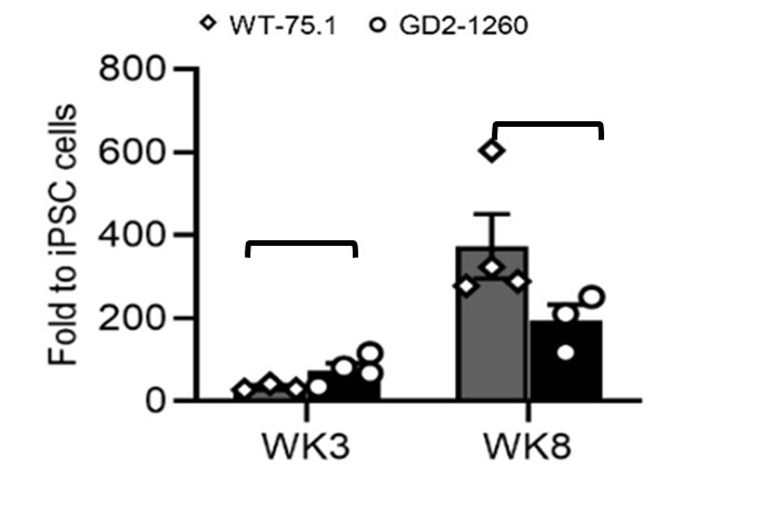
(b) Extracellular dopamine ELISA should be complemented with intracellular dopamine or TH+ neuron counts normalized per organoid or per total neurons.
We quantified TH expression at both the mRNA level (Fig. 3F) and the protein level (Fig. 3G/H) from whole-organoid lysates, which provides a more consistent and integrative measure across samples. These TH expression levels correlated well with the corresponding extracellular (medium) dopamine concentrations for each genotype. In contrast, TH<sup>+</sup> neuron counts may not reliably reflect total cellular dopamine levels because the number of cells captured on each organoid section varies substantially, making normalization difficult. Measuring intracellular dopamine is an alternative approach that will be considered in future studies.
(c) For CRISPR editing: the authors should report off-target analysis (GUIDE-seq or targeted sequencing of predicted off-targets) or at least in-silico off-target score and sequencing coverage of the edited locus. (off-target analysis (GUIDE-seq or targeted sequencing of predicted off-targets) or at least in-silico off-target score and sequencing coverage of the edited locus).
The off-target effect was analyzed during gene editing and the chance to target other off-targets is low due to low off-target scores ranked based on the MIT Specificity Score analysis. The related method was also updated as stated below:
“The chance to target other off-targets is low due to low off-target scores ranked based on the MIT Specificity Score analysis (Hsu, P., Scott, D., Weinstein, J. et al. DNA targeting specificity of RNA-guided Cas9 nucleases. Nat Biotechnol 31, 827–832 (2013). https://doi.org/10.1038/nbt.2647).”
(d) It should be clarified as to whether lipidomics normalization is to total protein per organoid or per cell, and include representative LC-MS chromatograms or method QC.
The normalization was to the protein of organoid lysate. This was clarified in the Methods section in the revision as stated below:
“The GluCer and GluSph levels in MLO were normalized to total MLO protein (mg) that were used for glycosphingolipids analyses. Protein mass was determined by BCA assay and glycosphingolipid was expressed as pmol/mg protein. Additionally, GluSph levels in the culture medium were quantified and normalized to the medium volume (pmol/mL).”
Representative LC-MS chromatograms for both normal and GD MLOs have been included in a new figure, Supplementary Figure 2.
(e) Figure legends should be improved in order to state the number of organoids, the number of differentiations, and the exact statistical tests used (including multiplecomparison corrections).
This was addressed above [see response to Weaknesses (1)-b and (5)-b].
(f) In the title, the authors state "reveal disease mechanisms", but the studies mainly exhibit functional changes. They should consider toning down the statement.
The title was revised to: Patient-Specific Midbrain Organoids with CRISPR Correction Recapitulate Neuronopathic Gaucher Disease Phenotypes and Enable Evaluation of Novel Therapies
(7) Recommendations
This reviewer recommends a major revision. The manuscript presents substantial novelty and strong potential impact but requires additional experimental validation and clearer, more conservative interpretation. Key items to address are:
(a) Strengthening genetic and biological replication (additional lines or replicate differentiations).
This was addressed above [see response to Weaknesses (1)-a, (1)-b, (1)-c].
(b) Adding functional mechanistic validation for major pathways (Wnt/mTOR/autophagy) and providing autophagy flux data.
(c) Including at least one neuronal functional readout (calcium imaging/MEA/patch) to demonstrate functional rescue.
As addressed above [see response to Weaknesses (2)], the suggested experiments in b) and c) would provide additional insights into this study and we will consider them in future work.
(d) Deepening therapeutic characterization (dose, biodistribution, durability) and including specificity controls.
This was addressed above [see response to Weaknesses (3)-a to e].
(e) Improving statistical reporting and explicitly stating biological replicate structure.
This was addressed above [see response to Weaknesses (1)-b, (5)-b].
Reviewer #2 (Public review):
Sun et al. have developed a midbrain-like organoid (MLO) model for neuronopathic Gaucher disease (nGD). The MLOs recapitulate several features of nGD molecular pathology, including reduced GCase activity, sphingolipid accumulation, and impaired dopaminergic neuron development. They also characterize the transcriptome in the MLO nGD model. CRISPR correction of one of the GBA1 mutant alleles rescues most of the nGD molecular phenotypes. The MLO model was further deployed in proof-of-principle studies of investigational nGD therapies, including SapC-DOPS nanovesicles, AAV9-mediated GBA1 gene delivery, and substrate-reduction therapy (GZ452). This patient-specific 3D model provides a new platform for studying nGD mechanisms and accelerating therapy development. Overall, only modest weaknesses are noted.
We thank the reviewer for the supportive remarks.
Reviewer #3 (Public review):
Summary:
In this study, the authors describe modeling of neuronopathic Gaucher disease (nGD) using midbrain-like organoids (MLOs) derived from hiPSCs carrying GBA1 L444P/P415R or L444P/RecNciI variants. These MLOs recapitulate several disease features, including GCase deficiency, reduced enzymatic activity, lipid substrate accumulation, and impaired dopaminergic neuron differentiation. Correction of the GBA1 L444P variant restored GCase activity, normalized lipid metabolism, and rescued dopaminergic neuronal defects, confirming its pathogenic role in the MLO model. The authors further leveraged this system to evaluate therapeutic strategies, including: (i) SapC-DOPS nanovesicles for GCase delivery, (ii) AAV9-mediated GBA1 gene therapy, and (iii) GZ452, a glucosylceramide synthase inhibitor. These treatments reduced lipid accumulation and ameliorated autophagic, lysosomal, and neurodevelopmental abnormalities.
Strengths:
This manuscript demonstrates that nGD patient-derived MLOs can serve as an additional platform for investigating nGD mechanisms and advancing therapeutic development.
Comments:
(1) It is interesting that GBA1 L444P/P415R MLOs show defects in midbrain patterning and dopaminergic neuron differentiation (Figure 3). One might wonder whether these abnormalities are specific to the combination of L444P and P415R variants or represent a general consequence of GBA1 loss. Do GBA1 L444P/RecNciI (GD2-10-257) MLOs also exhibit similar defects?
We observed reduced dopaminergic neuron marker TH expression in GBA1 L444P/RecNciI (GD2-10-257) MLOs, suggesting that this line also exhibits defects in dopaminergic neuron differentiation. These data are provided in a new Supplementary Fig. 4E, and are summarized in new Supplementary Table 2 in the revision.
(2) In Supplementary Figure 3, the authors examined GCase localization in SapC-DOPSfGCase-treated nGD MLOs. These data indicate that GCase is delivered to TH<sup>+</sup> neurons, GFAP<sup>+</sup> glia, and various other unidentified cell types. In fruit flies, the GBA1 ortholog, Gba1b, is only expressed in glia (PMID: 35857503; 35961319). Neuronally produced GluCer is transferred to glia for GBA1-mediated degradation. These findings raise an important question: in wild-type MLOs, which cell type(s) normally express GBA1? Are they dopaminergic neurons, astrocytes, or other cell types?
All cell types in wild-type MLOs are expected to express GBA1, as it is a housekeeping gene broadly expressed across neurons, astrocytes, and other brain cell types. Its lysosomal function is essential for cellular homeostasis and is therefore not restricted to any specific lineage. (https://www.proteinatlas.org/ENSG00000177628GBA1/brain/midbrain).
(3) The authors may consider switching Figures 2 and 3 so that the differentiation defects observed in nGD MLOs (Figure 3) are presented before the analysis of other phenotypic abnormalities, including the various transcriptional changes (Figure 2).
We appreciate the reviewer’s suggestion; however, we respectfully prefer to retain the current order of Figures 2 and 3, as we believe this structure provides the clearest narrative flow. Figure 2 establishes the core biochemical hallmarks: reduced GCase activity, substrate accumulation, and global transcriptomic dysregulation (1,429 DEGs enriched in neural development, WNT signaling, and lysosomal pathways), which together provide essential molecular context for studying the specific cellular differentiation defects presented in Figure 3. Presenting the broader disease landscape first creates a coherent mechanistic link to the subsequent analyses of midbrain patterning and dopaminergic neuron impairment.
To enhance readability, we have added a brief transitional sentence at the start of the Figure 3 paragraph: “Building on the molecular and transcriptomic hallmarks of GCase deficiency observed in nGD MLOs (Figure 2), we next investigated the impact on midbrain patterning and dopaminergic neuron differentiation (Figure 3).”
Recommendations for the authors:
Reviewing Editor Comments:
Your paper has been reviewed by three expert reviewers in the GBA field. Although they appreciate the work and its novelty, they raise several concerns. We suggest that you to address these concerns in the next version.
Reviewer #1 (Recommendations for the authors):
Statistical and presentation issues
(1) Missing or unclear sample sizes (n). For organoid-level assays, report the number of organoids and the number of independent differentiations.
This was addressed above [see response to Reviewer 1 Weaknesses (1)- b].
(2) Statistical assumptions not justified. Tests assume normality; where sample sizes are small, consider non-parametric tests and report exact p-values.
We have updated methods to describe the Statistical analysis details [see response to Reviewer 1 Weaknesses (5)-b].
(3) Quantification scope. Many image quantifications appear to be from selected fields of view, which are then averaged across organoids and differentiations.
This was addressed above [see response to Reviewer 1 Weaknesses (5)- c].
(4) RNA-seq QC and deposition. Provide mapping rates, batch correction details, and ensure the GEO accession is active. Include these in Methods/Supplement.
Our RNA-seq data were generated from a single batch of MLOs, with mapping rates exceeding 90%. The GEO accession will be made publicly available upon publication.
Reviewer #2 (Recommendations for the authors):
Please consider the following suggestions for revisions:
(1) Line 86: A bit more explanation/justification for the focus on midbrain-like organoids would be helpful, including introducing the nature of the midbrain pathology to better put some of the MLO findings in context. Is the nGD pathology for the midbrain significantly different / out of proportion to other affected brain regions?
nGD Patients often display impaired vertical gaze and movement disorders. These symptoms correlate with midbrain involvement due to the sensitivity of this region to neuroinflammatory and degenerative processes (Ref #7, #8). Both human and mouse studies indicate that the midbrain exhibits prominent substrate accumulation compared to other brain regions, suggesting a predisposition for greater pathological involvement in GD midbrain (Ref #8, #9, #10, #11). This rationale was added to Line 86 in the revision.
References:
(7) Goker-Alpan O, Ivanova MM. Neuronopathic Gaucher disease: Rare in the West, common in the East. J Inherit Metab Dis.(2024) 47(5):917-934. PMID: 38768609.
(8) Burrow TA, Sun Y, Prada CE, Bailey L, Zhang W, Brewer A, Wu SW, Setchell KDR, Witte D, Cohen MB, Grabowski GA. CNS, lung, and lymph node involvement in Gaucher disease type 3 after 11 years of therapy: clinical, histopathologic, and biochemical findings. Mol Genet Metab. (2015) 114(2):233-241. PMID: 25219293.
(9) Tamar Farfel-Becker, Einat B. Vitner, Samuel L. Kelly, Jessica R. Bame, Jingjing Duan, Vera Shinder, Alfred H. Merrill, Kostantin Dobrenis, Anthony H. Futerman. Neuronal accumulation of glucosylceramide in a mouse model of neuronopathic Gaucher disease leads to neurodegeneration, Human Molecular Genetics, (2014). Volume 23, Issue 4, Pages 843–854.
(10) E. Ellen Jones, Wujuan Zhang, Xueheng Zhao, Cristine Quiason , Stephanie Dale, Sheerin Shahidi-Latham, Gregory A. Grabowski, Kenneth D. R. Setchell, Richard R. Drake, and Ying Sun. High-Resolution MALDI Imaging Mass Spectrometry. SLAS Discovery (2017). Vol. 22(10) 1218–1228
(11) Xu YH, Xu K, Sun Y, Liou B, Quinn B, Li RH, Xue L, Zhang W, Setchell KD, Witte D, Grabowski GA. Multiple pathogenic proteins implicated in neuronopathic Gaucher disease mice. Hum Mol Genet. (2014) 23(15):3943-57. PMID: 24599400.
(2) Lines 359-360: Please specify the carbon-chain length of the sphingoid base of the GluCer species analyzed. Also, is there a citation for the statement that 18:0 and 16:0 are "brain-enriched species"?
The carbon-chain length analyzed ranges from 14:0 to 24:0. The sphingoid base for all GluCer species analyzed is d18:1. For example, the species referred to as GluCer 18:0 corresponds to GluCer(d18:1/18:0). Although both, 16:0 and 18:0 are enriched in the brain, 18:0 is the most abundant species in the brain (Ref #12, #13). We revised "brain-enriched species” to “brain-predominant species (18:0)”.
References:
(12) Nilsson, O., and Svennerholm, L. Accumulation of Glucosylceramide and Glucosylsphingosine (Psychosine) in Cerebrum and Cerebellum in Infantile and Juvenile Gaucher Disease. Journal of Neurochemistry (1982) 39, 709–718.
(13) Sun, Y., Zhang, W., Xu, Y.H., Quinn, B., Dasgupta, N., Liou, B., Setchell, K.D., and Grabowski, G.A. Substrate compositional variation with tissue/region and Gba1 mutations in mouse models--implications for Gaucher disease. PLoS One (2013). 8, e57560.10.1371/journal.pone.0057560.
(3) Figure 2: It would be interesting to compare the MLO findings to prior gene expression data. Are there previously published transcriptome analyses from nGD brain tissue (or other tissues) that the transcriptome data obtained from MLOs may be compared with? What about transcriptome analyses of mouse GD models?
We thank the reviewer for this valuable suggestion. To strengthen the biological context of our transcriptomic findings, we have added a new comparative table (new Supplementary Table 3) in the revised manuscript that summarizes key dysregulated pathways in our human nGD MLOs alongside previously published data from nGD mouse midbrain (Ref#14). The table highlights substantial overlap, including axon guidance, neuron differentiation, dopaminergic/glutamatergic/GABAergic synaptic signaling, lipid metabolism, apoptosis/cell death, and nervous system development, emphasizing the translational relevance of our model. We also note that our dataset uniquely reveals pronounced dysregulation of WNT signaling and anterior-posterior patterning (Fig. 2L and 2M), potentially reflecting human-specific early midbrain defects.
We added the following sentence to Discussion: “Comparative analysis with prior transcriptomic data from nGD mouse midbrain showed consistent dysregulation in axon guidance, synaptic signaling, lipid metabolism, and nervous system development (new Supplementary Table 3), supporting the fidelity of our human MLO model.”
Reference:
(14) Dasgupta N, Xu YH, Li R, Peng Y, Pandey MK, Tinch SL, Liou B, Inskeep V, Zhang W, Setchell KD, Keddache M, Grabowski GA, Sun Y. Neuronopathic Gaucher disease: dysregulated mRNAs and miRNAs in brain pathogenesis and effects of pharmacologic chaperone treatment in a mouse model. Hum Mol Genet. (2015) 24(24):7031-48. PMID: 26420838.
(4) Lines 402-405 & Figure 3D: Is it possible to include a merged image to better visualize the TH and FOXA2 co-staining / potential colocalization?
The merged images of TH (red) and FOXA2 (green) are shown in Fig. 3E. Yellow arrows indicate TH and FOXA2 co-stained cells, which appear yellow in the merged images. The results demonstrate that the number of co-stained cells is reduced in GD2-1260 MLOs compared with WT-75.1 MLOs at both, week 6 and week 8.
(5) Lines 447-448 & Figure 4F, G, J: It would be helpful to provide a direct analysis/visualization of MLO size between the WT-75.1, GD2-1260, and iso-GD2-1260 genotypes (allowing direct comparison of WT and iso). Similarly, the same 3-way analysis would be valuable for assessing dopamine levels.
We have included WT-75.1 in Fig. 4 F/G/J in the revision. All three genotypes, WT-75.1, GD2-1260, and iso-GD2-1260, are presented for analysis compared to WT-75.1. In new Figure 4F, MLO growth is presented by representative MLO images taken under wide field microscopy at day 2, Wk4 and Wk8 of differentiation. In new Fig. 4G, MLOs size was analyzed by NIS elements and presented as the area (µm<sup>2</sup>) of MLO in image (mean ± SEM). N≥10 MLOs were analyzed for each genotype. In new Fig. 4J. Dopamine levels in MLO culture medium from WT-75.1, GD2-1260 and iso- GD2-1260 MLOs at Wk12 cultured in 3 mL BGM medium for 72 hours were analyzed. Data are presented as mean ± SEM (n = 5 per group). Statistical analysis applied was described in the legend.
(6) Figure 4: What is the explanation/interpretation of the residual autophagy pathway dysfunction in CRISPR-corrected MLOs? nGD requires near-complete loss of GCase activity, so it is a bit curious that autophagic dysfunction would be observed with only ~50% GCase reduction? There is some discussion, but it doesn't fully capture the unexpected nature and implications of this result.
This phenomenon may be explained by a threshold effect in lysosomal function. Gaucher disease is an autosomal recessive disorder. The carriers with heterozygous GBA1 mutation, who retain approximately 50% of normal GCase activity, do not develop disease. This suggests that even partial restoration of GCase activity can reduce glucosylceramide accumulation below a pathological threshold, thereby restoring lysosomal integrity and autophagic flux. In addition, improved GCase activity may help normalize the lipid composition of lysosomal membranes, facilitating the fusion events required for effective autophagy.
(7) Lines 512-516 & Figure 5J: The data shown are inconclusive. Can these Western blot data be quantified, noting the number of replicates for each measurement? Without quantification and statistics, it is difficult to assess the claim that levels of LAMP1, LC3-I, LC3-II, 4E-BP1, and p-4E-BP1 in GD2-1260 treated with SapC-DOPS-fGCase are more similar to GD2-1260 treated following SapC-DOPS than to WT-75.1.
We performed quantitative analysis by comparing WT-75.1 and included the data in new Fig. 5J. The result was revised as:
Analysis of protein levels showed that decreased LAMP1 expression in GD2 1260 MLOs was not altered following SapC DOPS fGCase treatment (Figure 5J). The elevated LC3-II levels, an indicator of impaired autophagic flux, were reduced upon treatment, suggesting enhanced autophagic activity (Figure 5J). Moreover, phosphorylated 4E-BP1 (Thr37/46), but not total 4E-BP1, was improved in SapC-DOPS-fGCase–treated MLOs, reflecting a decrease in mTOR hyperactivation (Figure 5J). We anticipate that a longer duration of SapC-DOPS-fGCase exposure in nGD MLOs may produce a more robust therapeutic effect in rescuing nGD-associated phenotypes, which will be evaluated in future studies.
(8) Lines 518-520: The presented data support "effective restoration of GCase activity," but clarification is needed regarding "correction of GD-related disease phenotypes." Perhaps "selected molecular and biochemical phenotypes" would be more accurate. Data are not shown for several other phenotypes, including TH, FOXA2, and dopamine levels.
This was revised to “selected molecular and biochemical phenotypes “.
(9) Figure 5D-J: Please clarify whether all experiments were conducted 48 hours after treatment, as indicated for Figure 5C. If so, does this suggest that SapC-DOPS treatment exhibits only short-term effects? Were any data collected to evaluate the persistence of the treatment effect?
The treatment duration is specified in the Fig. 5 legend. Fig. 5D–J represent experiments conducted after two weeks of treatment, whereas Fig. 5C reflects a 48-hour treatment. In both Gaucher disease lines, two-week treatment restored GCase activity to wild-type levels and reduced GluSph substrate accumulation. These findings were intended as proof-of-principle to demonstrate therapeutic feasibility; evaluation of treatment persistence beyond two weeks was beyond the scope of this study.
Minor suggestions
(1) Line 80: "A brain organoid derived from hiPSCs of a healthy individual with GBA1 knockout and α-synuclein overexpression exhibited some PD features23." I would suggest enumerating what "PD features" are to distinguish from "clinical features", which I don't think is the intended meaning.
This was revised as “exhibited characteristic PD markers”.
(2) Figure 2I: The reported number of downregulated DEGs is incorrect. It should be 765, not 1429.
This was corrected in Figure 2I.
(3) Line 359: change "enrich" to "enriched".
This word was corrected.
Edit the synopsis directly in the row.
c'è qualche mandatorietà? come gestire auto-save?
AB_330248
DOI: 10.1016/j.isci.2026.115716
Resource: (Cell Signaling Technology Cat# 3671, RRID:AB_330248)
Curator: @scibot
SciCrunch record: RRID:AB_330248
AB_2534079
DOI: 10.1016/j.isci.2026.115716
Resource: (Thermo Fisher Scientific Cat# A-11012, RRID:AB_2534079)
Curator: @scibot
SciCrunch record: RRID:AB_2534079
AB_2249358
DOI: 10.1016/j.isci.2026.115716
Resource: (Cell Signaling Technology Cat# 3629, RRID:AB_2249358)
Curator: @scibot
SciCrunch record: RRID:AB_2249358
AB_561053
DOI: 10.1016/j.isci.2026.115716
Resource: (Cell Signaling Technology Cat# 2118, RRID:AB_561053)
Curator: @scibot
SciCrunch record: RRID:AB_561053
AB_2798136
DOI: 10.1016/j.isci.2026.115716
Resource: (Cell Signaling Technology Cat# 13166, RRID:AB_2798136)
Curator: @scibot
SciCrunch record: RRID:AB_2798136
AB_2800199
DOI: 10.1016/j.isci.2026.115716
Resource: (Cell Signaling Technology Cat# 93065, RRID:AB_2800199)
Curator: @scibot
SciCrunch record: RRID:AB_2800199
AB_2534069
DOI: 10.1016/j.isci.2026.115716
Resource: (Thermo Fisher Scientific Cat# A-11001, RRID:AB_2534069)
Curator: @scibot
SciCrunch record: RRID:AB_2534069
AB_10839118
DOI: 10.1016/j.isci.2026.115716
Resource: (Cell Signaling Technology Cat# 2500, RRID:AB_10839118)
Curator: @scibot
SciCrunch record: RRID:AB_10839118
AB_10013641
DOI: 10.1016/j.isci.2026.115716
Resource: (Cell Signaling Technology Cat# 6943, RRID:AB_10013641)
Curator: @scibot
SciCrunch record: RRID:AB_10013641
AB_2174466
DOI: 10.1016/j.isci.2026.115716
Resource: (Cell Signaling Technology Cat# 2541, RRID:AB_2174466)
Curator: @scibot
SciCrunch record: RRID:AB_2174466
AB_2160882
DOI: 10.1016/j.isci.2026.115716
Resource: (Cell Signaling Technology Cat# 3528, RRID:AB_2160882)
Curator: @scibot
SciCrunch record: RRID:AB_2160882
AB_477629
DOI: 10.1016/j.isci.2026.115716
Resource: (Sigma-Aldrich Cat# V9131, RRID:AB_477629)
Curator: @scibot
SciCrunch record: RRID:AB_477629
AB_2291558
DOI: 10.1016/j.isci.2026.115716
Resource: RRID:AB_2291558
Curator: @scibot
SciCrunch record: RRID:AB_2291558
AB_2128060
DOI: 10.1016/j.isci.2026.115716
Resource: (BD Biosciences Cat# 610467, RRID:AB_2128060)
Curator: @scibot
SciCrunch record: RRID:AB_2128060
AB_10891442
DOI: 10.1016/j.isci.2026.115716
Resource: (Cell Signaling Technology Cat# 8556, RRID:AB_10891442)
Curator: @scibot
SciCrunch record: RRID:AB_10891442
RRID:AB_2307391
DOI: 10.1016/j.isci.2026.115716
Resource: (Jackson ImmunoResearch Labs Cat# 111-035-144, RRID:AB_2307391)
Curator: @scibot
SciCrunch record: RRID:AB_2307391
AB_10694415
DOI: 10.1016/j.isci.2026.115716
Resource: (Cell Signaling Technology Cat# 4848, RRID:AB_10694415)
Curator: @scibot
SciCrunch record: RRID:AB_10694415
RRID:AB_2338505
DOI: 10.1016/j.isci.2026.115716
Resource: (Jackson ImmunoResearch Labs Cat# 115-035-068, RRID:AB_2338505)
Curator: @scibot
SciCrunch record: RRID:AB_2338505
AB_3698765
DOI: 10.1016/j.isci.2026.115716
Resource: RRID:AB_3698765
Curator: @scibot
SciCrunch record: RRID:AB_3698765
AB_476749
DOI: 10.1016/j.isci.2026.115716
Resource: (Sigma-Aldrich Cat# A5979, RRID:AB_476749)
Curator: @scibot
SciCrunch record: RRID:AB_476749
eLife Assessment
This study presents a valuable methodological contribution exploiting the DEER background decay to quantify supramolecular packing in amyloid fibrils. The evidence is incomplete: the observation of D < 1 is inconsistent with the theoretical lower bound of the model, and it remains unclear whether this reflects a genuine systematic limitation or falls within experimental uncertainty.
Reviewer #1 (Public review):
Summary:
Proteins' misfolding into amyloid fibrils is the hallmark of neurodegenerative disorders. Tau fibrils, in particular, exhibit subtle structural variations that distinguish different pathologies. Understanding the mechanism of amyloid formation requires structural characterization, usually done by NMR or cryo-EM, and insights into fibril packing order and homogeneity remain limited.
Here, the authors exploit DEER echo decays of singly spin-labeled proteins to quantify packing order. While DEER is most used to measure intramolecular distances between two spin labels within a single protein, it also provides access to intermolecular distance distributions through the so-called background decay. This background decay has been theoretically described and can be used to characterize the spatial distribution of spins in terms of local spin concentration and the dimensionality of their arrangement. In the case of singly labeled proteins, the DEER signal contains only this intermolecular information. The authors propose using the extracted dimensionality as a reporter of packing disorder along the fibril axis and demonstrate this approach on the tau protein.
The background decay follows an exponential form with a time constant proportional to alphaD, where D is the dimensionality of the spin distribution and ranges from 1 to 3. For a homogeneous frozen solution of singly spin-labeled proteins, D = 3, and alpha is proportional to pbCL, where pb is the probability of changing the orientation of the spins excited by the DEER pump pulse, and CL is the local spin concentration. In a homogeneous system, CL equals the spin bulk concentration. The parameter pb is instrument-dependent and can be experimentally determined. When 𝐷<3, alpha takes a more complex form (given by Eq. 3), but remains linear C with a pre-factor that depends on 𝑝𝑏 and a defined function of D. For known C and pb, a plot of alpha vs C yields a linear curve, the slope of which can be used to determine D.
This approach was applied to the tau fragment tau187, labeled with a nitroxide spin label at positions 272C, 313C, 322C, and 404C. DEER measurements were performed on mixtures of labeled and unlabeled proteins at different ratios, and D was determined. DEER measurements were performed on mixtures of labeled and unlabeled protein at varying ratios to determine D. Fibril formation was induced by heparin, and the resulting decrease in D was monitored over time, reaching a final value of ~1.5. The authors find that the final dimensionality (D) is reached within 12 minutes and is independent of concentration. Consistent values of D ≈ 1.5 are observed for residues 272C, 313C, and 322C located in the protein core, whereas residue 404C, positioned in the C-terminal "fuzzy" region, yields a higher value of D ≈ 2.
Comparisons across tau variants show that heparin-induced fibrils of longer constructs are mispacked, whereas shorter tau fragments form well-ordered, seeding-competent fibrils with lower conformational variability. Seeded aggregation further improves templating and packing, as indicated by reduced dimensionality. Finally, the authors demonstrate that the local spin density derived from the α parameter can be used to estimate the number of protofilaments.
With the method now established, its application to other amyloid systems may reveal correlations between fibril packing order and disease-related properties.
Strengths:
This study presents an original, conceptually clear method for quantifying fibril packing using a single parameter (dimensionality). The approach is experimentally accessible and straightforward to analyze, making it broadly applicable with standard pulse EPR instrumentation.
Weaknesses:
A discussion about the meaning of D<1 is missing. In addition, the treatment of multi-protofilament fibrils is limited. In particular, it remains unclear how increases in dimensionality arising from multiple protofilaments start to affect D and how it can be distinguished from packing disorder.
Reviewer #2 (Public review):
This manuscript by Tsay et al. reports an EPR (electron paramagnetic resonance) approach based on double electron electron resonance spectroscopy (DEER) to characterize the supramolecular packing of amyloid fibrils. The authors claim that this approach can "deliver an apparent dimensionality of the supramolecular organization of tau fibrils", "assess the amyloid core location and packing order, and track time-resolved formation of aggregation intermediates".
Specifically, the authors used the electron spin echo (ESE) decay to report the arrangement of spin labels in the amyloid fibrils. When the spin labels are arranged in a straight line, a planar surface, or a 3D space, the dimensionality of the ESE decay would be 1, 2, and 3, respectively. To demonstrate their methods, the authors used a singly spin-labeled tau protein, which is involved in several amyloid diseases, including Alzheimer's and other tauopathies. For the truncated 0N4R tau (residues 244-441, named tau187), four labeling sites were studied (272, 313, 322, and 404). Residues 272, 313, and 322 gave a dimensionality of ~1.5, while residue 404 gave a dimensionality of ~2.0. The authors explained that residues 272, 313, and 322 are expected to be part of the amyloid core, while 404 is part of the so-called fuzzy coat. However, the authors then explained that all three amyloid core sites are misaligned because their dimensionality is ~1.5 instead of 1. Using a short tau fragment of 16 amino acids (residues 295-313), the authors show that this peptide formed fibrils with a dimensionality of 0.8. Using the short tau fragment fibrils as seeds, the authors obtained tau187 fibrils with a dimensionality of 1.3. Furthermore, the α parameter (a fitting parameter used to obtain the dimensionality) was used to interpret the protofilament composition.
While this approach has great potential in providing structural insights into amyloid fibrils, there are several critical flaws in experimental design, data analysis, and interpretation in the current version.
(1) The authors didn't rigorously establish the central premise of the DEER approach to characterize the supramolecular structure of amyloid fibrils. The parallel in-register β-sheet structure of amyloid fibrils is supposed to give a dimensionality of 1 in the ESE decay analysis. For tau187 fibrils, the authors obtained 1.5. For tau16 fibrils, the authors obtained 0.8. Because the theoretical lower limit of dimensionality is 1, tau16 fibrils do not serve as evidence that this approach can identify a perfectly aligned parallel in-register β-sheets. A 20% deviation from the theoretical value suggests the low accuracy of using ESE decay to report amyloid core structures. The high-resolution structures of tau fibrils have been widely reported using cryo-EM methods; it shouldn't be difficult for the authors to identify a good protein candidate to obtain a dimensionality of 1 to establish their methods. With a good protein candidate, rigorous data analysis should be presented to show how reliable a core site can be distinguished from a supposedly disordered site.
(2) Regarding the claim of probing protofilament composition using the α parameter, the authors should prepare fibrils with defined protofilament composition. A number of amyloid fibril structures have been solved to show different numbers of protofilaments.
(3) Regarding the claim of tracking "time-resolved formation of aggregation intermediates", the authors need to show more than a couple of data points, and the real-time aggregation needs to be accompanied by characterizations with complementary methods such as TEM.
(4) The authors largely ignored progress that has been made on the previous spin labeling studies of amyloid fibrils. A lot of the claims, such as identifying amyloid core, real-time aggregation, and the effects of seeding on structures, have been characterized extensively using continuous-wave EPR. It would be to the benefit of the readers to show what additional values this approach provides over existing methods.
Reviewer #3 (Public review):
In this work, Tsay et al. examine the challenge of inferring the ordering of amyloid fibrils. There is a clear need for such methodology. In their work, they computationally analyze the case of the expected decay in the DEER signal for spins randomly distributed in one, two, and three dimensions and show that (not unexpectedly) the decay is sensitive to dimensionality for a range of spin label concentrations. More intriguingly, they measure the dimensionality of tau amyloid labeled at several positions. Intriguingly, they show uniform (but unexpected) dimensionality when the label is in the fibril core. Through further simulations, they show that this anomalous dimensionality cannot arise from label attraction or repulsion (which can lead to deviations from random positions). Instead, this dimensionality is interpreted (again using compelling simulations) to arise from mis-registering due to changes in packing. Taken together, this paper convincingly shows that the DEER signal can be used to get site-specific information on amyloid dimensionality and can discriminate between regions of fibril core vs the "fuzz coat". Overall, this paper moves forward the methodology and opens up the technique to attractive applications in the areas of amyloid formation. More substantively, the field of DEER has been fixated on the dipolar modulation, and it is only once in a while now that one comes across a paper with a fresh breath of air - this paper certainly is!
eLife Assessment
In this valuable study, the authors develop new approaches to investigate mRNA imprinting, a phenomenon in which RNA-protein complexes form in the nucleus to influence the fate of transcripts in the cytoplasm. They propose that the Pol II subunit Rpb4 serves as a key node in this pathway, recruiting proteins involved in cytoplasmic processes. Notably, some of the candidates identified in this study were previously thought to function exclusively in the cytoplasm. However, the evidence remains incomplete, as key controls are lacking and alternative explanations have not been fully addressed; additional validation would help strengthen the authors' conclusions.
Reviewer #1 (Public review):
Summary:
To understand the process of mRNA imprinting, the authors develop a series of unbiased methods to identify and follow proteins that associate with transcripts co-transcriptionally. The methods rely on RNA polymerase II pull-downs or proximity biotinylation to do so, and from these experiments, the authors identify some interesting candidate proteins, including Rpg1 / eIF3a, Ssa1/2, and Spt6. The authors characterize some of these proteins in follow-up experiments and show that Spt6 recruitment depends on Rpb4.
Strengths:
(1) The methods described in this study will be useful for the community beyond their immediate application.
(2) The topic of mRNA imprinting remains an open area in the field, and this paper provides hypothesis-generating datasets that may be of use.
(3) If correct, the idea that eIF3a binds co-transcriptionally would be of interest to the transcription and translation fields.
(4) The data showing the importance of Rpb4 for Spt6 binding are some of the strongest.
Weaknesses:
(1) Two main methods (PROFIT and BioPROFIT) are introduced in this study, both of which make use of a combination of tags, especially on RNA polymerase II subunits, to identify and track proteins that are potentially recruited co-transcriptionally. However, a more thorough characterization is needed to gain a sense of the false negatives and false positives. For instance, there are no direct experiments testing the requirement for transcription for the hits. This is a key experiment.
(2) Alternatives are also not robustly considered. For example, what is the evidence that the proteins remain bound to an RNA through its life cycle, as opposed to rebinding in the cytoplasm? For proteins with known cytoplasmic functions, like Rpg1/eIF3a, this conclusion needs more supporting evidence. This caveat is especially important to consider given the typical or known off-rates of many of these proteins.
(3) Showing direct evidence that biotinylated "target" proteins (like eIF3a) accumulate in the nucleus during short labeling or if nuclear export is blocked is an important control, as is an experiment inhibiting transcription and demonstrating that the signal decreases.
Reviewer #2 (Public review):
Summary:
The authors have provided valuable and solid evidence for the hypothesis, of which Choder is an early advocate, that transcription facilitates the assembly of an mRNA-protein complex that can affect the expression of mRNA (e.g., translation or degradation) in the cytoplasm.
Strengths:
In this work the authors have used two orthogonal approaches: an IP of a Flag labeled Pol II and RNAse digestion to release nascent chain associated proteins followed by mass spectrometry to identify cotranscriptional-associated proteins and then verifying this association with the transcriptional apparatus by proximity labeling technology using biotinylation of a specific sequence (Avi-tag) by the bacterial enzyme, BirA fused to a subunit of Pol II. Many of the proteins identified are thought to be exclusively cytoplasmic, for instance, those important for translation, such as the components of initiation factor EF3. The work represents a significant advance in support of the model where specific mRNAs can assemble proteins needed for their function in the cytoplasm during their transcription.
They also discover that a mutant Pol II subunit, Rbp4, which does not bind certain Avi-tagged proteins, does not facilitate their biotinylation. These results lend credible support to the hypothesis.
Weaknesses:
While the proximity labeling provides strong evidence that is consistent with the hypothesis, a proof is still lacking because it is inferred that the enzymatic labeling occurs at the site of transcription (a reasonable assumption). More definitive evidence could be provided by imaging the presence of the cytoplasmic proteins at the transcription site, although this may not be within the expertise of the investigator, so it would require a collaboration.
While not necessarily a significant weakness, it is worth considering that a remote possibility is that the cytoplasmic proteins discovered in this way were not tagged with biotin in the nucleus, but rather in the cytoplasm, where the Pol II-complex, either Flag or BirA tagged, may come in contact with the substrate before it is imported to the nucleus. The authors presumably rule out that the tagging could occur during translation of the Avi-tag on polysomes by inhibiting translation and showing that the tagging of the target protein is not inhibited (the data here is not totally convincing). Whether the Pol II-(BirA or Flag) could react with Avi-tagged proteins, even while briefly in the cytoplasm before nuclear import, is not completely resolved by these experiments since the Avi-tagged proteins could reside in the cytoplasm, not associated with polysomes, but complexed with Pol II subunits. The mutant Rpb does not rule out this possibility since it would not bind its substrate in the cytoplasm. In order to get into the nucleus in the first place, the cytoplasmic proteins would need to be transported there by a complex, possibly involving Pol II subunits, Rpbs. Perhaps the authors could address this possibility in the text.
One confusing issue in the protocol is the efficacy of the biotin-depleted media in which the cells are grown. Biotin is an essential cofactor for many reactions, so there are still endogenous biotin and biotin ligase needed that may add a background level of promiscuous biotinylation of some cytoplasmic proteins, for instance, those containing a universal biotin binding site.
Reviewer #3 (Public review):
Summary:
Various groups over the last several decades have provided many examples of proteins associating with nascent mRNA co-transcriptionally to influence gene expression at subsequent stages, including in the cytoplasm. In this and previously published works, the Choder group has described these events as "mRNA imprinting", which we know as a field that reflects the differential association of proteins with mRNAs in a gene-specific or environmentally induced fashion to regulate gene expression.
In this study, the authors use a proteomics-based approach termed PROFIT to identify factors associated with RNA Pol II in an RNA-dependent manner. The identified interactors have the potential to be part of mRNA-protein complexes (mRNPs) being formed co-transcriptionally with an "mRNA imprinting" function. PROFIT employs a pulldown of RNA Pol II via a tagged Rpb3 subunit, followed by RNase I-mediated elution to isolate proteins associated in an RNA-dependent manner. Proteomics analyses identified known mRNA-associated proteins that have previously been reported as imprinting factors, as well as other proteins involved in gene expression, including factors functioning in the cytoplasm. The authors suggest, based on the RNA-dependence and assumed formation of these interactions with RNA Pol II co-transcriptionally, that these novel hits could be mRNA imprinting factors. Although for most of these factors, it has not been determined whether they associate with RNA-Pol II in the context of transcription with nascent transcripts to contribute to the downstream regulations of these transcripts.
Strengths:
PROFIT successfully identified nuclear factors known to engage mRNA co-transcriptionally. This suggests that the method has the potential to detect imprinting factors. By employing a proximity-labeling technique, termed BioPROFIT, further evidence is provided for some of the novel interactors being in proximity to RNA Pol II. The authors further demonstrate that one of the factors, the eIF3 component Rpg1, exists in two fractions, with a soluble fraction that matures into a ribosome fraction, which is suggestive of Rpg1 traveling along the gene expression pathway with an mRNP to be engaged in translation. In addition, the authors showed that PROFIT detects changes in RNA Pol II associated factors in response to heat shock, consistent with gene expression reprogramming during stress. As such, these methods and proteomics data provide a starting point for a more detailed characterization of mRNP compositions formed in the nucleus and their impact on gene expression at later stages.
Weaknesses:
The authors interpret the interaction data from PROFIT and BioPROFIT under the assumption that this reflects interactions happening co-transcriptionally. There is no discussion of other ways these data may result, or more importantly, controls to prove these assumptions are true. Overall, these assays lack important controls and experimental validations by independent methods to demonstrate that the identified interactions occur co-transcriptionally within the nucleus and do not represent interactions occurring in the cytoplasm or artifacts related to experimental design. For example, the authors focus on Rpg1 as a potential imprinting factor, which would require this protein to shuttle and be localized at transcribing genes. Yet no evidence is presented that Rpg1 enters the nucleus or can be found in association with a transcribed gene, which leaves open the possibility that this interaction is occurring in the cytoplasm or forming post-lysis.
To the possibility of in vitro interactions, in the PROFIT assay, yeast collected from a 3L culture is cryo-ground and resuspended in 7 mL of lysis buffer. This ratio of cell material to buffer will create a highly concentrated cell lysate that is subsequently used over ~6.5 hours, which is the time for centrifugation, DNase I digestion, and immunoprecipitation. These conditions have a very high probability of promoting new interactions between RNA, RNA Poll II, other proteins, and/or RNA Pol II-associated nascent RNA complexes in vitro. Notably, the PROFIT assay detects many highly expressed proteins but does not identify many of the factors known to be loaded into nuclear mRNPs (e.g., Yra1, THO complex, Sub2, or Nab2). The BioPROFIT assay is used to try to address this issue, but biotinylation may occur post-lysis because the desalting process to remove biotin is performed just before the immunoprecipitation, providing ~2 hours for the reaction to happen in vitro. In addition, even if the biotinylation occurs in cells, nothing about this assay indicates this is occurring in the context of transcribing RNA Pol II or nascent transcripts. To address this major issue, the authors should add a mixing control to show that the detected interactions between RNA Pol II and the identified factors are produced in cells, not in the cell lysate. Specifically, mixing cell grindates from two independent yeast strains (e.g., RPB3-FLAG strain mixed with a TIF4631-HA strain) with the lysate used in the PROFIT assay with western blotting. In this case, if the interaction is detected, the interaction is produced in the cell lysate. To verify PROFIT hits associated with transcribing RNA Pol II and nascent transcripts, BIOPROFIT should be performed in cells treated with a transcription inhibitor (e.g., thiolutin) or mutants blocking transcription by Pol II. These types of verifications should be performed for the multiple novel hits reported in the manuscript.
Another in vitro issue must also be addressed. In the PROFIT assay, elution of RNA-associated factors from the immunoprecipitated material is performed by RNase I digestion, but the reaction time is very long (3 hours) at room temperature. During such a long incubation time and at higher temperature (i.e., above 4 Celsius), it is possible that non-RNA-mediated interactors dissociate from the beads and/or protein binding partners. This possibility is made more problematic by the fact that the authors define interactors using fold change over an Rpb3 no tag sample, where the sample does not contain isolated RNA Pol II complexes and their associated protein-binding partners. As such, even a small amount of non-RNA-mediated RNA Poll II interactors that elute would appear significantly enriched. For this point, a comparison of +/- RNase I elution in the Rpb3-FLAG pulldown sample should be performed using PROFIT.
Other points to address:
(1) The cartoon in Figure 1A should be corrected to present the PROFIT experiment as described in the text. Specifically, in the cartoon, UV is shown to be applied to cells, but this is done with cell grindate.
(2) The cartoon in Figure 2A should be corrected. In the cartoon, it shows the biotin ligase biotinylating proximal proteins during DNase digestion as well as on the Sepharose beads, but in theory, the majority of the biotinylation reaction occurs in cells. In addition, the cartoon depicts biotinylation of proximal proteins, but the system described uses wild-type BirA to specifically biotinylate an Avi-tag. To perform non-specific labeling of proximal proteins, BirA* would need to be used. Finally, the cartoon indicates mass spectrometry analysis of labeled proteins, but this is not done in the manuscript.
(3) In the text, the sentence "However, no bio-Spt6-Avi was released from the complexes containing Pol II mutants (Fig. 5C)" appears to have two errors. "Pol II mutants" should likely be "rpb4 mutant" and "Fig. 5C" is probably "Fig. 6C".
(4) In the Figure 6 legend, the sentence "The bulk Spt6 was detected by anti-HIS Abs that bound to (HIS)x6, which was placed upstream of the FLAG" suggests that "FLAG" should be "Avi-tag." Please correct it if necessary and accurately describe it in the strain list.
(5) On page 18, Npl3 is listed and discussed, but never mentioned anywhere prior in the paper. For example, the paragraph states "...our observation that it binds nascent RNA in an Rpb4-dependent manner...", but Npl3 is not listed in the supplemental Table 4, which lists PROFIT hits affected by rpb4∆. If Npl3 is to be discussed, the associated data needs to be properly presented.
eLife Assessment
This potentially important paper questions the evolutionary origin of the tunicate endoderm, using single-cell sequencing on a developmental series of the ascidian Styela clava that covers metamorphosis and gut development. The authors base their conclusions on a comparison with the development of mouse gut endoderm, where they point out similarities in the origin of tissues, perhaps representing a case of "deep homology". This work has the potential to make a significant contribution to the field of chordate evolution, but in its current form, the evidence it presents is incomplete and is limited by a problematic discussion of evolutionary implications and by major issues regarding the clarity and cogency of data presentation.
Reviewer #1 (Public review):
Summary:
The authors employ state-of-the-art single-cell sequencing technologies to map the gene expression profiles of the developing digestive tract in the ascidian Styela clava, a member of the invertebrate sister group to vertebrates. This data has the potential to provide a new perspective on the relationships between the guts of an invertebrate like this ascidian relative to vertebrate systems. Key findings include the elaboration of our understanding that the Styela gut arises from two distinct cellular origins, with this being comparable to the dual embryogenic origin of vertebrate guts (at least, as exemplified by the mouse digestive tract arising from both definitive and visceral endoderm).
Strengths:
The resolution that can be achieved from the series of developmental stages analysed by the authors through the metamorphosis and early gut specification and development is vital to the strength of this new dataset. This new scRNAseq data is likely to provide a useful foundation for future work that delves into the functions of various genes within regions of the ascidian gut.
Weaknesses:
The main weakness of the manuscript as it currently stands is the lack of clarity about the genetic comparisons between ascidian and mouse, and what the precise genetic underpinnings are for any statements of similarity.
Reviewer #2 (Public review):
This manuscript explores endodermal lineage specification during metamorphosis in Styela clava. As biphasic lifestyle organisms, the endoderm exists as a rudiment in the non-feeding larvae that differentiates throughout metamorphosis to build the digestive components of the adult body plan. The authors of this manuscript use scRNA sequencing of individuals throughout the metamorphic process, as well as maturing juveniles, to follow the trajectories of the endodermal precursors. They identify two distinct populations that give rise to the stomach and intestinal lineages, and they suggest that there are homologous relationships between tunicate & vertebrate dual-origin endodermal lineages. Additionally, the authors highlight the role of conserved FGF signal-dependent programs in digestive organ patterning and suggest that endodermal fate restriction occurs earlier in Styela in comparison with the mouse gut.
Overall, the paper is the first in-depth look at tunicate endodermal fate from a single-cell sequencing perspective and provides a robust framework for understanding the evolutionary origins of the deuterostome/chordate gut. The data is substantial and of great interest. However, we find their discussion of evolutionary implications to be highly problematic, and there are also numerous major issues regarding the clarity and cogency of their data presentation. Thus, we consider that substantial revision is required to provide a more accurate analysis of this data and its evolutionary implications. This revision would not require further experimentation.
eLife Assessment
This valuable study by Zhu et al. offers a high-resolution evolutionary framework for spider silk proteins (spidroins) through long-read transcriptomics across a broad phylogenetic range, with theoretical implications for protein family evolution, biomaterials, and silk biology. By identifying putative ancestral spidroin templates in early-diverging spiders, the authors make a significant contribution to understanding genetic innovations underlying silk diversification. The long-read sequencing approach is well-suited to these highly repetitive genes. However, the support is incomplete: key claims regarding direct ancestry between silk protein families, the independent origin of certain silk types, and the co-option of flagelliform spidroins in non-web-building spiders rely on absence-based inferences and indirect phylogenetic reasoning that the data cannot yet fully substantiate, and some gene family assignments overreach the available molecular evidence.
Reviewer #1 (Public review):
Summary:
In this study, Zhu et al. address spider silk spidroin evolution using long-read transcriptomics across 12 spider species. The study provides a novel evolutionary framework for spidroin diversification, proposing the existence of two ancient ancestral templates, i.e., AS and GS, and tracing how these templates diversified into major spidroin classes observed in radiated spiders. The manuscript further focused on the evolutionary history of multiple known spidroin proteins, with some previous hypotheses being revised.
Strengths:
A major challenge in silk biology, the highly repetitive content, was well addressed in this study by full-length transcriptome sequencing. Also, the authors performed very detailed analyses on sequence features across a wide range of species. I therefore think the study is supported by sound levels of sampling, technology, and analysis.
Weaknesses:
The manuscript presents a lot of detail regarding various sequence features and derived claims, but these features are sometimes not friendly to an audience not working with spider silks. Also, the current figures are not very helpful for understanding those described patterns. I found many colorful, trivial elements in almost every figure, but how their organization supported the corresponding statement was often unclear to me. I recommend that the authors further improve the figure design, including presenting a schematic evolutionary history for those spider silk proteins.
Reviewer #2 (Public review):
Summary:
This paper utilizes long-read transcriptomics across 12 representative spider species to propose a new evolutionary framework for spider silk proteins (spidroins). By identifying ancestral templates in the most basal spider lineages, the authors trace how simple genetic materials diversified into the high-performance fibers used by modern spiders.
Strengths:
(1) The authors utilized PacBio ISO-Seq (long-read transcriptomics), which is essential for resolving the massive, highly repetitive sequences of spidroin genes that often cause gaps in traditional short-read assemblies.
(2) The researchers sampled 12 species representing the major nodes of spider evolution, including the basal Mesothelae, the Mygalomorphae (tarantulas), and the highly diverse Araneomorphae.
(3) The study successfully identified two distinct primordial spidroins in basal spiders: the AS-type (alanine-serine-rich) and the GS-type (glycine-serine-rich) proteins.
Weaknesses:
(1) The GS-Type "Base Gene" Paradox
The paper proposes that the GS-type gene (Liphistius sp._5400) in Liphistius (the most ancient spider lineage) is the prototype for all modern dragline silk. However, the data presented significantly undermines this conclusion.
Every functional spider silk protein requires N-terminal and C-terminal domains to control fiber assembly. The authors admit that neither the N- nor the C-terminal of this GS-type protein shows homology to any known spidroins. Because it lacks these domains, the authors explicitly state that it "may not assemble into typical silk fibers". The authors are identifying this as a "base gene" solely because it contains poly-GS motifs. Their logic is that because GS motifs are found in modern silk and other silk-producing insects, this must be the ancestor.
In the same spider, the AS-type gene (Liphistius sp._6705) does have recognizable C-terminal sequences and motifs similar to modern eggcase silk. This proves that "real" spidroins existed in Liphistius, making the claim that the non-homologous GS-type is a "spidroin ancestor" look like a misidentification of a general repetitive protein.
(2) Overstated Classification of FLAG in RTA Spiders
The authors identified a transcript in the RTA spider Heteropoda davidbowie (H.dav_6495) and labeled it a "Flag-like spidroin". This label is based on the repetitive internal motifs, which contain "GPGGX" and "GPG"-the classic building blocks of flagelliform capture silk. However, both the N- and C-termini of this gene are highly homologous to ampullate spidroins (MaSp), not typical Flag proteins. By calling it a "Flag-like spidroin" rather than a "MaSp with GPG motifs," the authors are forcing an evolutionary narrative. It is equally possible that this is simply a divergent Major Ampullate spidroin that evolved capture-like motifs, rather than a capture silk gene that "moved" into the ampullate gland.
The authors explicitly state, "Its origin could not be traced through sequence analysis". This admission directly contradicts the confidence with which they propose a "revised evolutionary trajectory".
Appraisal and Impact
This study provides a high-resolution map of spider silk evolution by utilizing long-read transcriptomics to bridge the gap between basal and derived lineages. By identifying the earliest known genetic templates for silk, the paper offers a significant leap forward in understanding how complex biological materials originate, though it raises critical questions about the functional definition of a "spidroin".
Reviewer #3 (Public review):
Summary:
In this study, Zhu et al. use long-read transcriptomes, with correction using short-read RNA-seq, from 12 spider species that span the major evolutionary lineages to investigate the diversification of spider silk proteins (spidroins). Here, they identify 60 spidroin sequences and propose that two highly divergent sequences found in the basal Liphistius sp., where one is an alanine-serine-rich (AS-type), and one is a glycine-serine-rich (GS-type), represent ancestral templates from which all major spidroin families diversified. Using separate phylogenetic analyses for N-terminal domains, C-terminal domains, and repetitive domains, the authors argue that the AS-type lineage remained relatively conserved and gave rise to tubuliform spidroins (TuSp) used in eggcase silk, while the GS-type lineage evolved into minor ampullate spidroins (MiSp) and may have provided the substrate for major ampullate spidroins (MaSp). In addition, they describe a specific flagelliform-like (flag) transcript in a basal clade spider, with MaSp-like terminal domains, and propose that Flag was co-opted into ampullate silk glands before being progressively lost in more derived retrolateral tibial apophysis (RTA) lineages.
Strengths:
The taxon sampling is a strength of this study, covering representative species at key nodes across spider evolution, from the earliest-diverging Mesothelae through Mygalomorphae and into the most derived Araneomorphae lineages, which enables the authors to make comparative inferences about ancestral states. Also, the use of long-read sequencing is well-suited to the problem since spidroin genes contain highly repetitive coding sequences that would be very hard to resolve by short-read assembly alone. Thus, retrieving 30 full-length sequences in this context is notable, and the assembly quality appears reasonable for transcriptomic resources, with BUSCO completeness values reported between 85% and 93% across species.
The decision to analyse N-terminal, C-terminal, and repetitive domains in separate phylogenetic trees is methodologically sound and yields a biologically interesting result: terminal domains show greater diversification in basal lineages than repetitive regions, suggesting that specialisation of silk gland microenvironments preceded compositional innovation in the repetitive sequences.
Weaknesses:
While the paper has strengths in providing a useful comparative resource and generating interesting hypotheses, several of the central evolutionary conclusions are not directly supported by the current data. There are three main elements that require further attention:
(1) The GS-type Liphistius sequence (Liphistius sp._5400) is central to the manuscript's model for the origin of GA-rich ampullate spidroins, but the authors describe it as a spidroin-like transcript whose N- and C-terminal regions lack homology to known spidroins and may not support typical silk-fiber assembly. Since its terminal domains are excluded from the phylogenetic analyses, the proposed scenario, GS-type to MiSp to MaSp, rests largely on repeat-region similarity. Supplementary materials provided in this study further indicate no predicted signal peptide, although this feature alone is not unique among the annotated silk proteins. The manuscript should therefore either provide a stronger justification for treating Liphistius sp._5400 as an ancestral spidroin or more consistently frame it as a spidroin-like, repeat-based intermediate. The distinction between repeat-region clustering and full functional homology should also be made explicit.
(2) The whole-body transcriptome approach is an important sampling limitation that is acknowledged here, where the authors note that they were unable to recover complete spidroin repertoires for each species. Because the newly generated data are not silk-gland-specific, the absence of a transcript in a given species should be interpreted with caution and not equated directly with gene absence. This is particularly relevant to the manuscript's proposed loss of Flag during RTA evolution. In the focal taxa, the inference combines one positive transcript in H. davidbowie with non-detection in H. diardi, while broader support comes from limited synteny-based absence in a small number of external genomes. Therefore, while the Flag-loss scenario could be plausible, it remains suggestive rather than conclusive without more targeted silk-gland sampling or broader genomic validation.
(3) The Flag co-option model is interesting, but as presented now, it is based on limited evidence: a single Flag-like transcript in H. davidbowie, the absence of detection in H. diardi, restricted synteny comparisons, and terminal-domain similarity to ampullate spidroins. The manuscript does not present proteomic evidence that this Flag-like protein is incorporated into ampullate silk fibers, nor does it show a series of pseudogenized or truncated Flag loci across derived RTA lineages. This is a plausible and interesting scenario, but it should be framed more consistently as a testable hypothesis rather than as an established evolutionary pathway.
Through training, the models eventually learn that tokens 2339 and 588 can be conceptually identical, or they can have distinct meanings if “like” is a subwords (e.g., unlike vs. alike vs. businesslike).
Thats cool how tokens can be different depending on the context the word is used in
Here’s the thing: ChatGPT doesn’t know how many “r”s are in strawberry, because what we see as the word “ strawberry” GPT sees as token 41236. The letter “r” is token 81.
thats interesting I thought Chat was actually reading and processing the words we type
But on the other hand, English is by far the most well-represented language on the web, which is mostly what LLMs are trained on. There is simply more English in the training data than any other language. This means that tokenizers have a lot more data to find optimal chunking patterns in English compared to any other language.
I feel like they should try to build LLMs in other languages so people around the world can use them
having suffered from chronic underdevelopment since colonial times.
Suffered is a judgement based term. It projects a basis of living onto another that a rural lifestyle free from development is wrong, that the people who live on that land have the right to do with it as they see fit within universal being rights, but that resources should be provided from areas of greater development, or the proffering of such resources. That access to resources not be restricted, and if we can stop the accumulation of wealth. And this is a human reaction jumping to assumptions reading it. Waiting to post. reading more and being pleasantly surprised at the change in tone.
Lead Quality Crash-Test
Want better conversions from your leads? Use the lead quality crash-test to quickly filter high-value leads, improve sales conversions & save your sales team time.
You are right about the hair-splitting of my French friend [Dupin]: — that is all done for effect
The speaker suggests that Dupin’s “hair-splitting” logic is not only for solving the mystery but also for showing off his intelligence. It implies that his reasoning style is somewhat performative.
The difference is being intentional: coding with purpose follows the principles of deliberate practice.
Checkout principles of deliberate practice.
18 DO NO HARM GUIDEMALE, FEMALE, IN BETWEEN, AND BEYONDSometimes data are not collected at all about certain populations
Special interests group.
In other cases, however, such overrepresentation can be harmful, such as when parents with low incomes were overrepresented in a dataset that was used as an input to predict the risk of child abuse in Allegheny County, Pennsylvan
this can also be harmful in the sense that people might lean on those of the over representation to speak for the whole, even though they are not the whole
Researchers and analysts should also consider the root causes of disparities that are revealed in the data and frame their work and interpretations in
this is a great way to guide their work.
why”
multimodal.
Research questions should reflect the community’s values and account for cultural and historical context.
again emphasis on asking the community what it is that they need
FIGURE 03A tweet from Sarah Ibrahim demonstrates how people can see themselves in the data. Source: Sarah Ibrahim (@reina_sabah), “As an Indian woman, I can confirm that too much of my time is spent hiding behind a rock praying the terrifying gang of interna-tional giant ladies and their Latvian general don’t find me,” Twitter, August 6, 2020, 6:58 p.m., https://twitter.com/reina_sabah/status/1291509085855260672?lang=en.
haha. every time I see visuals like this it makes me feel some type of way.
3.
Numbers are concrete, the hit the logos argument, while the story telling will help with the pathos.
Every “we” implies a not-“we”. A group is constituted in part by who it excludes. Think back to the origin of humans caring about authenticity: if being able to trust each other is so important, then we need to know WHICH people are supposed to be entangled in those bonds of mutual trust with us, and which are not from our own crew. As we have developed larger and larger societies, states, and worldwide communities, the task of knowing whom to trust has become increasingly large. All groups have variations within them, and some variations are seen as normal. But the bigger groups get, the more variety shows up, and starts to feel palpable. In a nation or community where you don’t know every single person, how do you decide who’s in your squad? One answer to this challenge is that we use various heuristics (that is, shortcuts for thinking) like stereotypes and signaling to quickly guess where a person stands in relation to us. Sometimes wearing items of a certain brand signals to people with similar commitments that you might be on the same page. Sometimes features that are strongly associated with certain social groups—stereotypes—are assumed to tell us whether or not we can trust someone. Have you ever tried to change or mask your accent, to avoid being marked as from a certain region? Have you ever felt the need to conceal something about yourself that is often stereotyped, or to use an ingroup signal to deflect people’s attention from a stereotyped feature? There is a reason why stereotypes are so tenacious: they work… sort of. Humans are brilliant at finding patterns, and we use pattern recognition to increase the efficiency of our cognitive processing. We also respond to patterns and absorb patterns of speech production and style of dress from the people around us. We do have a tendency to display elements of our history and identity, even if we have never thought about it before. This creates an issue, however, when the stereotype is not apt in some way. This might be because we diverge in some way from the categories that mark us, so the stereotype is inaccurate. Or this might be because the stereotype also encodes value judgments that are unwarranted, and which lead to problems with implicit bias. Some people do not need to think loads about how they present in order to come across to people in ways that are accurate and supportive of who they really are. Some people think very carefully about how they curate a set of signals that enable them to accurately let people know who they are or to conceal who they are from people outside their squad.
The final section differentiates people that simply have a natural projection of "who they are" as individuals versus people that deliberately create curated signals to help them control how they present themselves. However, even though this differentiation is made in silence, it falls apart: regardless of whether or not these curated signals are created, these signals (like all other signals) can be misinterpreted, imitated, or decontextualized, just like Socrates stated regarding the written word in his Phaedrus.
Writing removes the ability for the person delivering the message to explain or correct the message delivered -- and curation of your identity is no exception. Ultimately, someone else can put on the same brand of clothing, use the same accent, mimic the same marker(s), etc. and the decision-making heuristics will have no way to distinguish between the two.
politics.
I entered this graphic at the end to show limitations. But do you think it could be done differently. Perhaps in a separate box? It is a powerpoint page.
live group sessions, access to select membership communities, and practical writing resources
bold
Live Events, Workshops & Experiences
I would remove the categories and simply list the resources in order of 'most valuable' to 'least valuable' so people can't miss the highest-profile bonuses. Also, list the value of each bonus in this section so it's crystal clear.
Check out the Premium Bonuses
I would give this a little more context with a short intro paragraph. Something along these lines: With these amazing resources, you can keep up the momentum and commit to your craft well beyond our time together during this summit.
You will have ongoing access to the presentations to revise anytime at your own pace.
I would give more detail around the length of the access here. Is it at least 12 months? Lifetime?
What’s Included:—> Bonus One Example—> Bonus Two Example—> Bonus Three Example
Make sure to also include more details around the ongoing access here on top of the bonuses
—together representing hundreds of dollars in added value.
I would drop this into a separate line, and specifically spell out the total value of the extra resources once they're finalised.
Together, these offerings represent several hundred dollars in value.
I would bold this line and drop it into a separate line so it stands out more. Plus, once you have the total value of all the extra resources, I'd make this more specific.
Get ongoing access to every conversation, plus a curated collection of bonus resources, tools, and live experiences from our speakers designed to help you move your story forward—together representing hundreds of dollars in added value.
I would centre this on the page
Many linguist who have studied AAVE-the words, sounds, dialects andlanguage structures- agree that it is a tangent of SAVE and Oakland’s use of it was“pedagogically sound” (Rickford 320). Understanding of the arrangement of AAVE, while notvilifying the incorrectness, allows students to appreciate the cultural value of AAVE while stillbeing able to communicate using the SAVE expected in the classroom.
scolars who have studied AAVE agree it is a tangent of SAE and understand not only the importance it culturaly but also its benifits in education aswell.
Even when you have conquered him, do not adopt hisvices. No animal must ever live in a house, or sleep in a bed, or wearclothes, or drink alcohol, or smoke tobacco, or touch money, or engagein trade. All the habits of Man are evil. And, above all, no animal mustever tyrannise over his own kind. Weak or strong, clever or simple, weare all brothers. No animal must ever kill any other animal. All animalsare equal
Orwell refers to the nature of mankind as malicious, however the reminder of Old Major implies that animals CAN adapt human vices--as exploitation and deceit is unlimited to anyone.
Rhetorical queestions are utilized to acknowledge exploitation as their own fault if they do not do something about it. Simplifies complex argument.
https://www.facebook.com/groups/1794856020751839/posts/4424498594454222/
Royal Canterbury pica - PC<br /> Royal Semi-Gothic pica - P SG
INVITATION LIST docx
Who is on this list?
In the first stanza, the person narrating the poem is describing a lot of things with a lot a imagery. They highlight that the sea is particularly calm tonight, suggesting that it's not always calm. The rhythm of the ebb and flow of the ocean is also presented to us in this first part of the poem where it says "gleam and is gone" and "waves draw back and fling" and "begin, cease, and then again begin". This sense of coming and going can represent the constant repetition of time and history. This evokes a feeling in the narrator which shifts the tone of the poem entirely. Suddenly, the poem becomes very reflective and saddening. In the second stanza, the narrator turns to history and thinks about the fact that they are probably not the only person in history that has ever felt this way. They point out the fact that Sophocles heard that very same noise of the ebb and flow of the waves on the AEgean and probably felt the same way. This shows that human nature is inherent and that even eras apart, people can find connections. In the third stanza, we start to see that the narrator believes that the reason humans don't learn from their past mistakes is because not enough people are faithful anymore. For some, the answers to everything could be found in religion, the same way for students the answers for everything is found at school, in our book and with our teachers. Although I don't agree with the author in saying that the problem is that people weren't faithful enough, I do believe that even now people are not educated enough. Lastly in the final stanza, the narrator illustrates how little faith he has in the world and it's future. At the very end he says "swept with confused alarms of struggle and flight, where ignorant armies clash by night." This affected me deeply, because I see our current situation in the world in these lines. If ignorant armies and clashing and they don't really know what for, we are losing the plot. To be confused in alarms, as in there is so much commotion and destruction that it is hard to keep up.
J2 = ˆL2 + ˆS2 + 2 ˆL · ˆ
tbh I dont know when we use the complex product((a + b)^2 = abs a ^2 + abs b ^2) Is this the only place we need to consider it?
⟨s, m′| ˆS±|s, m⟩ =√s(s + 1) − m(m ± 1) ̄h δm′,m±1
so this is for S+ and S-, up and down spin, and they only works on m with square root.
L2 |ℓ, m⟩ = ℓ(ℓ + 1) ̄h2 |ℓ, m⟩ˆLz |ℓ, m⟩ = m ̄h |ℓ, m⟩
so does s and m and j. I think mh-bar is only for z direction, and this rule is work for all other operators(with same square and z-direction)
L2 Yℓ,m(θ, φ) = ℓ(ℓ + 1) ̄h2 Yℓ,m(θ, φ)ˆLz Yℓ,m(θ, φ) = m ̄h Yℓ,m(θ, φ)
so Y actually is a eigenstate? which includes all about angular.
[ ˆL2, ˆLx] = [ ˆL2, ˆLy] = [ ˆL2, ˆLz ] = 0
A commutator property
Thus |Ψ(r, t)|2 is a probability
so the overall integration is equal to 1.
It ends when teachers interpret the evidence of their impact on students during the lesson(s) relative to their intended learning intentions and initial criteria of success —
I love this statement because it reminds me of a spiral curriculum, one where learning doesn’t stop until we know students have truly mastered it. It requires us to continuously examine evidence and data, monitor progress, and adjust our lessons to ensure students can transfer their learning and engage in deep, meaningful understanding.
Appendices
The appendices is here
detections
RAMSAR E3E NOT INCLUDED
of 44 e
number doesnt match total either of table 1 and intro
55 birds
numbers does not match table 1...........
However influential Europeans and Americans may have been in the making of this modern world, they did not make it themselves, and the West certainly did not “rise” over other parts of the world because of cultural (or racial) superiority, just as China’s recent “rise” cannot be attributed to its specific cultural traits
The modern world was shaped by many regions, not just the West, and no culture is naturally superior.
If globalization is understood as the process by which markets, politics, values, and environmental change are integrated across the globe, then globalization has a long history that is intertwined with the story told here of the emergence of the modern world.
Globalization is not new, it has existed for centuries through trade, empires, and cultural connections.
ce (2008) provided nine guidelines for using feedback to enhance learning m focus feedback on the task not the learner; one @ provide elaborated feedback (describing the ‘what’, “how ,and ‘why » a m present elaborated feedback in manageable units (for example, avoid cognitive load); w be specific and clear with feedback messages; . d m keep feedback as simple as possible, but no simpler (based on learner needs an instructional constraints); m reduce uncertainty between performance and goals; + + a ive unbiased objective feedback, written OF via COT puter (more trustwor thy sources 8 > are more likely to be received); i rmance to the Bm promote a learning goal orientation via feedback (move focus from perfo learning, welcome errors); and i i {f- m provide feedback after learners have attempted a solution (leading to more se re. gulation)
I found these guidelines helpful and specific. What I have realized is that we need to practice given feedback and these is important to preplan the feedback based on success criteria. So using these guideliness and practice how to craft feedback is important during the preplanning stages of lesson design.
Human-Artificial Intelligence (HAI) collaboration in writing offers opportunities to enhance efficiency and booststudent confidence; however, it also carries risks, such as reduced creativity, over-reliance on AI-generatedcontent, and academic integrity (Kim & Lee, 2023
El capital de los países centrales que no puede doblegar a su clase obrera se va.
IMPORTANTE.
fuera de, digamos, de de este de esta situación específica del tercer mundo, en general la burguesía salió victoriosa en los años 70 sin necesidad de un despliegue tan destructivo como el de la coyuntura 1445.
*1945
de moda que eh no había forma de de llamar a votar por Sergio Masa. No tiene no tiene cabeza llamar a votar por Sergio Maa
(Mi profe) Lila decia que entre Bullrich y Milei, votaría a Milei por como Bullrich había actuado como ministra en el gobierno de De la Rua y en el de Macri . Efectivamente si analizamos su pensamiento anarquista encontraremos que su posición respecto a Milei vs Massa estaría en lo correcto. Podemos imaginarnos que con Massa estaríamos mejor que con Milei porque en nuestro imaginario hubiera continuado con las politicas sociales propias del Peronismo, sin embargo su plan politico implicaba dos ejes propios del gobierno de Milei. 1. Ajuste (En el presupuesto para 2024 ya buascaba alcanzar superavit recortando en politicas de estado
Debemos tener en cuenta además que el Peronismo no esta conformado solamente por la centroizquierda y que ya le ha hecho el juego a gobiernos social-democratas y de derecha como el de Alberto Fernandez o Menem.
The Hypothes.is extension can be used on any webpage. Likely, on some of the more popular webpages, the community using Hypothes.is has already made comments about that page. We can even use the extension to do a scavenger hunt of sorts (see below).
Additionally, technically the extension can "replace" the LMS integration. However, the LMS integration allows us to automatically grade a posts and replies, use D2L groups to break students into smaller reading groups, etc.
The answer to the bonus question in the quiz is:
thistle
When Scott returned to Missouri, he attempted to buy his freedom. After his owner refused, he sued in the state courts, arguing that because he had lived in areas where slavery was banned, he should be free.
He should have been set free, if slavery was banned in some areas then it should be banned in all.
Hey! This is a public annotation. Private groups can also be created. However, the LMS integration allows for automatic grading of posts/replies as well as direct integration with JSTOR and YouTube video transcripts.
Want an extra five points? In the quiz will be a "bonus" question asking for a word. The word is:
thistle
onlyfans.com/clariceex/c2
OF shill.
World War I was a pivotal event in human history.
How come
Los programadores cometen errores. Los errores de programación se denominan bugs y el proceso para rastrearlos se denomina debugging o depuración.
Algo relacionado a la depuración que funciona más como resultado y que es importante mencionar es la optimización del código, en referencia a la actividad de pulir las líneas o las dinámicas por formas que optimicen el uso de los recursos o que funcionen más rápido, ya que mejora en un alto nivel el funcionamiento del código y puede retirar lentitud o algún error que se haya presentado, también me gustaría relacionarlo a la discusión sobre la optimización de los algoritmos de los modelos de IA actuales para que dejen de demandar recursos de hardware como memorias RAM y procesadores de cada vez más potencia y explotar su potencial desde sus límites
proceso de dividir una tarea compleja y larga, en pequeñas subtareas lo suficientemente simples como para realizarse con una de estas instrucciones básicas.
No muchas personas tienen en cuenta este proceso al momento de adentrarse en la programación y por eso les resulta tan complejo, a mi me paso en mis inicios con HTML ya que consideraba una página web como un único ecosistema de código donde cualquier cambio puede afectar a el resto y sería imposible de editar. Ahora se que teniendo claridad en la representación del texto y sus respectivos detalles se puede crear la página bloque a bloque.
Numerical data that are normally distributed can be analyzed with parametric tests, that is tests which are based on the parameters that define a normal distribution curve. Parametric tests assume that: Data are numerical The distribution in the underlying population is normal Observations within a group are independent of one another The samples have been drawn randomly from the population The samples have the same variance (“homogeneity of variances”).
Normal distributions are shown via the bell curve, creating a uniform shape typically seen with large quantities of individuals participating in a study.
Last year, I pulled my thirteen-year-old daughter out of 8th grade and let her finish the year educating herself. At her middle school, she had been assigned half of one novel in two-and-a-half years - and I knew that she'd be better educated if she stayed home to read novels and poetry collections, study math, read a book on physics, watch Bill Nye science videos, examine history, and memorize Spanish verbs and nouns. Basically, I trusted her to challenge herself and examine complex material.
his 13 yr old is more mature than my 16 year old
I'm not saying that bigotry and violence and rape are good things. Clearly they're terrible. But they're also common occurrences in contemporary media and secondary schools. So is protection even possible?
I don't think this is a strong argument for those wanting to protect. This would be the reason they would want to do more protection
As my favorite librarian, Julie Vignol of South Eugene High School, says, "If teens are going to be able to vote at 18, shouldn't they be reading the most controversial and interesting books as teenagers so they learn to think and discuss and debate and change minds? Isn't thinking a big part of becoming a responsible voter?"If we censor and limit teens' reading material, don't we then stunt their intellectual development?
This is a solid argument.
el receptor tipo Toll 4 actúa como un objetivo molecular predominante para los ácidos grasos saturados en el hipotálamo, desencadenando la red de señalización intracelular que induce una respuesta inflamatoria y determina la resistencia a las señales anorexigénicas.
.
Las ratas alimentadas con una dieta rica en grasas monoinsaturadas no desarrollan resistencia hipotalámica a la leptina, mientras que la mutación de pérdida de función del receptor tipo Toll 4 y la inhibición inmunofarmacológica de dicho receptor protegen a los ratones de la obesidad inducida por la dieta.
.
En modelos animales de obesidad inducida por la dieta, la activación de una respuesta inflamatoria en el hipotálamo produce resistencia molecular y funcional a las hormonas anorexigénicas insulina y leptina
Intro
Libraries are recognized for their firm belief in the right to access in-formation, and this naturally extends to the right to access and understandtreatment and disease information.
This made me realize how important libraries are in helping people access and understand reliable health information.
The result of these stresses and omissions is that crucial information isnever given or, if it is given, it often gets lost
This helped me realize how easy it is for people to misunderstand health information, which shows why evaluating information is so important.
Authority of health information is critical in the environment of theResource Centre.
This source is useful because it emphasizes evaluating reliable information, although it mainly focuses on a library setting.
Strategiesfor effective collaboration will be outlined in the context of health literacypromotion efforts.
This is a strong academic source because it outlines strategies and is based on research in a real-world case study.
A working definition of health information literary is “the set of abilitiesneeded to: recognize a health information need; identify likely informationsources and use them to retrieve relevant information; assess the qualityof the information and its applicability to a specific situation; and analyze,understand, and use the information to make good health decisions”
This section defines health information literacy as the ability to find, evaluate, and use health information to make decisions.
Information literacy is a setof abilities enabling individuals to “recognize when information is neededand have the ability to locate, evaluate, and use effectively the neededinformation”
This explains that health information literacy involves recognizing, finding, evaluating, and using information effectively.
For a historical perspective on the importance of health literacy, listen to a presentation from Dr. Rima Rudd. Dr. Rudd is a health literacy expert at the Harvard T.H. Chan School of Public Health. The title of her presentation is "Health Literacy Research Findings and Insights: Increasing Organizational Capacity for Shaping Public Health Messages."
Reading this changed how I think about responsibility, because it showed that organizations also play a role in helping people understand health information. This is important for real life since not everyone has the same level of knowledge or access.
Using health literacy best practices can build trust and advance health equity. Trust is an important part of a person's willingness to engage in care and behaviors that promote health.
This source helped me understand that health literacy is more than just reading or understanding information. It also involves actually using it to make decisions. It connects to the "Evaluate" concept because people need to thing critically about health information before acting on it.
New definitions
This acticle is useful because it provides updated definitions and explains why health literacy matters. It is not biased but it is focused on educating the public rather debating the topic which makes it strong for understanding basics.
The resources on this site will help you learn about health literacy and what you, your organization, or community can do to improve it. The goals and strategies in the National Action Plan to Improve Health Literacy are a good place to begin.
This souce is reliable because it comes from the CDC, a trusted public health organization. The information is clearly explained and based on updated research, althought it is more information and educational rather than presenting different viewpoints or arguments.
The definition of health literacy was updated in August 2020 with the release of the U.S. government's Healthy People 2030 initiative. The update addresses personal health literacy and organizational health literacy and provides the following definitions: Personal health literacy is the degree to which individuals have the ability to find, understand, and use information and services to inform health-related decisions and actions for themselves and others. Organizational health literacy is the degree to which organizations equitably enable individuals to find, understand, and use information and services to inform health-related decisions and actions for themselves and others.
This section explains that health literacy is the ability to find, understand, and use health information to make decisions. It also introduces the idea that organizations play a role in helping people access and understand this information, not just individuals.
The new definitions: Emphasize people's ability to use health information rather than just understand it Focus on the ability to make "well-informed" decisions rather than "appropriate" ones Acknowledge that organizations have a responsibility to address health literacy Incorporate a public health perspective
The updated definition focuses more on using information rather than just understanding it. It also emphasizes making well informed decisions and recognizes that health literacy is connected to public health and equity
e self-attributes
there are so many components for plannign that everytime I have a new teacher I realized all the work you add to one lesson! all the components you need to attend to to be able to support students! How!? How did I do this every day for so many years! how can we help teachers realized that it will be better, yet we have students that have different needs! how do we provide more time for teachers to plan and be ready for every diverse students we have in the classroom. The self attributes supported the thinking I have been having with teachers who are new to the resource of HMH.
En conjunto, estos datos sugieren que TLR4 es un vínculo molecular entre la nutrición, los lípidos y la inflamación, y que el sistema inmunitario innato participa en la regulación del equilibrio energético y la resistencia a la insulina en respuesta a los cambios en el entorno nutricional.
.
la cual activa la transcripción de muchos genes proinflamatorios que codifican moléculas proinflamatorias, incluidas citocinas, quimiocinas y otros efectores de la respuesta inmunitaria innata
.
Los TLR son una familia de receptores de reconocimiento de patrones que desempeñan un papel fundamental en el sistema inmunitario innato al activar vías de señalización proinflamatorias en respuesta a patógenos microbianos
.
los mecanismos moleculares primarios mediante los cuales los AGL activan estas redes de señalización
.
Se ha planteado la hipótesis de que los AGL, cuyos niveles están elevados en la obesidad debido a una mayor liberación del tejido adiposo expandido, causan resistencia a la insulina
.
TLR4 es el receptor de LPS y desempeña un papel fundamental en la inmunidad innata. La estimulación de TLR4 activa vías proinflamatorias e induce la expresión de citocinas en diversos tipos celulares. Las vías inflamatorias se activan en los tejidos de animales y humanos obesos y desempeñan un papel importante en la resistencia a la insulina asociada a la obesidad. Aquí mostramos que los ácidos grasos nutricionales, cuyos niveles circulantes suelen estar aumentados en la obesidad, activan la señalización de TLR4 en adipocitos y macrófagos, y que la capacidad de los ácidos grasos para inducir la señalización inflamatoria en células o tejido adiposo y macrófagos se ve atenuada en ausencia de TLR4.
TRL 4 - Acidos grados
The second aspect of teacher as collaborator is deeper collaboration with professional colleagues. Teachers gravitate toward greater transparency as they collaborate to assess starting points, design learning experiences, and reflect on student progress. The common language and knowledge build- ing about practices is a powerful catalyst for change and a vehicle for forg- ing new relationships within grades and departments and across schools, regions, and globally.
This part resonated with my current goal of getting teachers to collaborate more and have deeper conversations and planning. My struggle is to have dual language teacher who need to plan in Spanish focusing only in English during collaboration time. I wonder what else can we do to support all teachers.

install webpack
x
here is an emphasis on planning, being clear about the purposes and outcomes of lessons (both by the teachers and students), having expectations or targets of what the impact should be, and then continually evaluating the impact of the teacher on the learner. It is important, however, to note that while the emphasis in this book is very much on the teacher, this does not mean that students cannot learn via other sources (such as the Internet, peers, family) or that they cannot become their own teachers. Such self-learning is surely a goal of our teaching efforts.
Planning with students in mind support the whole child, when teachers plan with all its components in mind they are making sure they are targetting different needs, to be able to do this there's need to be a time and opporutnities for teachers to plan together, understand the lesson and plan for access and challenge so every student has access to the target for the day.
💻/asus/🧊/me/📓/2026/04/19/test.orbitb

feedback
As we prepare to engage in our final professional learning of the year, feedback is part of this learning. These check list has helped connect with my research and what I will be presenting. I will be using this checklist to have teachers evaluate themselves on where they are within feedback.
IndexedDB i
need that too to-IndexDB API
blockstore-idb - store blocks in IndexedDB in the browser
that's what we need
keys are CIDs and the values are Uint8Arrays.
blockstore is a key/value store
Además de la disfunción metabólica en los órganos periféricos, la obesidad y la inflamación asociada a la obesidad también se han relacionado con alteraciones en la función cerebral, particularmente en áreas que regulan la homeostasis energética y el metabolismo sistémico. El hipotálamo controla circuitos neuroendocrinos, incluido el sistema de melanocortina, que regulan el comportamiento alimentario y el gasto energético. Se ha demostrado que las dietas ricas en grasas y calorías inducen procesos inflamatorios en el hipotálamo antes de la inducción de estos eventos en los tejidos periféricos
Que descandena la infl inducida por por la obesidad
También hay evidencia de que la hipoxia se desarrolla a medida que el tejido adiposo se expande debido a la hipoperfusión relativa del tejido adiposo en crecimiento o al aumento del consumo de oxígeno ( 18 ), y la hipoxia celular puede a su vez iniciar la inflamación al inducir el programa genético HIF1
Que descandena la infl inducida por por la obesidad
Una vez activadas, estas vías pueden aumentar la síntesis y secreción de quimiocinas como la proteína quimioatrayente de monocitos-1 (MCP1) de adipocitos o hepatocitos, lo que lleva a la infiltración de macrófagos proinflamatorios.
Que descandena la infl inducida por por la obesidad
AGL
Diferentes especies lipídicas que se elevan debido a la dieta o la obesidad también pueden contribuir a la inflamación. Los ácidos grasos libres pueden promover la inflamación al unirse indirectamente a TLR4 y TLR2
Que descandena la infl inducida por por la obesidad
Ver mas del tema de lo AGL
n embargo, el LPS derivado del intestino es un factor circulante sistémico, por lo que probablemente sea un amplificador de los procesos inflamatorios, en lugar de un mecanismo desencadenante específico del tejido.
Que descandena la infl inducida por por la obesidad
Se han investigado numerosos mecanismos en modelos de roedores de obesidad dietética y genética (12 ,13 ). Por ejemplo, la obesidad da lugar a una mayor permeabilidad intestinal, lo que resulta en niveles circulantes más altos de LPS de especies bacterianas Gram positivas intestinales (14 ). Este LPS derivado del intestino puede iniciar una cascada inflamatoria a través de la activación de receptores de reconocimiento de patrones (PRR) como TLR4 en las células adiposas
Que descandena la infl inducida por por la obesidad
Los desencadenantes precisos de la inflamación asociada a la obesidad no se comprenden bien y también pueden diferir de un tejido a otro. Si bien la obesidad está estrechamente asociada con un aumento de los marcadores inflamatorios en el hígado, el tejido adiposo, el músculo esquelético, los islotes pancreáticos y el cerebro, las relaciones temporales precisas entre estos eventos en roedores o humanos obesos siguen siendo inciertas
Que descandena la infl inducida por por la obesidad
esta reacción inmunitaria inicia una respuesta crónica en el tejido adiposo que involucra una variedad de células T efectoras, células B, células NK y otras que producen citocinas que gobiernan la acumulación y actividad de los macrófagos M1 proinflamatorios
Activacion del sistema inmunitario innato en la obesidad
configure Helia with permanent block storage
permanent blockstorge for helia
snarfed from https://github.com/orbitdb/orbitdb/blob/main/docs/GETTING_STARTED.md
need to open in its own browser window so that it can be seen
💻/asus/🧊/me/📓/2026/04/19/test.orbitb orbitdb_docs_GETTING_STARTED.md at main · orbitdb_orbitdb (19_04_2026 23:41:49).html
As we have noted, the collaborative learning phase of instruction is too often neglected. If used at all, it tends to be a special event rather than an established instructional routine. When done right, collaborative learning is a way for students to consolidate their thinking and understanding. Negotiating with peers, discussing ideas and information, and engaging in inquiry with others gives students the opportunity to use what they have learned during focused and guided instruction. Collaborative learning is not the time to introduce new infor- mation to students. This phase of instruction is a time for stu- dents to apply what they already know in novel situations or engage in a spiral review of previous knowledge. It is important, too, that you allow collaborative learning to be a little experimental, a little messy. In order for students to consol- idate their thinking and interact meaningfully with the content and one another, they need to encounter tasks that will reveal their partial understandings and misconceptions as well as confirm what they already know. In other words, wrestling with a problem is anecessary condition of collaborative learning. If you are pretty
When we plan for opportunities for students to collaborate we are uplifting possibilities. We are letting students learn from eachother, hear different ideas and perspectives and make sense of the learning or skill. It is part of 21 century skill and we need to preplan these times. Gradual release is part of these learning and you do it together is what is missing in the amount of minutes we have to teach curriculum.
Resources Limited health literacy: Reports and evidence Several reports document that limited health literacy affects many types of health conditions, diseases, situations, and outcomes, including health status and costs.
I think this article works for researching health literacy because it provides great information from a reliable source. It also provides more reliable sources that have additional information on the topic, allowing you to continue your research even more from this article.
Limited health literacy and limited literacy are not the same, but they are related. Strong literacy and numeracy skills help people understand and use health information and services, but research shows that most people say that information and services are unfamiliar, complicated, and too technical.
This article is a good source because it has a lot of helpful information. It also is from the CDC, so it is a reliable source to use for correct information when it comes to health.
Health literacy is important for everyone because, at some point in our lives, we all need to be able to find, understand, and use health information and services. Taking care of our health is part of everyday life, not just when we visit a doctor, clinic, or hospital. Health literacy can help us prevent health problems, protect our health, and better manage health problems when they arise.
This article provides information on what health literacy is and how health literacy affects people. It also provides information on how to develop one's skills.