Another part of your research plan should include the type of sources you want to gather.
Knowing what type of source, you are looking for can help you narrow down the information you are looking for.
Another part of your research plan should include the type of sources you want to gather.
Knowing what type of source, you are looking for can help you narrow down the information you are looking for.
Find a happy medium between a too-broad or too-specific topic to research.
Balance your research
If you narrow your focus, however, you can find targeted resources that can be synthesized into a new argument.
Narrowing down your topic can help you know what to research without overwhelming yourself.
After all, researching and writing a long paper requires time, effort, and organization.
Dedication
If you begin researching without a plan, you could find yourself wasting hours reading sources that will be of little or no help to your paper. To save time and effort, decide on a research plan before you begin.
Plan your research to help you stay organize and not waste time on irrelevant information.
Writing a research paper is an ideal way to organize thoughts, craft narratives, or make arguments based on research, and share your newfound knowledge with the world.
great way to express your knowledge about a certain topic.
The text Successful Writing stresses that when you perform research, you are essentially trying to solve a mystery—you want to know how something works or why something happened. In other words, you want to answer a question that you (and other people) have about the world. This is one of the most basic reasons for performing research.
You are looking for answers to your questions.
No matter what field of study you are interested in, you will most likely be asked to write a research paper during your academic career. Boundless Writing explains that a research paper is an expanded essay that relies on existing discourse to analyze a perspective or construct an argument. Because a research paper includes an extensive information-gathering process in addition to the writing process, it is important to develop a research plan to ensure your final paper will accomplish its goals. As a researcher, you have countless resources at your disposal, and it can be difficult to sift through each source while looking for specific information. If you begin researching without a plan, you could find yourself wasting hours reading sources that will be of little or no help to your paper. To save time and effort, decide on a research plan before you begin.
Most students will have to write a research paper at some point, no matter what they study. A research paper is a longer essay that uses information from other sources to analyze ideas or make an argument. It's important to make a clear plan before you start. Without a plan, you might waste time reading sources that don't actually help your paper.
The text Successful Writing stresses that when you perform research, you are essentially trying to solve a mystery—you want to know how something works or why something happened. In other words, you want to answer a question that you (and other people) have about the world. This is one of the most basic reasons for performing research. But the research process does not end when you have solved your mystery. Imagine what would happen if a detective collected enough evidence to solve a criminal case, but she never shared her solution with the authorities. Presenting what you have learned from research can be just as important as performing the research. Research results can be presented in a variety of ways, but one of the most popular—and effective—presentation forms is the research paper. A research paper presents an original thesis, or purpose statement, about a topic and develops that thesis with information gathered from a variety of sources.
Research is like solving a mystery. You're trying to figure out how something works or why something happened. Writing research is important because it presents your main ideas and supports them with information from different sources.
thrift or praise
thirst or praise
rtifical
artificial
there there
2 there
priti
spirits
prefent
present
lefs
Headless
bufy
Busy
LLM
Highlight: LLM
LLM Document
Highlight: LLM Document
Graph
Highlight: Graph
Graph Visualization
Highlight: Graph Visualization
We present
Highlight: We present
The assignment’s purpose, audience, and tone dictate what the paragraph covers and how it will support one main poin
This is important because the tone and audience can complete change the circumstances of the writing and how it comes across.
Set due dates for the stage of your writing process,
Creating personal timeline will help ensure I will stay on track for "the writing process" and the 9 steps that come with it.
However, PIW predominantly and most generously enables white people to dismiss race and promote colorblindedness.
This is very typical and common. Anything white people do it is always just swept under the rag with no problem and zero consequences all soothes don't look bad.
This includes the lack of access to the levels of generational wealth that whites have, and being denied privileges associated with freedom, safety and security, which are typically afforded to whites.
This is such a common problem. White people tend to have more privileges financially and it is typically what makes it harder for people of different races to be equal to them and that's why we see such a major inequality.
Your thesis will probably change as you write, so you will need to modify it to reflect exactly what you have discussed in your essay.
It is okay to change your thesis if needed.
A thesis is weak when it makes an unreasonable or outrageous claim or insults the opposing side.
Do not insult different ideas from yours.
For any claim you make in your thesis, you must be able to provide reasons and examples for your opinion.
Show evidence for your thesis statement
A thesis statement must present a relevant and specific argument.
Not a fact but an opinion?
As you may recall, the creation of a thesis statement begins when you choose a broad subject and then narrow down its parts until you pinpoint a specific aspect of that topic
Be as specific as possible in order to not confuse the reader what your main idea is.
Remember that a thesis statement does not summarize an issue but rather dissects it.
NOT A SUMMARY
“What do I want to write about it?”
Asking yourself this question will help you come up with your thesis statement.
The textbook Successful Writing explains that writers need a thesis statement to provide a specific focus for their essay and to organize what they will discuss in the body of their writing.
Helps you organize your thoughts and ideas.
Relatedly, white settlers would see Africans as dark and therefore opposite of them, having protruding lips and often created caricatures with images of dark people with huge lips.
This is an example of stereotype as well as an example of racism by saying only black people have protruding lips.
he Prussian explorer Alexander von Humboldt visited the islands around 1802, and publicized guano’s value as a fertilizer throughout Europe. Seeing a lucrative business opportunity, Europeans and Americans fell on the area in a guano rush, and by the middle of the century several nations had enlisted the work of Chinese peasants in a Pacific labor system that has been compared with the slavery of the Atlantic world. Although the Chinese workers were technically free, many were debtors who had been tricked into labor contracts promising work in California. Once they reached the guano islands and realized they had been duped, there was no way off.
Alexander von Humboldt promoted guano as a valuable fertilizer, sparking a “guano rush” in the 19th century. Europeans and Americans exploited Chinese laborers, many tricked into contracts and trapped on islands, creating a system compared to slavery despite their technical freedom.
by moo htoo
The last two hundred years of human history is also the story of the Industrial Revolution and its affects. The life of a peasant living in France, Mexico, China, India, or Ethiopia in 1100 CE was not that different from that of a similar peasant living in the same place 200 years earlier or later.
Before the Industrial Revolution, peasant life across the world stayed largely the same for centuries, with little social or economic change.
by moo htoo
But too much had changed to return to the past. Liberals tried to distance themselves from the social leveling and economic redistribution the Jacobins had attempted, identifying instead with ideas like free trade and a limited franchise. But radicals pushed for greater equality and more rights for regular people.
After Napoleon, a full return to the past was impossible. Liberals favored moderate reforms like free trade and limited voting, avoiding radical redistribution. Radicals pushed for greater equality and more rights for ordinary people.
by moo htoo
At the Congress of Vienna, which ran from November 1814 through June 1815, the old ruling families of Europe got together to try to restore what they thought of as peace and order. To a large extent their priority was trying to restore the status quo ante: the borders that had existed before Napoleon’s conquests and the types of social organization that had prevailed before the French Revolution.
The Congress of Vienna (1814–1815) was a meeting of European monarchs to restore stability after Napoleon. They aimed to return borders and social systems to their pre-revolutionary state and preserve traditional power.
by moo htoo
Gasunie stelt dat bestaand gasnetwerk ook geschikt is voor waterstoftransport. Duikt op in meerdere lokale programma's voor GR 2026 in Amersfoort.
Origo Folder for my hyperpost Peergos Account
? - filename=hyperpost-0.html - urn=/💻/asus/🧊/♖/hyperpost/0/
No Graon Zome

[[The Means of Production by Maximilian Kasy]] overview in slides by author https://maxkasy.github.io/home/files/slides/meansofprediction_slides_long.pdf
Report 'Verantwoord vooruit, AP visie op generatieve AI verantwoord-vooruit-ap-visie-op-generatieve-ai2026 in Zotero
Convergence appeared on the horizon some 3 month ago
the Groan Zone
ord itself imilieu in which records are creattermined by all these factors: fustructures, as well as records-creaobservation I am not abandoninggrounding in the evidence, structuway. I am asserting, however, thatcircumstances of creation a
When I think about this passage alongside the rise of artificial intelligence (AI), Cook’s emphasis on context feels even more urgent. Terry Cook argues that records are shaped by the functional and structural environments in which they are created. In an AI-driven world, where systems generate, sort, and analyze massive volumes of data automatically, understanding that broader context becomes essential. AI can process content at scale, but without contextual grounding, it risks misinterpreting records or reinforcing surface-level patterns. I see AI as both an opportunity and a challenge for appraisal theory. On one hand, AI tools can help identify patterns across enormous bureaucratic systems, making macro-level analysis more feasible. They can cluster records, detect trends, and even suggest appraisal priorities. This could strengthen Cook’s top-down approach by giving archivists analytical support in mapping institutional functions. On the other hand, AI systems are trained on existing data, which may already reflect institutional biases and power imbalances. If archivists rely too heavily on AI-driven selection, we risk automating those biases. Cook stresses that archivists must actively and consciously shape the archival record. AI does not remove that responsibility—it arguably heightens it. I cannot simply defer judgment to an algorithm.
ds no longer needednow asserting that archivists should be active, probinand disposes of information and, even more importantlythese acts of recording were meant to serve.6 Archivistappraisal of functions b
When I think about Terry Cook’s macro-appraisal approach, I appreciate how it forces me to step back and look at the bigger picture. Instead of just asking which individual records might be useful someday, I have to consider which institutions, functions, and power structures actually shape society. I like that this approach acknowledges that archives are not neutral and that archivists play an active role in shaping collective memory. It feels more honest and socially aware. At the same time, I find Cook’s model demanding. It requires deep knowledge of organizations and social systems, which can be time-consuming and complex. I also worry about the level of subjectivity involved. If I am deciding which societal functions matter most, my own biases could influence what gets preserved. That level of responsibility is powerful, but also risky.
Diving in caves along
Test
Wealth Inequality in America.
problematic chatbot anthropomorphism
For sure! :'(
From that first rush to surround the soon-to-be-broken- down APC, we find a fraternization performance that amounts to a ritualized trespassing.
In Egypt, military space is usually strictly guarded and "separated" from the public. By climbing onto tanks (trespassing), protestors symbolically broke the army's "right of separateness" and forced the soldiers into personal, face to face interactions that made it harder for them to fire on the crowd.
Fraternization is an intuitively familiar idiom of contentious street politics that dates back to at least the eighteenth century.
Ketchley puts the Egyptian protests in a historical relation (citing the 1848 French Revolution) where protestors use physical proximity to break down the military’s discipline and force soldiers to choose a side.
However, sans-serif fonts are recommended for people with visual and other disabilities (Kitchel, 2011/2019), and research suggests these fonts might increase comprehension as well as readability for people with certain disabilities (Wilson & Read, 2016)
This is an important reminder that even small design choices, such as font type, can significantly impact the accessibility of materials for students with disabilities. In my future classroom, I want to be mindful of formatting so all students can read comfortably.
Learning happens when people can identify and access the important information in a lesson, process that information, and relate it to their prior knowledge. Good design can facilitate active processing.
I like the emphasis on connecting new information to prior knowledge. This makes me think about how important it is to start lessons by asking what students already know or have experienced.
Perhaps the most important thing to consider when delivering information in multiple formats simultaneously is cognitive load, or the strain on learners as they try to hold information in memory while also trying to process new information.
This makes me think about how easily students can get overwhelmed if we present too much at once. In my future classroom, I want to be intentional about not combining too many directions, visuals, and sounds all at the same time.
Many have anecdotal experiences with their own mental health and those they talk to. For example, cosmetic surgeons have seen how photo manipulation on social media has influenced people’s views of their appearance:
This passage serves as an effective introductory hook to the topic of social media and body image. However, it functions more as a starting point for discussion than a fully developed argument. To be more impactful, it would benefit from additional evidence, deeper analysis, and a more complete conclusion.
Some people view internet-based social media (and other online activities) as inherently toxic and therefore encourage a digital detox, where people take some form of a break from social media platforms and digital devices. While taking a break from parts or all of social media can be good for someone’s mental health (e.g., doomscrolling is making them feel more anxious, or they are currently getting harassed online), viewing internet-based social media as inherently toxic and trying to return to an idyllic time from before the Internet is not a realistic or honest view of the matter.
Rather than seeing the internet as the problem itself, it might be more productive to think critically about platform design, algorithms, and personal habits. The goal probably shouldn’t be total withdrawal, but learning how to engage more intentionally and sustainably.
Many have anecdotal experiences with their own mental health and those they talk to. For example, cosmetic surgeons have seen how photo manipulation on social media has influenced people’s views of their appearance: People historically came to cosmetic surgeons with photos of celebrities whose features they hoped to emulate. Now, they’re coming with edited selfies. They want to bring to life the version of themselves that they curate through apps like FaceTune and Snapchat. Selfies, Filters, and Snapchat Dysmorphia: How Photo-Editing Harms Body Image
Overall, this phenomenon highlights how platform design and digital tools can shape mental health in subtle but powerful ways. It raises important ethical questions about responsibility—both for users and for the companies that create and promote these technologies.
While taking a break from parts or all of social media can be good for someone’s mental health (e.g., doomscrolling is making them feel more anxious, or they are currently getting harassed online), viewing internet-based social media as inherently toxic and trying to return to an idyllic time from before the Internet is not a realistic or honest view of the matter.
Taking a break from social media doesn't always necessarily address mental health problems
Many have anecdotal experiences with their own mental health and those they talk to. For example, cosmetic surgeons have seen how photo manipulation on social media has influenced people’s views of their appearance: People historically came to cosmetic surgeons with photos of celebrities whose features they hoped to emulate. Now, they’re coming with edited selfies. They want to bring to life the version of themselves that they curate through apps like FaceTune and Snapchat.
With people editing themselves to look better, it really creates this environment that is built on lies, which can sometimes discourage other users when they can't really tell if the post is edited or not. This also applies to not just appearances but also lifestyles, where some people will edit them by buying expensive stuff or going on expensive trips.
browser game running your own solar energy company
test
test
Wikipedia itself suggests it be used as a starting point and not an end.
Wikipedia is a nice resource to use when you want to jump start into a topic. You can find some nice resources on the topic you are looking for, as a lot of what they can write about is cited at the bottom of the entry.
An antecedent-opportunity-propensity framework is a well-validated theory of achievement growth (Byrnes, 2020) hypothesizing that a relatively small set of student, family, and school factors explain racial and ethnic disparities in STEM achievement
theoretical framework used.
Addressing racial and ethnic underrepresentation in the sci-ence, technology, engineering, and mathematics (STEM) workforce is a national priority (American Society of Mechanical Engineers, 2021; National Academies of Sciences, Engineering, and Medicine [NASEM], 2011; National Science Foundation [NSF], 2021). Less than 10% of the U.S. STEM workforce is Black or Hispanic1 (Funk & Parker, 2018; National Science Foundation [NSF], 2019). White or Asian students are more likely to complete STEM college degrees (Steenbergen-Hu & Olszewki-Kubilius, 2017). Less than 1% of those with a bachelor’s degree in sci-ence or engineering are American Indian, Native American, or Pacific Islanders (AINAPI). The contrasting percentages for those who are White are 57% and 64% (NSF, 2021). The nation’s economic competitiveness and scientific innovation is constrained by racial and ethnic underrepresentation in the STEM workforce (Bell et al., 2019; NASEM, 2011). The earning potential of high-achieving adults of color is also constrained. High-achieving college students of color major-ing in STEM report early career earnings that are 26% to 40% higher than closely matched counterparts majoring in other fields (Melguizo & Wolniak, 2012).
Bigger issue the research is trying to address.
Research Question 1: Are Black, Hispanic, or AINAPI students less likely than White students to display advanced science or mathematics achievement during elementary school? If so, how large are the observed gaps?Research Question 2: Do antecedent, opportunity, and propensity factors explain the lower likelihoods that Black, Hispanic, or AINAPI students display advanced science or mathematics achievement during elementary school?
Research questions. Both questions are related to showing achievement and looking for an explanation as to why they are not.
motivation to engage in tasks, such as course assignments, is based on the individual’s perceived levels of the innate psychological needs of autonomy, competence, and relatedness (Deci and Ryan Citation2000; Ryan and Deci Citation2017). Autonomy refers to feelings of independence and choice (often to pursue one’s interests and values) and freedom from external control (Liu, Wang, and Deci Citation2016). Competence refers to feelings of skill and a sense that one can improve and succeed (Deci et al. Citation1991).
As a psychologist I find myself thinking about motivation, autonomy, and competence as very useful; the relatedness I could see varying based on students' perceived psychological safety.
The first thing you need to do is determine the scope of your research.
I often do this when I am researching. It helps me gets a better understanding of what I am talking about and what I am looking for when I start researching.
rofiterez
verbes
studio
Noms
petit
Adjectif
Research is an ongoing cycle of questions and answers, which can quickly become very complex.
When I research subjects, it often leads me to have more questions as time goes on. There is always constant discussion and different ways of going about a topic, so finding something that has one consistent thing that everyone agrees on is very hard!
Are there ways social media sites can be designed to be better for the mental health of its users?
I think social media platforms hae the responsibility of making sure that their reported accounts get looked into and taken care of. The ability to report for different reasons ex. Harassment, catfishing is great. I think them keeping up with these accounts and holding users accountable for behavior in this way will help with the toxicity in online spaces.
Munchausen Syndrome (or Factitious disorder imposed on self) is when someone pretends to have a disease, like cancer, to get sympathy or attention. People with various illnesses often find support online, and even form online communities. It is often easier to fake an illness in an online community than in an in-person community, so many have done so (like the fake @Sciencing_Bi fake dying of covid in the authenticity chapter). People who fake these illnesses often do so as a result of their own mental illness, so, in fact, “they are sick, albeit […] in a very different way than claimed.”
This is still happening on the internet to this day. There is a community of people that have this syndrome where they validate each other on their "illnesses" even though people irl do not believe them, they ofen choose illnesses that are hard to diagnose or they self diagnose themselves.
People with various illnesses often find support online, and even form online communities. It is often easier to fake an illness in an online community than in an in-person community, so many have done so (like the fake @Sciencing_Bi fake dying of covid
I have noticed this a lot in movies in film as literature class I took in high school, this stuff is taken seriously, I have seen this before on Instagram, where someone pretended to have cancer, and I thought "They shouldn't be doing this, this could legally get them in trouble." I don't know why this is growing, people need to realize that this is indeed asking for money, which should stop at all costs because people should have a sense of intelligence that this is not right ethically.
Trauma dumping can be bad for the mental health of those who have this trauma unexpectedly thrown at them, and it also often isn’t helpful for the person doing the trauma dumping either:
This action simply doesn't benefit anyone
Doomscrolling is: “Tendency to continue to surf or scroll through bad news, even though that news is saddening, disheartening, or depressing. Many people are finding themselves reading continuously bad news about COVID-19 without the ability to stop or step back.” Merriam-Webster Dictionary
This issue became especially severe during the COVID-19 pandemic. Large-scale prevention and control measures intensified people’s sense of uncertainty and anxiety, which in turn led many to lose trust in governments and other perceived “authorities,” weakening public credibility overall. At the same time, heightened stress seemed to push people toward more extreme positions, fostering a kind of defensive aggression in public discourse. As a result, hostility and resentment became more visible in everyday interactions.
Venting is done with the permission of the listener and is a one-shot deal, not a recurring retelling or rumination of negativity.
This statement emphasizes two boundaries of "healthy venting": the other person's consent and avoiding repeated repetition. On social media, the audience is often mixed (classmates, strangers, friends), making it difficult to ascertain who is willing to accept your emotions, thus making it easier to slip into "trauma dumping." Furthermore, "repeatedly retelling/ruminating" only intensifies the emotions, like repeatedly tearing open a wound instead of processing and resolving the problem. Shifting venting from "seeking attention" to "seeking understanding and a way out" often requires a more specific target audience, setting, and frequency.
The incel worldview is catastrophizing.
This statement highlights a typical psychological mechanism: magnifying limited setbacks to the point that "the worst outcome is bound to happen." In social media/forum environments, this mindset is easily reinforced by echo chambers—when others use the same language to explain pain, it becomes even harder to break free from that framework. It may seem like "analyzing reality," but it's actually feeding anxiety and despair with extreme narratives. Prolonged immersion in this can make people more inclined to choose content that validates pain rather than actions that can help bring about change.
Por lo tanto,
Faltó término b^2
termino
término
hipotesis
hipótesis
inical
inicial
État des Lieux et Mécanismes des Inégalités Scolaires en France
Ce document de synthèse analyse les interventions de Sébastien Goudot, chercheur en psychologie sociale, concernant les mécanismes de reproduction des inégalités sociales au sein du système éducatif français.
Il examine les données statistiques récentes, les processus d'interaction en classe et les leviers d'action pour les professionnels de l'éducation.
La France figure parmi les pays de l’OCDE où l’origine sociale pèse le plus lourdement sur la réussite scolaire.
Loin d'être une fatalité, ces inégalités se construisent dès le plus jeune âge (3 ans) et se nichent dans les détails infimes de la vie scolaire.
Si l'école maternelle est bénéfique pour tous, elle ne parvient pas à gommer les différences initiales de capital culturel.
Le système français se caractérise par une "démocratisation quantitative" (davantage d'élèves issus de milieux populaires accèdent au supérieur) qui masque une ségrégation qualitative persistante, où les filières d'élite restent quasi inaccessibles aux plus défavorisés.
--------------------------------------------------------------------------------
Les données produites par la DEPP (Direction de l’évaluation, de la prospective et de la performance) mettent en lumière l'ampleur du phénomène :
| Indicateur | Données Clés | | --- | --- | | Poids de l'origine sociale | En France, l'origine sociale explique 20 % de la variance de réussite (contre 15 % en moyenne dans l'OCDE). | | Précocité des écarts | Dès l'âge de 3 ans (Petite Section), des différences marquées apparaissent en langage, mathématiques et fonctions exécutives. | | Entrée en 6ème | 95 % des élèves favorisés maîtrisent les compétences fondamentales en français, contre 75 % pour les milieux populaires. En mathématiques, l'écart est plus violent : seulement 50 % de réussite pour les élèves défavorisés. | | Évolution CP-CM2 | 50 % des élèves en grande difficulté en CP ne le sont plus en CM2. Cependant, cette ascension profite majoritairement aux élèves favorisés grâce au recours massif au tutorat privé extérieur. | | Orientation post-3ème | Malgré une mixité maintenue jusqu'en 3ème, les trajectoires divergent radicalement après le collège (voie générale vs voie professionnelle/décrochage). |
Autres facteurs d'inégalité identifiés :
• Le mois de naissance : Un enfant né en décembre est statistiquement plus en difficulté qu'un enfant né en janvier en raison de l'écart de maturation biologique (presque un an).
• Le genre : Si les filles réussissent mieux globalement jusqu'au CP, une inversion s'opère en mathématiques et dans certains domaines scientifiques plus tard dans la scolarité.
--------------------------------------------------------------------------------
Sébastien Goudot souligne que les inégalités ne résultent pas d'un manque de bienveillance des enseignants, mais de mécanismes souvent inconscients qui se déploient lors des interactions quotidiennes.
Les recherches utilisant des laboratoires portables (caméras à 360°) révèlent qu'à niveau scolaire égal, les élèves issus de milieux favorisés :
• Prennent plus souvent la parole spontanément.
• Sont interrogés nommément plus fréquemment par l'enseignant.
• Coupent davantage la parole aux autres.
• Produisent des interventions plus longues.
Congruence et décalage culturel
Ce phénomène s'explique par la socialisation familiale :
• Milieux favorisés : L'enfant est invité tôt à exprimer son avis et ses projets.
L'école est le prolongement naturel de la maison ("pédagogisation de la vie quotidienne").
• Milieux populaires : L'éducation valorise souvent le respect des règles et la discrétion ("ne pas faire son intéressant").
L'élève cherche à se fondre dans la masse, ce qui peut être interprété à tort comme un manque d'intérêt.
L'école place en permanence les élèves en situation de comparaison. Les élèves qui maîtrisent déjà certains codes (ex: savoir lire avant le CP) réussissent plus vite et avec moins d'effort apparent.
• Conséquence psychologique : L'élève en difficulté finit par se percevoir comme "moins intelligent" ou "pas fait pour l'école". Ce manque de sentiment d'auto-efficacité réduit sa persévérance et son engagement.
• Conséquence systémique : L'école valide ainsi une hiérarchie qui préexistait à l'entrée en classe.
--------------------------------------------------------------------------------
La croyance méritocratique (réussir par le seul talent et l'effort) remplit une fonction psychologique rassurante mais occulte la réalité sociale :
• Elle masque le "travail invisible" réalisé dans les familles favorisées lors des loisirs ou des repas.
• Elle rejette la responsabilité de l'échec sur l'élève ou sa famille, interprétant la difficulté comme un manque d'effort.
Les études montrent que l'évaluation n'est pas neutre. À travail rigoureusement identique :
• Les enseignants trouvent statistiquement plus d'erreurs dans les copies d'élèves perçus comme issus de milieux populaires.
• Les garçons sont davantage orientés vers les filières scientifiques que les filles à niveau égal.
--------------------------------------------------------------------------------
Sébastien Goudot insiste sur la distinction entre ce qui relève du contrôle des acteurs et ce qui dépend du système national.
• Réduire la comparaison sociale : Éviter de rendre les notes à voix haute ou de classer les élèves publiquement.
• Réguler la parole : Veiller activement à une répartition équitable du temps de parole, au-delà de la spontanéité des élèves.
• Formation des personnels : Sensibiliser les enseignants et chefs d'établissement à la psychologie sociale des inégalités pour déconstruire les stéréotypes.
• Questionner les pratiques : Réfléchir collectivement à la place des devoirs à la maison (source majeure d'inégalité) et à l'usage de l'enseignement explicite.
• Lutter contre la ségrégation : Agir sur la mixité sociale et scolaire entre les établissements.
• Répartition des moyens : Aligner les ressources (enseignants expérimentés, budgets) sur les besoins réels des territoires les plus précaires.
• L'égalité des places : Selon le concept de François Dubet, réduire les écarts de salaire et de prestige entre les métiers "à l'arrivée" permettrait de diminuer la pression sélective "au départ" et de rendre l'échec scolaire moins tragique socialement.
Citation clé : "Les inégalités ne sont pas une fatalité mais elles se nichent dans les détails parfois même les plus infimes de la vie de l'écolier dans la classe." — Sébastien Goudot.
his is the first chapter in the main body of the text. You can change the text, rename the chapter, add new chapters, and add new parts.
This is a test of Hypothesis :)
Larger efforts at trying to determine emotions or mental health through things like social media use, or iPhone or iWatch use, have had very questionable results, and any claims of being able to detect emotions reliably are probably false.
Larger efforts to determine emotions or mental health through social media or devices like the iPhone or Apple Watch have produced iffy results, and any claims of reliably detecting emotions are probably false. This is interesting because people today are trying to assess mental health online, but social media can affect moods in ways that are misleading, so we can’t assume everything we see is accurate.
For example, Facebook has a suicide detection algorithm, where they try to intervene if they think a user is suicidal (Inside Facebook’s suicide algorithm: Here’s how the company uses artificial intelligence to predict your mental state from your posts). As social media companies have tried to detect talk of suicide and sometimes remove content that mentions it, users have found ways of getting around this by inventing new word uses, like “unalive.”
I would like to add that not just facebook who has taking noticable actions, but also TikTok, where they seem to be more transparent about it. TikTok has this algorithm where they can detect harmful words in a search bar, and on top of the results, instead of getting what you searched, they will provide links that allow people to seek help.
There, they saw students speaking to each other and looking at one another during break time instead of their phones.
I think this highlights the importance of no phones in schools, kids can speak to one another and build lasting relationships. This shows us that students can get back to regular interactions.
“You should see how bad it is,” Torigian said. “It’s great to say no phones, but I don’t think people realize the addiction of the phones and what students will go to to tell you ‘No, you’re not taking my phone.’”
I think it's really scary that students are so addicted to their phones and there needs to be better systems put in place to stop this dependence with phones. Parents should give their kids devices till they are older.
eLife Assessment
This study presents useful findings on the molecular mechanisms driving female-to-male sex reversal in the ricefield eel (Monopterus albus) during aging, which would be of interest to biologists studying sex determination. The manuscript describes an interesting mechanism potentially underlying sex differentiation in M. albus. However, the current data are incomplete and would benefit from more rigorous experimental approaches.
Reviewer #1 (Public review):
Summary:
This preprint investigates the molecular mechanism by which warm temperature induces female-to-male sex reversal in the ricefield eel (Monopterus albus), a protogynous hermaphroditic fish of significant aquacultural value in China. The study identifies Trpv4 - a temperature-sensitive Ca<sup>2+</sup> channel - as a putative thermosensor linking environmental temperature to sex determination. The authors propose that Trpv4 causes Ca<sup>2+</sup> influx, leading to activation of Stat3 (pStat3). pStat3 then transcriptionally upregulates the histone demethylase Kdm6b (aka Jmjd3), leading to increased dmrt1 gene expression and ovo-testes development. This work aims to bridge ecological cues with molecular and epigenetic regulators of sex change and has potential implications for sex control in aquaculture.
Strengths:
(1) This study proposes the first mechanistic pathway linking thermal cues to natural sex reversal in adult ricefield eel, extending the temperature-dependent sex determination paradigm beyond embryonic reptiles and saltwater fish
(2) The findings could have applications for aquaculture, where skewed sex ratios apparently limit breeding efficiency
Weaknesses:
Although the revised manuscript represents an improvement over the original version, substantial weaknesses remain.
Scientific Concerns
(1) Western blot normalization and exposure: The loading controls (GAPDH) in Fig. S3C appear overexposed, as do several Foxl2 blots. Because these signals are likely outside the linear range, I am not convinced that normalization is reliable. This raises concerns about the validity of the quantified results.
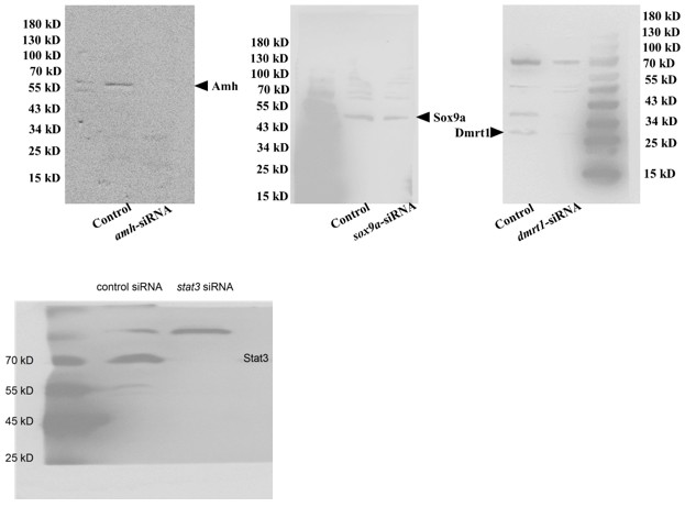
(2) Antibody validation and referencing (Line 776): The authors need to refer explicitly to figures demonstrating antibody validation. At present, these data are provided only as a supplementary file that is not cited in the manuscript. In addition, the Sox9a antibody appears to yield indistinguishable signals in control and RNAi conditions, suggesting that it may not recognize eel Sox9a. This issue is not addressed by the authors. Furthermore, antibody validation Western blots should be quantified.
(3) Unclear sample sizes (N values): Sample sizes remain unclear for several figures:
(a) Fig. 3F - No N value is provided. Each graph shows three data points; does this indicate that only three samples were quantified? If ten samples were collected, why were all not quantified?
(b) Fig. 4 - No N values are reported.
(c) Fig. 5A - Again, only three data points are shown per group, despite the apparent availability of twelve samples. The rationale for this discrepancy is not explained.
(4) qRT-PCR normalization: The manuscript does not specify the reference gene(s) used for qRT-PCR normalization. Although expression levels are reported as "relative," neither the identity of the reference gene(s) nor the justification for their selection is provided.
(5) Specificity of key antibodies: While the authors have made some effort to validate anti-Amh, anti-Sox9, and anti-Dmrt antibodies, the results remain incomplete. The Amh and Dmrt antibodies detect reduced protein levels following knockdown of their respective targets, which is encouraging. However, the Sox9a antibody shows no difference between control and RNAi conditions, suggesting it does not recognize eel Sox9. This is not acknowledged in the manuscript. In addition, no validation data are presented for Foxl2. Antibody validation data must be clearly referenced in the main text and presented in an interpretable and quantitative manner.
(6) Immunofluorescence data quality: The immunofluorescence images remain difficult to interpret. I strongly encourage the authors to enlarge the image panels and to present monochrome images (white signal on black background). The current presentation severely limits interpretability.
(7) Unreferenced supplementary figure: Fig. S4 is included in the submission but is not referenced anywhere in the manuscript text.
(8) Fig. 5B image resolution: The micrographs in Fig. 5B are too small to allow meaningful evaluation of the data.
(9) Unexplained data inclusion (Fig. 5E): Fig. 5E includes a pERK blot that is not mentioned in the Results section. The rationale for including these data is unclear.
(10) Poor blot quality (Fig. S3C): The blots in Fig. S3C exhibit high background and overexposure. I am concerned about the reliability of the quantification shown in panel D.
(11) Poor blot quality (Fig. S5G): The Stat3 blots in Fig. S5G contain numerous white artifacts, raising concerns about their suitability for normalization in panel H.
(12) Missing controls (Fig. 6E): Fig. 6E lacks controls for HO-3867 and Colivelin treatments alone. Without these controls, it is not possible to determine whether the reported effects are meaningful.
(13) Graphical presentation: The use of a light blue-to-pink gradient in bar graphs throughout the manuscript does not aid interpretation. I recommend using more distinct colors (e.g., red, orange, green, blue, purple, gray, black) to improve clarity. In summary, the interpretation of the study remains limited by persistent issues related to data presentation, image quality, and reagent specificity.
Author response:
The following is the authors’ response to the original reviews.
Public Reviews:
Reviewer #1 (Public review):
Summary:
This study investigates the molecular mechanism by which warm temperature induces female-to-male sex reversal in the ricefield eel (Monopterus albus), a protogynous hermaphroditic fish of significant aquacultural value in China. The study identifies Trpv4 - a temperature-sensitive Ca<sup>2+</sup> channel - as a putative thermosensor linking environmental temperature to sex determination. The authors propose that Trpv4 causes Ca<sup>2+</sup> influx, leading to activation of Stat3 (pStat3).pStat3 then transcriptionally upregulates the histone demethylase Kdm6b (aka Jmjd3), leading to increased dmrt1 gene expression and ovo-testes development. This work aims to bridge ecological cues with molecular and epigenetic regulators of sex change and has potential implications for sex control in aquaculture.
Strengths:
(1) This study proposes the first mechanistic pathway linking thermal cues to natural sex reversal in adult ricefield eel, extending the temperature-dependent sex determination paradigm beyond embryonic reptiles and saltwater fish.
(2) The findings could have applications for aquaculture, where skewed sex ratios apparently limit breeding efficiency.
We thank you for the encouraging comments of our work, and answering your questions has greatly improved the quality of the manuscript.
Weaknesses:
(A) Scientific Concerns:
(1) There is insufficient replication and data transparency. First, the qPCR data are presented as bar graphs without individual data points, making it impossible to assess variability or replication. Please show all individual data points and clarify n (sample size) per group. Second, the Western blotting is only shown as single replicates. If repeated 2-3 times as stated, quantification and normalization (e.g., pStat3/Stat3, GAPDH loading control) are essential. The full, uncropped blots should be included in the supplementary data.
We thank you for the critical comments. Now we have remade the bar graphs with individual data points, and added the sample size per group if possible. Quantification and/or normalization of the WB data based on at least two replicates were included. The representative uncropped blots have also been loaded as the supplementary data.
(2) The biological significance of the results is not clear. Many reported fold changes (e.g., kdm6b modulation by Stat3 inhibition, sox9a in S3A) are modest (<2-fold), raising concerns about biological relevance. Can the authors define thresholds of functional relevance or confirm phenotypic outcomes in these animals?
We thank you for the inspiring comments. Most of the experiments were transient in nature, for instance, warm temperature treatment of fish for 3-4 days, the fold change of gene expression were modest.
We admit that there are some shortcomings in this work. The major one is lacking of data showing that Trpv4 inhibition/activation,or pStat3 inhibition/activation can cause a gonadal phenotype change, for instance, from ovary to ovotestis or causing females to intersex fish. We only showed that pharmacological or RNAi can lead to change in sex-biased gene expression or affect temperature-induced gene expression, but not gonadal transformation.
In natural population, the sex change of ricefield eel may take several months to one year or even longer. We propose that the magnitude and duration of temperature exposure promote sex change of ricefield eel by driving the accumulation of testicular differentiation genes in sufficient quantities. In experimental condition, to realize the gonadal phenotype change, animals may need to be under repeated pharmaceutical treatment (3 day interval treatment) for longer time to reach a threshold. However, long term treatment significantly increases the death rate of the animals, caused by stress or frequent manipulation.
Inspired by your comment, we are optimizing the experimental conditions in order to cause some phenotypic outcomes, thanks.
(3) The specificity of key antibodies is not validated. Key antibodies (Stat3, pStat3, Foxl2, Amh) were raised against mammalian proteins. Their specificity for ricefield eel proteins is unverified. Validation should include siRNA-mediated knockdown with immunoblot quantification with 3 replicates. Homemade antibodies (Sox9a, Dmrt1) also require rigorous validation.
We thank you for the comments about the specificity of the antibodies. First,when choosing the commercial antibodies, we have compared the immunogen of the animal with the corresponding amino acids of ricefield eel, making sure that it was conserved to some extent (at least> 85% similarity). Second, we have referred the published work, where the antibodies have been proven to work in zebrafish, frogs, and turtles et al. This was true for pStat3 and Stat3 antibodies (Weber et al. 2020; Ge et al., 2024). Third, the specificity for each antibody was assessed using WB, based on the predicted size of the protein and the correct control setting.
For instance, we are very confident for the specificity for Dmrt1 antibody. First, Dmrt1 protein was readily detected in testes of males but barely detected in ovaries of females (Author response image 1). Second, Dmrt1 protein was not detected in ovary of fish at cool temperature, but clearly detected in nuclei of follicles in warm temperature-treated fish (Figure 3C, 4B), in line with our qPCR results. Third, by performing IF, Dmrt1 was not detected in females reared at lower temperature. By contrast, after warm temperature treatment or Trpv4 activation, it was detected in the nuclei in specific cell types but not everywhere (Figure 3E, 6C).
Author response image 1.

Although we have carefully evaluated the antibodies before experiments as described above, in response to your concerns, we went on to validate Amh, Dmrt1, Sox9a, and Stat3 antibodies using the corresponding siRNAs (Author response image 2). The results indicated that the antibodies, although not perfect, can be used in this work, as the expected band was gone or reduced in intensity. The experiments were repeated two times, and shown were representative.
Author response image 2.
(4) Most of the imaging data (immunofluorescence) is inconclusive. Immunofluorescence panels are small and lack monochrome channels, which severely limits interpretability. Larger, better-contrasted images (showing the merge and the monochrome of important channels) and quantification would enhance the clarity of these findings.
We apologize for the poor quality of the IF images. At your suggestion, we have repeated the majority of the IF experiments, and imaging data with better quality were presented in the revised manuscript. Quantification of WB and IF was also included to enhance the clarity. Please see the revised manuscript, Thanks.
(B) Other comments about the science:
(1) In S3A, sox9a expression is not dose-responsive to Trpv4 modulation, weakening the causal inference.
We have repeated the experiments, and new data was included for the replacement of the old one in the revised manuscript.
(2) An antibody against Kdm6b (if available) should be used to confirm protein-level changes.
We thank you for the nice suggestion. Unfortunately, current commercial antibody for Kdm6b is for mammals, which was not working in ricefield eel. At your suggestion, we are going to make one in future.
In sum, the interpretations are limited by the above concerns regarding data presentation and reagent specificity.
Reviewer #2 (Public review):
Summary:
This study presents valuable findings on the molecular mechanisms driving the female-to-male transformation in the ricefield eel (Monopterus albus) during aging. The authors explore the role of temperature-activated TRPV4 signaling in promoting testicular differentiation, proposing a TRPV4-Ca<sup>2+</sup>-pSTAT3-Kdm6b axis that facilitates this gonadal shift.
We thank you for the encouraging comments. Answering your questions has greatly improved our understanding of Trpv4 function in ricefield eel, and the quality of the manuscript.
Strengths:
The manuscript describes an interesting mechanism potentially underlying sex differentiation in M. albus.
Weaknesses:
The current data are insufficient to fully support the central claims, and the study would benefit from more rigorous experimental approaches.
(1) Overstated Title and Claims:
The title "TRPV4 mediates temperature-induced sex change" overstates the evidence. No histological confirmation of gonadal transformation (e.g., formation of testicular structures) is presented. Conclusions are based solely on molecular markers such as dmrt1 and sox9a, which, although suggestive, are not definitive indicators of functional sex reversal.
We thank you for pointing out this. The title has been changed to “Trpv4 links environmental temperature to testicular differentiation in hermaphroditic ricefield eel.”
(2) Temperature vs Growth Rate Confounding (Figure 1E):<br /> The conclusion that warm temperature directly induces gonadal transformation is confounded by potential growth rate effects. The authors state that body size was "comparable" between 25C and 33C groups, but fail to provide supporting data. In ectotherms, growth is intrinsically temperature-dependent. Given the known correlation between size and sex change in M. albus, growth rate-rather than temperature per se-may underlie the observed sex ratio shifts. Controlled growth-matched comparisons or inclusion of growth rate metrics are needed.
We thank you for the critical comments. We have repeated the experiments, and have carefully compared the body length and weight, and results showed that there is no big difference between 25 and 33 degree groups. Please see Figure S1D-E, and the text in the last paragraph of “Warm temperature promotes gonadal transformation” section in the Results part.
(3) TRPV4 as a Thermosensor-Insufficient Evidence:<br /> The characterisation of TRPV4 as a direct thermosensor lacks biophysical validation. The observed transcriptional upregulation of Trpv4 under heat (Figure 2) reflects downstream responses rather than primary sensor function. Functional thermosensors, including TRPV4, respond to heat via immediate ion channel activity-typically measurable within seconds-not mRNA expression over hours. No patch-clamp or electrophysiological data are provided to confirm TRPV4 activation thresholds in eel gonadal cells.
We thank you for the critical comments. The patch-clamp or electrophysiological experiments require special equipment and well-trained expert, unfortunately, our lab members and nearby collaborators have no experience in performing the kind of experiments. The Trpv4 is a well-known cation channel protein that is activated by moderate heat (> 27 degree). And a body of published work has demonstrated its role in the regulation of Ca<sup>2+</sup> signals via change its configuration in response to temperature (J Physiol. 2017 Oct 25;595(22):6869–6885. doi: 10.1113/JP275052; Cell Death Dis 11, 1009 (2020). https://doi.org/10.1038/s41419-020-03181-7; Cell Death Dis 10, 497 (2019). https://doi.org/10.1038/s41419-019-1708-9; Cell calcium, https://doi.org/10.1016/j.ceca.2026.103108).
Consistently, warm temperature increased calcium influx within an hour, similar to the Trpv4 agonist treatment (Figure 2E, 5D), and addition of ion channel Trpv4 inhibitor prevents the calcium signals by war temperature treatment. Moreover, calcium signaling activity is closely linked with pStat3 activity and expression of sex-biased genes (Figures 5G, 6F). Although we did not show biophysical data, these results implied that Trpv4 is a thermosensor, and regulate the downstream pathway via the regulation of calcium signals, in line with it functions as an ion channel.
Additionally, the Ca<sup>2+</sup> imaging assay (Figure 2F) lacks essential details: the timing of GSK1016790A/RN1734 administration relative to imaging is unclear, making it difficult to distinguish direct channel activity from indirect transcriptional effects.
We have added more information for Ca<sup>2+</sup> imaging assay (now Figure 2E and the corresponding text in Figure 2 legend, in the revised manuscript). In particular, we added the timing of treatment to better show that it was a direct effect.
(4) Cellular Context of TRPV4 Activity Is Unclear:<br /> In situ hybridisation suggests TRPV4 expression shifts from interstitial to somatic domains under heat (Figures. 2H, S2C), implying potential cell-type-specific roles. However, the study does not clarify: (i) whether TRPV4 plays the same role across these cell types, (ii) why somatic cells show stronger signal amplification, or (iii) the cellular composition of explants used in in vitro assays. Without this resolution, conclusions from pharmacological manipulation (e.g., GSK1016790A effects) cannot be definitively linked to specific cell populations.
We thank you for the inspiring comments. We have performed IF experiments using Trpv4 specific antibodies (antibody specificity was confirmed). It was clearly shown that Trpv4 was expressed in a portion of follicle cells. To explore the identity of Trpv4-expressing somatic cells, we have performed double IF experiments using Trpv4 and Foxl2, a granulosa cell marker. The results (Figure 2H) clearly showed that Trpv4-expressing cells are a portion of Foxl2-positive granulosa cells. We propose that Trpv4-expressing granulosa cells may play an important role in sensing the temperature, and that Trpv4-expressing granulosa cells transdifferentiate into Sertoli cells by warm temperature exposure, because Dmrt1, a Sertoli cell marker, started within follicles in a typical granulosa cell location. Unfortunately, current Dmrt1/Trpv4 antibodies are both produced from rabbit. To overcome this, we are ordering mouse Dmrt1 antibodies, and in future we will perform Trpv4/Dmrt1 double IF to show if Dmrt1 positive cells co-localize with Trpv4 expressing cells. We would like to update the results to you once the antibody was available.
As our animal experiments (Figure 2H) have clearly shown the identify of Trpv4 expressing somatic cells, we did not repeat the experiments using explants, to explore the cellular composition of explants used in in vitro assays.
(5) Rapid Trpv4 mRNA Elevation and Channel Function:<br /> The authors report a dramatic increase in Trpv4 mRNA within one day of heat exposure (Figures 4D, S2B). Given that TRPV4 is a membrane channel, not a transcription factor, its rapid transcriptional sensitivity to temperature raises mechanistic questions. This finding, while intriguing, seems more correlational than functional. A clearer explanation of how TRPV4 senses temperature at the molecular level is needed.
We appreciate you for your inspiring comments. Actually, we are also wondering about how trpv4 mRNA was regulated by warm temperature. First of all, the up-regulation of trpv4 mRNA is true, as evidenced by multiple pieces of data using qPCR and ISH experiments. It appears that ovarian cells respond to the temperature changes by increasing calcium influx via Trpv4 ion channel,as well as by increasing trpv4 mRNA expression levels.
Then, how trpv4 mRNA is regulated by heat? It is well-known that gene expression can be regulated by subtle temperature change via some direct temperature sensing genes (Haltenhof et al., 2020). We hypothesized that trpv4 is a downstream target of these thermosensors, displaying a mechanism similar to mammals. Actually, we have performed some experiments, and the preliminary data were obtained, which support our hypothesis.
Because the mechanistic explanation study is undergoing and not published, we chose not to discuss it in detail in the revised manuscript. We wish to report it by the end of this year, and by then are pleased to update you with the progress.
(6) Inconclusive Evidence for the Ca<sup>2+</sup>-pSTAT3-Kdm6b Axis: Although the authors propose a TRPV4-Ca<sup>2+</sup>-pSTAT3-Kdm6b-dmrt1 pathway, intermediate steps remain poorly supported. For example, western blot data (Figures 3C, 4B) do not convincingly demonstrate significant pSTAT3 elevation at 34C. Higher-resolution and properly quantified blots are essential. The inferred signalling cascade is based largely on temporal correlation and pharmacological inhibition, which are insufficient to establish direct regulatory relationships.
We thank you for the critical comments. In response to your concerns, we have repeated experiments, and better resolution WB data with proper quantification were included in the revised manuscript. In particular, we convincingly demonstrate that 34 degree caused significant pStat3 elevation.
To directly establish regulatory relationship of the members, at your suggestion, we provided some genetic and molecular biology data to support our conclusion in the revised manuscript. For instance, we have knockdown the stat3 gene by using siRNAs, and as shown in Figure 6F, we further showed that pStat3 is functionally downstream of Trpv4. Moreover, ChIP and luciferase assays were performed to show that pStat3 directly binds and activate kdm6b (Figure 7B-C). We have also performed various pharmacological inhibition to further strength our conclusion (Figures 6B-E).
(7) Species-Specific STAT3-Kdm6b Regulation Is Unresolved:<br /> The proposed activation of Kdm6b by pSTAT3 contrasts with findings in the red-eared slider turtle (Trachemys scripta), where pSTAT3 represses Kdm6b. This divergence in regulatory direction between the two TSD species is surprising and demands further justification. Cross-species differences in binding motifs or epigenetic context should be explored. Additional evidence, such as luciferase reporter assays (using wild-type and mutant pSTAT3 binding motifs in the Kdm6b promoter) is needed to confirm direct activation.
We thank you for the inspiring comments. At your suggestion, we have performed luciferase assay using kdm6b promotor that is intact or mutated. The results were in favor of our statement. Please see Figure 7C and the related text.
A rescue experiment-testing whether Kdm6b overexpression can compensate for pSTAT3 inhibition-would also greatly strengthen the model.
We thank you for the nice suggestion. It is technically challenging to perform kdm6b overexpression or any Kdm6b gain of function experiments (we have tried to make lentivirus, however, it was not working). There is no Kdm6b-specific agonists.
Inspired by you, we are establishing constitutive kdm6b transgenic ricefield eel. Although it require at least a year to allow the fish to grow up for functional experiments, once it was established, we can directly answer some important questions.
(8) Immunofluorescence-Lack of Structural Markers: <br /> All immunofluorescence images should include structural markers to delineate gonadal boundaries. Furthermore, image descriptions in the figure legends and main text lack detail and should be significantly expanded for clarity.
We thank you for the critical comments. At your comments, we have first performed IF using beta-catenin as structural marker. However, the results were not good for some unknown reasons. Then, we used Vimentin as a structural maker, as it can label all the cells in gonads. Foxl2 was used as granulosa cell marker. Dmrt1 was used as Sertoli cell marker.
Some essential description was added in the figure legend as requested. Please see detail in the revised manuscript.
(9) Pharmacological Reagents-Mechanisms and References: <br /> The manuscript lacks proper references and mechanistic descriptions for the pharmacological agents used (e.g., GSK1016790A, RN1734, Stattic). Established literature on their specificity and usage context should be cited to support their application and interpretation in this study.
These pharmacological agents have been used by others (Ge et al., 2017; Liu et al., 2021; Weber et al., 2020; Wu et al.,2024), and they are properly cited in the manuscript.
(10) Efficiency of Experimental Interventions: <br /> The percentage of gonads exhibiting sex reversal following pharmacological or RNAi treatments should be reported in the Results. This is critical for evaluating the strength and reproducibility of the interventions.
We thank you for the critical and important comments. Actually another reviewer has asked the same question. We admit that this was the big shortcoming of the work, as we did not provide data demonstrating that Trpv4 inhibition/activation, or pStat3 inhibition/activation can cause a gonadal phenotype change, for instance, from ovary to ovotestis or causing sex reversal of fish. We only showed that pharmacological or RNAi can lead to alteration of sex-biased gene expression or affect temperature induced gene expression.
In wild population, the entire sex change of ricefield eel may take months to one year or even longer. We propose that the magnitude and duration of temperature exposure promote sex change of ricefield eel by driving the accumulation of testicular differentiation genes in sufficient quantities. In experimental condition, to realize the gonadal phenotype change, animals may need to be under repeated pharmaceutical treatment (3 day interval treatment) for longer time to reach a threshold, however, long term treatment significantly increases the death rate of the animals, caused by stress or frequent manipulation. Actually, my students have tried the experiments, unfortunately, either the number of sex-versing animals were small or the experiments lacked of repeat. So no percentage of gonadal transformation after treatment can be provided at this time, but we have indicated the number of samples when performing molecular experiments (showing expression of sex-biased genes).
Inspired by your important comment, we are optimizing the experimental conditions in order to cause some phenotypic outcomes. By then, the percentage of gonads exhibiting sex reversal following pharmacological or RNAi treatments can be calculated, showing the biological significance.
Recommendations for the authors:
Reviewer #1 (Recommendations for the authors):
Editorial Concerns:
(1) The term "sex reversal" should be clearly defined upfront as female-to-male, and the developmental consequences (e.g., increase in body size post-transition) should be explicitly stated early in the introduction.
We thank our editorial for pointing out this. We have added those in the introduction Part. It reads “The species begins life as a female and then develops into a male through an intersex stage, thus displaying a female-to-male sex reversal during aging. Females are small in size (< 25 cm), and during and after sex change, there is a gradual increase in body size (> 55 cm for the majority of males).”
Additional information was shown in the first and second paragraph in the results Part.
(2) The manuscript references skewed sex ratios in cultured ricefield eel but fails to specify the direction (e.g., too many males or females). This should be clarified to contextualize the biological and commercial problem.
According to your suggestion, we now added additional information, and it reads “The reproductive mode of ricefield eel, which leads to much more females than males in spawning season, severely affects the sex ratio, and decreases the productivity of broodstock. Moreover, adult females lay limited eggs (~200) due to its small size.”
(3) Define TSD (temperature-dependent sex determination) upon first use, not at the second mention.
We have checked this, and make sure it was properly done.
(4) The phrase "quality fries for aquaculture" should be reworded or defined; it is unclear to non-specialists.
We thank you for pointing out this. Now it reads “adult females lay limited eggs (~200) due to its small size, which is a limiting factor for massive production of seedling for aquaculture industry”.
(5) Several in-text citations (e.g., Weber 2020, Wu 2024) are absent from the bibliography. ]
We have double checked the reference, thanks.
(6) The inclusion of page and line numbers would facilitate peer review.
We have now shown the page and line.
(7) The discussion is written vaguely. Clarify species names when discussing comparative biology and consider breaking down complex sentences to aid comprehension for a broad audience, such as that of eLife.
We have added the species name, and try our best to use concise expression. Thanks.
Thank you for this research, interrogating trans people would give a nice perspective. Make sure you provide a safe environment for them please.
Comets and Asteroids: Debris of the Solar System
Skim in this chapter for lecture on Friday, Feb. 20th...
parasitize
to live on, in, or with another organism (the host) while feeding off it and causing harm
Educational Resources
Open Educational Resources?
Hotspot für Openness in den Anwendungsdomänen Digital Humanities
Viele Fremd- und Fachwörter, für Personen, die aus den Geisteswissenschaften kommen vielleicht schwer zu verstehen
Digital Humanities,
Bei Geisteswissenschaften ist es Digital Humanities, bei Verwaltungswissenschaft Verwaltungswissenschaft
Beschreibung des Datenkompetenzzentrums QUADRIGA
Übergang deutlich machen. Erklären, dass QUADRIGA hinter den OERs steht
(Förderkennzeichen: 16DKZ2034A)
Warum ist das kleingedruckt?
7.4. QUADRIGA#
QUADRIGA lieber am Anfang vorstellen
So you might find a safe space online to explore part of yourself that isn’t safe in public (e.g., Trans Twitter and the beauty of online anonymity). Or you might find places to share or learn about mental health (in fact, from seeing social media posts, Kyle realized that ADHD was causing many more problems in his life than just having trouble sitting still, and he sought diagnosis and treatment). There are also support groups for various issues people might be struggling with, like ADHD, or having been raised by narcissistic parents.
Online spaces can offer a powerful sense of safety and belonging, especially for people who feel unable to express certain parts of themselves in public. Anonymity can create room for exploration, honesty, and connection that might not otherwise be possible offline. At the same time, social media can also serve as an entry point to self-understanding, as people encounter language and experiences that help them recognize patterns in their own lives. Support groups and online communities show how digital platforms, despite their flaws, can meaningfully reduce isolation and encourage people to seek help.
Kontakt: Universität Potsdam Potsdam Graduate School QUADRIGA Datenkompetenzzentrum Am Kanal 47 14467 Potsdam Tel.: +49 331 977-4595 Fax: +49 331 977-4555 E-Mail: robin.moeser@uni-potsdam.de Impressum der Universität Potsdam
Das ist richtig so?
1h 15min
Wirklich?
In diesem Kapitel wurde durch eine quantitative Analyse von Worthäufigkeiten des semantischen Felds “Grippe” die Forschungsfrage untersucht,
Haben wir untersucht...
Inzwischen lassen sich zahlreiche weitere Beispiele finden, die zeigen, wie aufschlussreich n-Gramm-Analysen sein können. Betrachtet man etwa im englischen Google-Books-Korpus alle 2-Gramme, die mit dem Verb “to hate” (hassen) beginnen und mit einem Substantiv enden, so gehört 2-Gramme “hate war” (den Krieg hassen) zu den häufigsten Treffern. Auffällig sind dabei zwei sehr ausgeprägte Häufigkeitsspitzen, die zeitlich mit dem Ersten und dem Zweiten Weltkrieg zusammenfallen.
Spannend
Who could possibly have predicted this?
lol ^^
dazu in der Lage sind, semantisch ähnliche Wörter zu erzeugen,
sehr gut semantisch ähnliche Wörter erzeugen
jedoch
Jedoch, aber usw. immer rausnehmen wenn geht
Die Grundlage unserer Analyse besteht darin, die Textstellen zu identifizieren
Die Analyse hat das Ziel Textstellen... Sehr komplizierter Satz gerade
n …
Welche Kapitel
untersucht
2 Mal untersucht hintereinander
In der Korpusanalyse kehren wir wieder zu unserer Fragestellung und auf die Operationalisierung der Fragestellung zurück. Unsere Fragestellung lautet:
Kehren wir zu unserer Fragestellung zurück, die lautet...
wir
Sie
kann
zu oft kann
Ihrer
zur, zu oft Ihrer
Diese
Die
Mit spaCy
Ich würde lieber von NLPs sprechen am Beispiel von spaCy
4.3. Resümee#
Wollt ihr nicht etwas dazu schreiben wie KI beim Code erstellen helfen kann und was zu beachten ist?
Im Folgenden wird exemplarisch der Roman “Feldblumen” von Adalbert Stifter (txt-Datei) mit der Bibliothek spaCy annotiert. Es werden folgendene Schritte durchgeführt:
ganz oft folgend
Dieses
Das
Korpusverarbeitung – Annotation mit spaCy
Warum nutzt ihr spaCy und nicht Stanza? Stanza ist deutlich stärker bei alten Sprachen https://stanfordnlp.github.io/stanza/ Reflektiert, was es für Alternativen gibt. Es gibt auf eine gute veröffentlichtung zu NLPs allgemein von Hiltmann et al https://arxiv.org/abs/2502.04351
können
Zu oft können Sie lernen...
Natural Language Processing
(NLP)
Lösungen
Finde ich super
folgenden
wieder folgend, das verfolgt mich ; )
Welche Aussagen beschreiben die verschiedenen Metadatenschemata korrekt?
Eine vierte Auswahl überlegen
Zu welchem Metadatenschema gehört das Element "teiHeader"?
Wird in der Frage eigentlich schon veraten
folgenden
wieder folgend
helfen Ihnen
unterstützen Sie dabei
Diese
Die
können
werden verschiedene Strategien... gewählt
Key points des Kapitels
Überlegt, ob ihr mit Anglizismen arbeiten möchtet
Resümee
Fazit
# --- Create Plotly figure ---
einheitlich auszeichnen, mit oder ohne ----
3.4.4. Option 1. ELTeC-DEU corpus#
Sehr schön visualisiert
e folgenden S
wieder etwas mit folgend
Im Folgenden
Es wird oft im Folgenden geschrieben
↓
Besseres Icon wählen
dies
das
Bereits in dieser Übersicht zeigt sich
Die Übersicht zeigt,
Health Check EndpointsEvery microservice must implement health endpoints:# Liveness: Is service alive?GET /healthz → 200 OK if process running# Readiness: Ready to handle traffic?GET /ready → 200 if database connected, caches warm, dependencies available → 503 if not ready yet# Startup: Has service completed initialization?GET /startup → 200 once initialization completeConfigure probes:
vorgestellten
hier beschriebenen
man
man ist kein schönes Wort
Eckert, Penelope(1997). Age as a sociolinguistic variable. In: FlorianCoulmas (ed.), The handbook of sociolinguistics. Oxford: Blackwell, 151–167.
Age variation
Crystal, David(2001). Language and the internet. Cambridge: Cambridge University Press.
Online communication
Coates, Jennifer(1993). Women, men and language: A sociolinguistic account of gender differences in language. London/New York: Longman.
Gender variation
On ai, productivity and shorter work weeks and why that will not happen
This extracts light on the evolving social and economic dynamics within Palanpur. The discussion of mechanization reveals that investment decisions are not driven only by productivity gains. Tractors function not only as agricultural tools but also as prestige goods, embodying social status andupward mobility. Their use for freight and passenger transport further illustrates how local actors creatively adapt private assets to compensate for insufficient formal infrastructure. In this sense, economic change is deeply embedded in social meanings and institutional constraints. At the same time, these developments raise broader questions about the future of caste and hierarchy in a context of gradual industrialization and market expansion. As access to alternative income sources, improved markets, and new technologies expands, traditional caste-based occupational boundaries may weaken, offering greater opportunities for mobility. Yet sociocultural norms often are resilient and adapting. Economic growth may transform the material foundations of caste distinctions without fully erasing their symbolic and social power. The key issue, then, is whether sustained structural transformation will ultimately render caste hierarchies economically irrelevant, or whether they will persist in reshaped forms within an increasingly diversified rural economy.
He was powerless to harm the enemy or tohelp his friends.
This quote shows how soldiers often have no control over what happens. This make us wonder if solders are really heroes or if they just have to do what they're told.
It included the iron triangle, local governance arrangements, civic associa-tions, and most importantly unions.
Will this be in danger as we move forawrd in the half life?
The layering of multiple dimensions of decline andmarginalization is distinct to the region and has produced cultural distancebetween it and the rest of the country.
Still confused why republicans aren't balmed as well
Trump has already ‘made America great again’ becausehe has conclusively demonstrated that the white privilege of denigrating minor-ities without consequence is alive and well
Jeez
autarky
Economic independence
presidential politics, a capacity that had previ-ously been grounded on their unions, civic associations and their party
Which had been granted by the dems
Itwas also rooted in expectations that are a legacy of white supremacy
Explains why he did well in the south
ignores the role of lost working-class power and voicein the Democratic Party
Gives too much credit to trump and rep.
If we interpret Trump’s ability tosecure votes as his ability to channel white revanchism against a morediverse society then it is possible to see the loss of relative status in theRust Belt as an important explanatory factor.
Thesis here
withholding their vote
Exit/voice
the Democratic Party does not appear to be particularly con-cerned with the well-being of either
Burn
newly won and briefly held material afflu-ence
Material affluence whihc brings privelage they did not have before, leaving with half-life of industrialization
polemical
critical
The opposition between the credentialled and the uncreden-tialled had its purest partisan expression in 2016
Old incomomy = industrial, new econonmy = tech/finance
myopia
Nearsightedness
Under "Image and shape" → click "Change image" → select Ubuntu → choose Canonical Ubuntu 22.04 (aarch64/ARM64) → click "Select image".
Müsste eigenen schritt haben und nicht versteckt sein in schritt 2
Medieval Christian pilgrims assiduously acquired, from the sites they visited, pilgrim badges that signified the protection of the saints they had visited, and that served as mementos of their visits and as signs of their status as pilgrims. Such badges were also advertising devices for their shrines, for which the production and marketing of pilgrim badges was a major commercial undertaking and an important source of income
just testing
over de belastingschuld die uit de wet voortvloeit heeft de inspecteur bij het vaststellen van de schuld geen ruimte om te bepalen of de regel goed tot zijn recht komt en ook geen ruimte om de schuld hoger of lager vast te stellen.
Bij een gebonden beschikking heeft de inspecteur bij het vaststellen van de materiele belastingschuld 1) geen ruimte om te bepalen of dit regel goed tot zijn recht behoort en 2) ook geen ruimte om de schuld hoger of lager vast te stellen.
articles
fdfdf
Guide de Référence Parcoursup 2026 : Stratégies et Mécanismes de Formulation des Vœux
La procédure Parcoursup 2026 s'inscrit dans une volonté de simplification et de transparence accrue pour les lycéens et leurs familles.
S'appuyant sur une offre diversifiée de 25 000 formations, la plateforme centralise un calendrier unique et un dossier de candidature commun. Les points critiques à retenir pour cette session incluent :
• Calendrier charnière : La formulation des vœux s'étend du 19 janvier au 12 mars 2026, avec une date limite de finalisation des dossiers fixée au 1er avril.
• Souveraineté pédagogique : Contrairement aux idées reçues, ce n'est pas un algorithme qui analyse les candidatures, mais les équipes pédagogiques (enseignants) de chaque établissement.
• Outils de décision : Le simulateur de chances, basé sur les données réelles des trois dernières années, devient un outil central pour lutter contre l'autocensure et la surconfiance.
• Sécurisation : La plateforme garantit la gratuité des démarches (hors frais de concours spécifiques) et interdit toute demande d'acompte financier avant l'admission définitive.
--------------------------------------------------------------------------------
Parcoursup est conçu comme un outil de simplification administrative regroupant la quasi-totalité de l'offre d'enseignement supérieur en France.
Le système repose sur trois piliers d'unification :
• Dossier unique : Un seul dossier à constituer quel que soit le nombre d'établissements visés.
• Calendrier unique : Des échéances identiques pour tous, évitant la multiplication des calendriers spécifiques.
• Cadre de présentation unique : Toutes les "fiches formations" utilisent la même structure pour faciliter la comparaison objective (statut public/privé, taux d'accès, frais de scolarité).
La plateforme offre des protections spécifiques :
• Liberté de choix : Aucune pression ne peut être exercée sur l'ordre des vœux des candidats.
• Interdiction des acomptes : Les établissements ne peuvent exiger de paiement pour "réserver" une place avant l'obtention du baccalauréat et l'inscription administrative finale.
• Transparence : Les critères de sélection et les chiffres des années précédentes doivent être explicitement affichés.
--------------------------------------------------------------------------------
Il est crucial de distinguer les catégories de formations pour adapter sa stratégie de vœux.
| Type de formation | Exemples | Capacité de refus | | --- | --- | --- | | Non-sélectives | Licences (L.AS, PPPE), PASS | Admission possible dans la limite des places ; si saturation, classement des dossiers. | | Sélectives | CPGE, BTS, BUT, Écoles d'infirmiers, Écoles de commerce/ingénieurs | Possibilité de refuser un candidat si son profil ne correspond pas aux critères. |
L'apprentissage permet d'alterner formation théorique (CFA) et pratique (employeur).
• Double compteur : Un candidat peut formuler jusqu'à 10 vœux en apprentissage en plus de ses 10 vœux sous statut étudiant.
• Condition d'admission : La proposition d'admission n'est validée que par la signature d'un contrat d'apprentissage avec un employeur.
• Conseil stratégique : Il est recommandé de postuler à la fois sous statut étudiant et en apprentissage pour un même diplôme afin de sécuriser sa rentrée.
--------------------------------------------------------------------------------
L'examen des vœux est une prérogative humaine exercée par les commissions pédagogiques des établissements.
Chaque formation définit sa propre pondération. À titre d'exemple, une fiche formation peut afficher :
• Résultats scolaires : Jusqu'à 70 % de la note finale.
• Méthode de travail : Environ 20 %.
• Savoir-être / Motivation : Entre 5 % et 30 % (notamment pour les filières de santé).
• Engagement et activités : Souvent entre 2 % et 5 %.
Le dossier remonte automatiquement les notes du lycée via l'Identifiant National Élève (INE).
• Étudiants à l'étranger (AEFE) : L'identifiant est fourni par l'établissement (souvent le numéro Cyclade).
• Fiche Avenir : Remplie par les enseignants pour les lycéens de terminale.
• Fiche de suivi : Pour les étudiants en réorientation, permettant d'expliciter leur nouveau projet.
• Frais de candidature : Certaines écoles (IEP, ingénieurs) peuvent demander des frais de dossier (ex: 150€), à régler avant le 1er avril.
--------------------------------------------------------------------------------
Pour la session 2026, Parcoursup met en avant des outils de visualisation basés sur l'historique 2023-2025.
Chaque fiche formation propose une rubrique détaillant l'admission de l'année précédente :
• Nombre total de candidats.
• Nombre de propositions d'admission envoyées.
• Nombre d'étudiants ayant finalement intégré la formation.
• Taux d'accès par type de baccalauréat (Général, Technologique, Professionnel).
Cet outil permet de tester son profil (spécialités choisies et moyenne générale) :
• Objectif : Lutter contre l'autocensure (notamment chez les jeunes filles pour les filières sélectives) et la surconfiance (inciter à diversifier les vœux même pour les dossiers brillants).
• Indicateurs : Le simulateur indique si des profils similaires ont été admis "régulièrement" (20 % à 50 % de chances) ou "très fréquemment" au cours des trois dernières années.
• Interprétation : Ce sont des données statistiques et non une garantie d'admission.
--------------------------------------------------------------------------------
Il est impératif de ne pas se limiter à un seul vœu, même avec un excellent dossier. Une stratégie équilibrée doit alterner :
1. Vœux d'ambition : Formations très sélectives.
2. Vœux de raison : Formations correspondant au profil.
3. Vœux de précaution : Formations avec un taux d'accès élevé (licences non-sélectives).
• Coordonnées : Il est fortement conseillé de renseigner un numéro de téléphone portable pour recevoir les alertes SMS.
• Accompagnement parental : Les parents peuvent ajouter leur adresse mail dans le dossier de leur enfant pour recevoir les notifications en double, assurant ainsi le respect des délais.
• Contact humain : L'information numérique ne remplace pas les Journées Portes Ouvertes (JPO) et le dialogue avec les professeurs principaux ou les conseillers d'orientation.
• 19 janvier - 12 mars : Création du dossier et saisie des vœux.
• Jusqu'au 1er avril : Finalisation des dossiers et confirmation des vœux.
• 2 juin : Début de la phase de réponses des établissements.
• 2 juin - 11 juillet : Phase de décision et choix final pour les candidats.
the vulnerability of individual objects to water can be affected (i.e. increased) significantly by the state of the degradation of the materials.
depends on the state of the material
Many custodians underestimate the likelihood and effects of sporadic events such as water leaks. There is a tendency to use basements for collection and archival storage and to leave boxes of material on the floor, perhaps only "temporarily." Many containers for paper documents and small artifacts purchased by museums are acid-free, but few institutions consider acquiring watertight boxes made from fluted polypropylene.
underestimating water
3.2.2. Trauma Dumping# While there are healthy ways of sharing difficult emotions and experiences (see the next section), when these difficult emotions and experiences are thrown at unsuspecting and unwilling audiences, that is called trauma dumping [m11]. Social media can make trauma dumping easier. For example, with parasocial relationships, you might feel like the celebrity is your friend who wants to hear your trauma. And with context collapse, where audiences are combined, how would you share your trauma with an appropriate audience and not an inappropriate one (e.g., if you re-post something and talk about how it reminds you of your trauma, are you dumping it on the original poster?). Trauma dumping can be bad for the mental health of those who have this trauma unexpectedly thrown at them, and it also often isn’t helpful for the person doing the trauma dumping either: Venting, by contrast, is a healthy form of expressing negative emotion, such as anger and frustration, in order to move past it and find solutions. Venting is done with the permission of the listener and is a one-shot deal, not a recurring retelling or rumination of negativity. A good vent allows the venter to get a new perspective and relieve pent-up stress and emotion. While there are benefits to venting, there are no benefits to trauma dumping. In trauma dumping, the person oversharing doesn’t take responsibility or show self-reflection. Trauma dumping is delivered on the unsuspecting. The purpose is to generate sympathy and attention not to process negative emotion. The dumper doesn’t want to overcome their trauma; if they did, they would be deprived of the ability to trauma dump.
This passage introduces “trauma dumping,” which means sharing traumatic experiences and negative emotions with unwilling or unprepared audiences without their consent. Social media increases this behavior through parasocial relationships and context collapse. Trauma dumping harms recipients’ mental health and does not truly help the sharer, while venting is healthy because it is consensual and constructive. Trauma dumping raises important ethical concerns. From a care ethics perspective, it ignores others’ emotional well-being, and from a utilitarian view, it causes more harm than good. Therefore, users should be mindful of emotional boundaries and share personal trauma responsibly.
13.2.2. Trauma Dumping# While there are healthy ways of sharing difficult emotions and experiences (see the next section), when these difficult emotions and experiences are thrown at unsuspecting and unwilling audiences, that is called trauma dumping [m11]. Social media can make trauma dumping easier. For example, with parasocial relationships, you might feel like the celebrity is your friend who wants to hear your trauma. And with context collapse, where audiences are combined, how would you share your trauma with an appropriate audience and not an inappropriate one (e.g., if you re-post something and talk about how it reminds you of your trauma, are you dumping it on the original poster?). Trauma dumping can be bad for the mental health of those who have this trauma unexpectedly thrown at them, and it also often isn’t helpful for the person doing the trauma dumping either: Venting, by contrast, is a healthy form of expressing negative emotion, such as anger and frustration, in order to move past it and find solutions. Venting is done with the permission of the listener and is a one-shot deal, not a recurring retelling or rumination of negativity. A good vent allows the venter to get a new perspective and relieve pent-up stress and emotion. While there are benefits to venting, there are no benefits to trauma dumping. In trauma dumping, the person oversharing doesn’t take responsibility or show self-reflection. Trauma dumping is delivered on the unsuspecting. The purpose is to generate sympathy and attention not to process negative emotion. The dumper doesn’t want to overcome their trauma; if they did, they would be deprived of the ability to trauma dump.
This passage introduces “trauma dumping,” which means sharing traumatic experiences and negative emotions with unwilling or unprepared audiences without their consent. Social media increases this behavior through parasocial relationships and context collapse. Trauma dumping harms recipients’ mental health and does not truly help the sharer, while venting is healthy because it is consensual and constructive. Trauma dumping raises important ethical concerns. From a care ethics perspective, it ignores others’ emotional well-being, and from a utilitarian view, it causes more harm than good. Therefore, users should be mindful of emotional boundaries and share personal trauma responsibly.
“Tendency to continue to surf or scroll through bad news, even though that news is saddening, disheartening, or depressing. Many people are finding themselves reading continuously bad news about COVID-19 without the ability to stop or step back.”
It is frustrating to know that even if people are looking at negative news as suggested, dishearten or saddening, people just hard to resist of stopping viewing, instead they continue to watch for more. This happens to me as well, not just watching sad news but also listen to sad music when feeling low.
Übersicht Digitale Produkte
Ich nutze auch folgende KI Software für Psychotherapie und bin damit sehr zufrieden:
BerichtBiber für PTV3 Berichte DokuDachs für tägliche Therapiedokumentation
the model can inspect existing elisp, propose changes, and iterate directly inside the document. It turns into an interesting and (dare I say it) fun form of agentic, REPL-driven co-development
Making ai agent "mold" your environment for the task by creating supporting mappings, configurations and plugins. Something like: "Create an optimal marks and window layout for this project"
The idea that social media can create these fake, one-sided relationships really resonated with me.
It’s really eye-opening to see how social media algorithms are designed to keep us scrolling, even when it’s bad for our mental health. I’ve definitely felt the pressure to keep up with others’ posts, and it’s helpful to understand that this isn’t just my own issue—it’s a feature of the platforms we use.
eLife Assessment
This manuscript details important findings that DNA polymerase kappa shows age-related changes in subcellular localization within different cell types in the brains of mice, from the nucleus in young cells to the cytoplasm in old cells. The authors' findings suggest that age-related alterations in POLK localization could drive mechanistic and functional changes in the aging brain. The authors provide solid evidence for their study, with data broadly supporting their claims with minor weaknesses.
Reviewer #1 (Public review):
Summary:
Abdelmageed et al. investigate age-related changes in the subcellular localization of DNA polymerase kappa (POLK) in the brains of mice. POLK has been actively investigated for its role in translesion DNA synthesis and involvement in other DNA repair pathways in proliferating cells, very little is known about POLK in a tissue-specific context or let alone in post-mitotic cells. The authors investigated POLK subcellular distribution in the brains of young, middle-aged, and old mice via immunoblotting of fractioned tissue extracts and immunofluorescence (IF). Immunoblotting revealed a progressive decrease in the abundance of nuclear POLK, while cytoplasmic POLK levels concomitantly increased. Similar findings were present when IF was performed on brain sections. Further IF studies of cingulate cortex (Cg1), motor cortex (M1, M2), and somatosensory (S1) cortical regions all showed an age-related decline in nuclear POLK. Nuclear speckles of POLK decrease in each region, meanwhile the number of cytoplasmic POLK granules decreases in all four regions, but granule size is increasing. The authors report similar findings for REV1, another Y-family DNA polymerase.
The authors then investigate the colocalization of POLK with other DNA damage response (DDR) proteins in either pyramidal neurons or inhibitory interneurons. At 18 months of age, DNA damage marker gH2AX demonstrated colocalization with nuclear POLK, while strong colocalization of POLK and 8-oxo-dG was present in geriatric mice. The authors find that cytoplasmic POLK granules colocalize with stress granule marker G3BP1, suggesting that the accumulated POLK ends up in the lysosome.
Brain regions were further stained to identify POLK patterns in NeuN+ neurons, GABAergic neurons, and other non-neuronal cell types present in the cortex. Microglia associated with pyramidal neurons or inhibitory interneurons were found to have higher abundance of cytoplasmic POLK. The authors also report that POLK localization can be regulated by neuronal activity induced by Kainic acid treatment. Lastly, the authors suggest that POLK could serve as an aging clock for brain tissue, but POLK deserves further characterization and correlation to functional changes before being considered for a biomarker.
Strengths:
Investigation of TLS polymerases in specific tissues and in post-mitotic cells is largely understudied. The potential changes in sub cellular localization of POLK and potentially other TLS polymerases opens up many questions about DNA repair and damage tolerance in the brain and how it can change with age.
Weaknesses:
The work is quite novel and interesting, and the authors do suggest some potentially interesting roles for POLK in the brain, but these are in of themselves a bit speculative. The majority of the findings of this paper draw upon findings from POLK antibody and its presumed specificity for POLK. However, this antibody has not been fully validated and would benefit from further validation of the different band sizes. More mechanistic investigation is needed before POLK could be considered as a brain aging clock but does not preclude the potential for using POLK as a biological "dating" system for the brain.
Comments on revisions:
The revised manuscript is suitably improved and addresses reviewer comments.
Reviewer #2 (Public review):
Summary:
Abdelmageed et al., demonstrate POLK expression in nervous tissue and focus mainly on neurons. Here, they describe an exciting age-dependent change in POLK subcellular localization, from the nucleus in young tissue to the cytoplasm in old tissue. They argue that the cytosolic POLK associates with stress granules. They also investigate cell-type specific expression of POLK, and quantitate expression changes induced by cell autonomous (activity) and cell nonautonomous (microglia) factors.
Comments on revisions:
Do the authors have any explanation or reason for why they weren't able to achieve a higher knockdown of POLK using siRNA in Figure 1A2? It does not seem statistically different by eye, as all values in the KD overlap with the control. This does not seem like strong evidence that their antibody works.
Author response:
The following is the authors’ response to the original reviews.
Public Reviews:
Reviewer #1 (Public review):
Summary:
Abdelmageed et al. investigate age-related changes in the subcellular localization of DNA polymerase kappa (POLK) in the brains of mice. POLK has been actively investigated for its role in translesion DNA synthesis and involvement in other DNA repair pathways in proliferating cells, very little is known about POLK in a tissue-specific context, let alone in post-mitotic cells. The authors investigated POLK subcellular distribution in the brains of young, middle-aged, and old mice via immunoblotting of fractioned tissue extracts and immunofluorescence (IF). Immunoblotting revealed a progressive decrease in the abundance of nuclear POLK, while cytoplasmic POLK levels concomitantly increased. Similar findings were present when IF was performed on brain sections. Further, IF studies of the cingulate cortex (Cg1), the motor cortex (M1, M2), and the somatosensory (S1) cortical regions all showed an age-related decline in nuclear POLK. Nuclear speckles of POLK decrease in each region, meanwhile, the number of cytoplasmic POLK granules decreases in all four regions, but granule size is increasing. The authors report similar findings for REV1, another Y-family DNA polymerase.
The authors then investigate the colocalization of POLK with other DNA damage response (DDR) proteins in either pyramidal neurons or inhibitory interneurons. At 18 months of age, DNA damage marker gH2AX demonstrated colocalization with nuclear POLK, while strong colocalization of POLK and 8-oxo-dG was present in geriatric mice. The authors find that cytoplasmic POLK granules colocalize with stress granule marker G3BP1, suggesting that the accumulated POLK ends up in the lysosome.
Brain regions were further stained to identify POLK patterns in NeuN+ neurons, GABAergic neurons, and other non-neuronal cell types present in the cortex. Microglia associated with pyramidal neurons or inhibitory interneurons were found to have a higher abundance of cytoplasmic POLK. The authors also report that POLK localization can be regulated by neuronal activity induced by Kainic acid treatment. Lastly, the authors suggest that POLK could serve as an aging clock for brain tissue, but POLK deserves further characterization and correlation to functional changes before being considered as a biomarker.
Strengths:
Investigation of TLS polymerases in specific tissues and in post-mitotic cells is largely understudied. The potential changes in sub-cellular localization of POLK and potentially other TLS polymerases open up many questions about DNA repair and damage tolerance in the brain and how it can change with age.
Weaknesses:
The work is quite novel and interesting, and the authors do suggest some potentially interesting roles for POLK in the brain, but these are in and of themselves a bit speculative. The majority of the findings of this paper draw upon findings from POLK antibody and its presumed specificity for POLK. However, this antibody has not been fully validated and needs further work. Further validation experiments using Polk-deficient or knocked-down cells to investigate antibody specificity for both immunoblotting and immunofluorescence should be performed. More mechanistic investigation is needed before POLK could be considered as a brain aging clock.
We are thankful for the overall enthusiasm and positive comments.
(a) Concern over POLK antibody characterization in mouse:
We performed siRNA and shRNA knock downs in mouse primary cortical neurons as well as efficiently transfectable murine lines like 4T1 and Neuro-2A showing knock down of 99kDa and 120kDa bands recognized by sc-166667 anti-POLK antibody (exact figure number Figure 1 and S1). We show that in IF sc-166667 and A12052 (Figure S1G) shows similar immunostaining patterns and we used sc-166667 in all reported figures and western blots.
(b) More mechanistic investigation is needed before POLK could be considered as a brain aging clock:
We sincerely appreciate the valuable suggestion. We agree as a terminal assay POLK nucleo-cytoplasmic status is not practical for longitudinal studies. However, we believe it may serve an investigative/correlative endogenous signal for determining tissue age, that may be useful to "date" brain sections, since not many such cell biological markers exist. We have added clarification texts to address this.
Reviewer #2 (Public review):
Summary:
Abdelmageed et al., demonstrate POLK expression in nervous tissue and focus mainly on neurons. Here they describe an exciting age-dependent change in POLK subcellular localization, from the nucleus in young tissue to the cytoplasm in old tissue. They argue that the cytosolic POLK is associated with stress granules. They also investigate the cell-type specific expression of POLK, and quantitate expression changes induced by cell-autonomous (activity) and cell nonautonomous (microglia) factors.
I think it is an interesting report but requires a few more experiments to support their findings in the latter half of the paper. Additionally, a more mechanistic understanding of the pathways regulating POLK dynamics between the nucleus and cytosol, what is POLK doing in the cytosol, and what is it interacting with; would greatly increase the impact of this report. However, additional mechanistic experiments are mostly not needed to support much of the currently presented results, again, it would simply increase the impact.
(a) Concern on more mechanistic understanding of the pathways regulating POLK dynamics between the nucleus and cytosol:
We sincerely appreciate the reviewer’s enthusiasm and valuable guidance in helping us better understand the mechanism of nuclear-cytoplasmic POLK dynamics. Previously, we developed a modified aniPOND (accelerated native isolation of proteins on nascent DNA) protocol, which we termed iPoKD-MS (isolation of proteins on Pol kappa synthesized DNA followed by mass spectrometry), to capture proteins bound to nascent DNA synthesized by POLK in human cell lines (bioRxiv https://www.biorxiv.org/content/10.1101/2022.10.27.513845v3). In this dataset, we identified potential candidates that may regulate nuclear/cytoplasmic POLK dynamics. These candidates are currently undergoing validation in human cell lines, and we are preparing a manuscript on these findings. Among these, some candidates, including previously identified proteins such as exportin and importin (Temprine et al., 2020, PMID: 32345725), are being explored further as potential POLK nuclear/cytoplasmic shuttles. We are also conducting tests on these candidates in mouse cortical primary neurons to assess their role in POLK dynamics. In the revised version of the manuscript, we have included a discussion of our current understanding.
(b) Question on “… what is POLK doing in the cytosol, and what is it interacting with …”: Our data so far indicate that POLK accumulates in stress granules and lysosomes. We are very grateful for the reviewer’s insightful suggestions and will make every effort to incorporate them in the revised manuscript. We characterized POLK accumulation in the cytoplasm using six additional endo-lysosomal markers, as recommended by the reviewer. This data is now part of entirely new Figure 3.
Reviewer #3 (Public review):
Summary:
In this study, the authors show that DNA polymerase kappa POLK relocalizes in the cytoplasm as granules with age in mice. The reduction of nuclear POLK in old brains is congruent with an increase in DNA damage markers. The cytoplasmic granules colocalize with stress granules and endo-lysosome. The study proposes that protein localization of POLK could be used to determine the biological age of brain tissue sections.
Strengths:
Very few studies focus on the POLK protein in the peripheral nervous system (PNS). The microscopy approach used here is also very relevant: it allows the authors to highlight a radical change in POLK localization (nuclear versus cytoplasmic) depending on the age of the neurons.
The conclusions of the study are strong. Several types of neurons are compared, the colocalization with several proteins from the NHEJ and BER repair pathways is tested, and microscopy images are systematically quantified.
Weaknesses:
The authors do not discuss the physical nature of POLK granules. There is a large field of research dedicated to the nature and function of condensates: in particular numerous studies have shown that some condensates but not all exhibit liquid-like properties (https://www.nature.com/articles/nrm.2017.7, https://pubmed.ncbi.nlm.nih.gov/33510441/ https://www.mdpi.com/2073-4425/13/10/1846). The change of physical properties of condensates is particularly important in cells undergoing stress and during aging. The authors should discuss this literature.
We highly appreciate the reviewer bringing up the context of biomolecular condensates. Our iPoKD-MS data referenced above suggests candidates from various biomolecular condensates that we are currently investigating. We appreciate the reviewer providing important literature cited these articles in text and potential biomolecular condensates are discussed in the revised version.
Recommendations for the authors:
Reviewer #1 (Recommendations for the authors):
The work is quite novel and interesting, and the authors do suggest some potentially interesting roles for POLK in the brain, but these are in of themselves a bit speculative. The majority of the findings of this paper rely upon the POLK antibody and its specificity for POLK, which is not fully characterized and needs further work (validation of antibodies using immunoblots of Polk KO cells or siRNA KD of POLK in murine cells) to provide confidence in the authors' findings.
Points
siRNA knockdown of Polk in primary neurons showed a dramatic reduction in signal by IF even though qPCR analysis showed a reduction of only ~35% at the transcript level. Typically many DNA repair genes need to be knocked down by 80% or more to see discernable differences at the protein level. siRNA knockdown in a murine cell line (MEFs, neurons, or some other easily transfectable cell type) needs to be performed with immunoblotting with whole cell and fractionated (nuclear/cytoplasmic) lysates in order to better validate the anti-POLK antibodies and which bands that are visualized during immunoblotting are specific to POLK.
We performed siRNA and shRNA knock downs in mouse primary cortical neurons as well as efficiently transfectable murine lines like 4T1 and Neuro-2A showing knock down of 99kDa and 120kDa bands recognized by sc-166667 anti-POLK antibody (exact figure number Figure 1 and S1). We show that in IF sc-166667 and A12052 (Figure S1G) shows similar immunostaining patterns and we used sc-166667 in all reported figures and western blots.
Figure 1B and C, it is not clear which antibody(ies) are used for the immunoblotting of nuclear and cytoplasmic fractions and for a blot with whole tissue lysates. Please place the antibody vendor or clone next to the corresponding blot or describe it in the figure legend. Bands of varying sizes are present in 1B (and Figure S1) but only a band at 99 kDa was shown in 1C. Because there are no bands of equivalent size present in the nuclear and cytoplasmic fractions in Figure 1B, please describe or denote which bands were used for quantification purposes for nuclear and cytoplasmic POLK.
This has been clarified by using only one antibody throughout the manuscript sc-166667. We observed in whole cell lysate an intense ~99kDa and a faint ~120kDa band, which gets intense in nuclear fraction and is absent in cytoplasmic fraction. We have noted this in multiple human cell lines and hiPSC-derived neurons, which is our ongoing work. We do not know yet if the ~120kDa is a modification or isoform of POLK. We have hints from our proteomics data that it may be SUMOylated or ubiquitinylated or other post translational modifications. We added this in the discussion section.
Figure 1I, is there a quantification beyond just the representative image? There is no green staining pattern outside the cytoplasm in the 1-month-old M1 images that is present in all the other images in the panel.
Fig 1I is now Fig S1G in the revised manuscript. Since REV1 and POLH were not central to the study that focused on POLK, they were meant to be exploratory data panels and as such we did not quantify beyond the qualitative evaluation, which broadly resembled POLK’s disposition with age. We have noted there are some sample to sample variability in the background signal. In general, outside the cytoplasm as subcellularly segmented by fluorescent nissl expression, tends to be variable by brain areas but also higher in older brains
"Association with PRKDC further suggests POLK's role in the "gap-filling" step in the NHEJ repair pathway in neurons." There is no strong evidence in the literature for mammalian POLK playing a role in NHEJ. Some description of a role in HR has been described, however. The reference regarding the iPoKD-MS data set that provides evidence of POLK associating with BER and NHEJ factors is listed as Paul, 2022 but is in the reference list as Shilpi Paul 2022.
We removed this speculative statement and citation fixed.
Figure 4A, what is the age of the mouse for the representative images?
19 months and now mentioned in the figure legend
Figure 4C, Could the data from the different ages be plotted side by side to better evaluate the differences for each cell type/region?
Data is plotted side by side
Why was the one-month time point chosen as this could still represent the developing and not mature murine brain?
Reviewer correctly noted that a 1 month brain is still developing, but mostly from the behavioral and circuit maturation standpoint. However, from cell division and neurogenesis perspective, that is considered to be complete by first postnatal month, with neuron production thereafter largely restricted to specialized adult niches in the dentate gyrus and subventricular zone–olfactory bulb pathway; these adult neurogenic stem cells are embryonically derived and are regulated in ways that are distinct from the early, expansionary developmental waves of neurogenesis. In our study we performed our measurements in the cortical areas only. (Caviness et al., 1995, PMID: 7482802; Ansorg et al., 2012, PMID: 22564330; Ming & Song, 2011, PMID: 21609825; Bond et al., 2015, PMID: 26431181; Bond et al., 2021, PMID: 33706926; Bartkowska et al., 2022, PMID: 36078144). Also, in Figure 6A it was incorrectly mentioned to be just 1month, we rechecked our metadata and noted that young brains were comprised of 1 and 2 month old brains and now it has been corrected.
Furthermore, can the authors describe which sex of mice was used in these experiments and the justification if a single sex was used? If both sexes were used, were there any dimorphic differences in POLK localization patterns?
This is an important aspect, but in the beginning to keep mice numbers within manageable limits, we were focusing more on the age component. While both males and female brains were assayed but due to uneven sample distribution between sexes, we could not estimate if there were any statistically significant sexual dimorphic differences in IN, PN and NNs. Future studies will investigate the sex component as a function of age.
The suggestion of POLK as a brain aging clock may be a bit premature as the functional and behavioral consequences of cytoplasmic POLK sequestration are not fully known. Furthermore, investigation of POLK levels in other genetic models of neurodegeneration or with gerotherapeutics would be needed to establish if the POLK brain clock is responsive to changes that shift brain aging. Lastly, this clock may be impractical and not useful for longitudinal studies due to the terminal nature of assessing POLK levels.
We agree as a terminal assay POLK nucleo-cytoplasmic status is not practical for longitudinal studies. However, we believe it may serve an investigative/correlative endogenous signal for determining tissue age, that may be useful to "date" brain sections, since not many such cell biological markers exist. We have added clarification text.
Some discussion of the Polk-null mice is warranted, as they only have a slightly shortened lifespan, and any disease phenotypes were not reported. This stands in contrast to other DNA repair-deficient mice that mimic premature aging and show behavioral and motor deficits. This calls into question the role of POLK in brain aging.
Discussion statements on Polk-null mice has been added.
Please correct the catalog number for the SCBT anti-POLK antibody to sc-166667
Typographical error has been corrected
Reviewer #2 (Recommendations for the authors):
Results:
Figure by figure
(1) A progressive age-associated shift in subcellular localization of POLK The authors state that POLK has not been studied in nervous tissue before and they want to see if it is expressed, and if it changes subcellular location as a function of age. The authors argue age = stress like that seen in previous models using genotoxic agents and cancer cells. Indeed, POLK seems to convincingly change subcellular location from the nucleus to larger cytosolic puncta.
(2) Nuclear POLK co-localizes with DNA damage response and repair proteins This was a difficult dataset for me to decipher. To me, it appears as though POLK colocalizes with these examined proteins in the CYTOSOL, not the nucleus. Especially, in the oldest mice.
We added in the discussion that DNA repair proteins were observed to be present in the cytoplasm and biomolecular condensates citing relevant reviews and primary references.
(3) POLK in the cytoplasm is associated with stress granules and lysosomes in old brains LAMP1 has some issues as a lysosome marker. The authors even state it can be on endosomes. It would be nice to use a marker for mature lysosomes, some fluorescent reporter that is activated only by lysosomal proteases or pH. It is also of interest if POLK is localized to the membrane or the inside of these structures. The authors have access to an airyscan which is sufficient to examine luminal vs membrane localization on larger organelles like lysosomes.
We thank the reviewer for pushing us to investigate the nature of cytoplasmic POLK in endo-lysosomal compartments. We have now added a full-page figure on the cell biological results from six different markers, subset (Cathepsin B and D) are known to present in the lumens of endo-lysosomes, in Figure 3. Further high-resolution membrane vs lumen was not pursued, which is perhaps better suited in cultured neurons rather than thick fixed tissues.
(4) Differentially altered POLK subcellular expression amongst excitatory, inhibitory, and nonneuronal cells in the cortex.
This seems fine. I don't see anything wrong with the author's statement that there is more POLK in neurons vs non-neuronal cells.
(5) Microglia associated with IN and PN have significantly higher levels of cytoplasmic POLK I don't see really any convincing evidence of the author's claim here. They find a difference at early-old age, but not at old-old, or other ages. This is explained by "However, this effect is lost in late-old age (Figure 5D), likely due to the MG-mediated removal of the INs.". But no trend being observed, no experiment to show sufficiency, and no experiment to uncover a directional relationship; this is a tough claim to stand by.
Changes made in text to reflect speculative nature of this observation
(6) Subcellular localization of POLK is regulated by neuronal activity
Interesting and fairly difficult experiment. Can the authors talk more about what these values mean? I am confused as to why there is a decline in nuclear puncta at 80 min. Also, why are POLK counts in 6c similar at baseline between young and early-old? In Figures 5 and 6 I also worry about statistical analysis. Are all assumptions checked to use t-tests? Why not always use a test that has fewer assumptions?
We have explained in the text the artificial nature of few hour long acute slice preparations is very different and inherently a stressful environment, especially for the old brains, compared to the vascular perfused PFA fixed brain tissues tested between young and old ages.
We don’t have a proper explanation for the initial dip in nuclear puncta in both young and old brains at 80min of very similar magnitude. It could be a separate biological phenomenon that occurs at much shorter time scales that would not otherwise be captured in a fixed tissue assay and needs careful investigation using live tissue fluorescence imaging that is beyond the scope of this manuscript.
We apologize for the typographical error in the figure legend. We rechecked our R code and the tests were all Wilcoxon rank-sum (Mann–Whitney U) two-sided nonparametric.
Figure 6B & E had absurdly small p values due to large sample numbers. So, we implemented random sampling of 100 cells repeating for 200 times and presented the distribution of p values and Cohen’s d in the supplement and reported the median p value and Cohen’s in the main plot.
(7) POLK as an endogenous "aging clock" for brain tissue
Trainable model. What are the criteria for the model, and how does it work? The cutoffs it uses to classify each age group might be interesting in that the model may have identified a trait the researchers were unaware of. Otherwise, it is not especially useful. Maybe as an independent 'blind' analysis of the data?
We have added a better description of the models, assumptions and how two different unsupervised approaches converge on the same set of features with high AUROCs.
Minor questions:
The cartoons (1a, 2a-b, 5a, 6a) help a lot. However, I still had to work a bit to understand some of the graphs (e.g., 5d, 6b-e, fig 7). Is there a simpler way to present them? Maybe simply additional labelling? I'm not sure.
A more thorough discussion of statistical tests is warranted I think. I am not very clear why some were chosen (t-test vs nonparametric with fewer assumptions). Infinitesimally small p values also make me think maybe incorrect tests were done or no power analysis was performed beforehand. A fix for this is just discussing what went into the testing methods and why they were chosen.
Statistical analysis for Fig2 (using Generalized Estimating Equations), and Fig6 (with random repeated subsampling; method explained in text, figure legend updated and supplementary data on the distribution of p values and cohen’s d are added) to address the very small p values. Descriptions rewritten in relevant text.
In the absence of further mechanistic experiments, it would still be interesting to hear what the authors think is going on and what the significance of this altered subcellular location means. How do the authors think this is occurring? I think they are arguing that cytosolic localization of POLK is 100% detrimental to the neuron. ("The reduction of nuclear POLK in old brains is congruent with an increase in DNA damage markers") Do they have any idea what the 'bug' is in the POLK system then?
Statements in the discussion has been added.
Reviewer #3 (Recommendations for the authors):
POLK is detected as small " as small "speckles" inside the nucleus at a young age (1-2 months) and larger "granules" can be seen in the cytoplasm at progressively older time points (>9 months). In the nucleus, is POLK bound to DNA? In the cytoplasm, how are the POLK molecules organized: are they bound to a substrate or are they just organized as a proteins condensate without DNA?
In human U2OS cell line Dnase1 treatment leads to loss of POLK from the nucleus as well as its activity as reported in Fig5 of Paul, S. et. al. 2023 bioRxiv. While we haven’t reproduced these results in mouse primary neurons, we anticipate a similar situation which will be tested in the future. We have addressed limited aspects of the POLK in the cytoplasm in all new Fig3 with six endo-lysosomal markers, and added text.
When POLK proteins accumulate in the cytoplasm in aging cells, do they also repair condensates in the cytoplasm? What is the function of cytoplasmic POLK granules? More generally, is it known if other granules or foci, such as repair foci are found in the cytoplasms in aging cells, or in cells under stress?
Six markers for endo-lysosomes were tested to characterize the cytoplasmic granules now shown in Fig3.
While the authors quantify the number and sizes of the POLK signal, they don't discuss their physical nature. Some membrane-less condensates exhibit liquid-like properties, such as stress granules, P-bodies, or in the nucleus some repair condensates. In some diseased tissues, some condensates lose their liquid properties and become solid-like. Is it known if POLK condensates behave like liquid condensates or they are simply formed by bound molecules on DNA? Since they are larger and fewer in the cytoplasm, is it because several small puncta fused together to form a larger one? It would be worthwhile to discuss these points.
Discussion statements on the nature of condensates in context of the POLK cytoplasmic signal has been added.
Immigration status refers to the way in which a person is present in the United States.
Considering the time that we are in, How can I create a safe space for clients to share their immigration status without fear, while being mindful of the risks and barriers they may be facing?
Language is one of the earliest and most profound ways that we demonstrate this client-centered trust.
How can I be more mindful of the language I use with clients so that I build trust instead of unintentionally reinforcing power or bias?
Social workers understand how racism and oppression shape human experiences
One important connection I am making is both personal and academic. Working in healthcare, I see how racism and oppression shape patients’ access to care, trust in providers, and overall health outcomes. Many individuals from marginalized communities face barriers tied to housing, income, and insurance, which reflects what we learn in social work about systemic inequality. This reminds me that challenges clients face are often rooted in larger systems, not personal failure.
I also see a political connection. Policies around healthcare, education, and criminal justice continue to disproportionately impact communities of color. As social workers, understanding these systems is essential so we can advocate not only for individuals but also for broader structural change.
eLife Assessment
This structural biology study provides insights into the assembly of the GID/CTLH E3 ligase complex. The multi-subunit complex forms unique, ring shaped assemblies and the findings presented here describe a "specificity code" regulates formation of subunit interfaces. The data supporting the conclusions are convincing, both in thoroughness and rigor. This study will be valuable to biochemists, structural biologists, and could lay foundation for novel designed protein assemblies.
Reviewer #1 (Public review):
Summary:
GID/CTLH-type RING ligases are huge multi-protein complexes that play an important role in protein ubiquitylation. The subunits of its core complex are distinct and form a defined structural arrangement, but there can be variations in subunit composition, such as exchange of RanBP9 and RanBP10. In this study, van gen Hassend and Schindelin provide new crystal structures of (parts of) key subunits and use those structures to elucidate the molecular details of the pairwise binding between those subunits. They identify key residues that mediate binding partner specificity. Using in vitro binding assays with purified protein, they show that altering those residues can switch specificity to a different binding partner.
Strengths:
This is a technically demanding study that sheds light on an interesting structural biology problem in residue-level detail. The combination of crystallization, structural modeling, and binding assays with purified mutant proteins is elegant and, in my eyes, convincing.
Weaknesses:
I mainly have some suggestions for further clarification, especially for a broad audience beyond the structural biology community.
(1) The authors establish what they call an 'engineering toolkit' for the controlled assembly of alternative compositions of the GID complex. The mutagenesis results are great for the specific questions asked in this manuscript. It would be great if they could elaborate on the more general significance of this 'toolkit' - is there anything from a technical point of view that can be generalized? Is there a biological interest in altering the ring composition for functional studies?
(2) Along the same lines, the mutagenesis required to rewire Twa1 binding was very complex (8 mutations). While this is impressive work, the 'big picture conclusion' from this part is not as clear as for the simpler RanBP9/10. It would be great if the authors could provide more context as to what this is useful for (e.g., potential for in vivo or in vitro functional studies, maybe even with clinical significance?)
(3) For many new crystal structures, the authors used truncated, fused, or otherwise modified versions of the proteins for technical reasons. It would be helpful if the authors could provide reasoning why those modifications are unlikely to change the conclusions of those experiments compared to the full-length proteins (which are challenging to work with for technical reasons). For instance, could the authors use folding prediction (AlphaFold) that incorporates information of their resolved structures and predicts the impact of the omitted parts of the proteins? The authors used AlphaFold for some aspects of the study, which could be expanded.
Reviewer #2 (Public review):
Summary:
This is a very interesting study focusing on a remarkable oligomerization domain, the LisH-CTLH-CRA module. The module is found in a diverse set of proteins across evolution. The present manuscript focuses on the extraordinary elaboration of this domain in GID/CTLH RING E3 ubiquitin ligases, which assemble into a gigantic, highly ordered, oval-shaped megadalton complex with strict subunit specificity. The arrangement of LisH-CTLH-CRA modules from several distinct subunits is required to form the oval on the outside of the assembly, allowing functional entities to recruit and modify substrates in the center. Although previous structures had shown that data revealed that CTLH-CRA dimerization interfaces share a conserved helical architecture, the molecular rules that govern subunit pairing have not been explored. This was a daunting task in protein biochemistry that was achieved in the present study, which defines this "assembly specificity code" at the structural and residue-specific level.
The authors used X-ray crystallography to solve high-resolution structures of mammalian CTLH-CRA domains, including RANBP9, RANBP10, TWA1, MAEA, and the heterodimeric complex between RANBP9 and MKLN. They further examined and characterized assemblies by quantitative methods (ITC and SEC-MALS) and qualitatively using nondenaturing gels. Some of their ITC measurements were particularly clever and involved competitive titrations and titrations of varying partners depending on protein behavior. The experiments allowed the authors to discover that affinities for interactions between partners is exceptionally tight, in the pM-nM range, and to distill the basis for specificity while also inferring that additional interactions beyond the LisH-CTLH-CRA modules likely also contribute to stability. Beyond discovering how the native pairings are achieved, the authors were able to use this new structural knowledge to reengineer interfaces to achieve different preferred partnerings.
Strengths:
Nearly everything about this work is exceptionally strong.
(1) The question is interesting for the native complexes, and even beyond that, has potential implications for the design of novel molecular machines.
(2) The experimental data and analyses are quantitative, rigorous, and thorough.
(3) The paper is a great read - scholarly and really interesting.
(4) The figures are exceptional in every possible way. They present very complex and intricate interactions with exquisite clarity. The authors are to be commended for outstanding use of color and color-coding throughout the study, including in cartoons to help track what was studied in what experiments. And the figures are also outstanding aesthetically.
Weaknesses:
There are no major weaknesses of note, but I can make a few recommendations for editing the text.
Reviewer #3 (Public review):
Summary:
Protein complexes, like the GID/CTLH-type E3 ligase, adopt a complex three-dimensional structure, which is of functional importance. Several domains are known to be involved in shaping the complexes. Structural information based on cryo-EM is available, but its resolution does not always provide detailed information on protein-protein interactions. The work by van gen Hassend and Schindelin provides additional structural data based on crystal structures.
Strengths:
The work is solid and very carefully performed. It provides high-resolution insights into the domain architecture, which helps to understand the protein-protein interactions on a detailed molecular level. They also include mutant data and can thereby draw conclusions on the specificity of the domain interactions. These data are probably very helpful for others who work on a functional level with protein complexes containing these domains.
Weaknesses:
The manuscript contains a lot of useful, very detailed information. This information is likely very helpful to investigate functional and regulatory aspects of the protein complexes, whose assembly relies on the LisH-CTLH-CRA modules. However, this goes beyond the scope of this manuscript.
eLife Assessment
This important study provides mechanistic evidence that tea-adapted two-spotted spider mite overcomes green tea catechin defenses via the horizontally transferred dioxygenase TkDOG15, supporting a two-step adaptation model, combining enzyme refinement and inducible upregulation. The evidence is convincing because multi-omics signals converge with functional validation (RNAi knockdown and recombinant enzyme assays) and well-controlled behavioral/toxicity assays to link TkDOG15 activity and expression to survival and feeding on tea.
Reviewer #1 (Public review):
Summary:
This study investigates the molecular mechanisms allowing the KSM mite to infest tea plants, a host that is toxic to the closely related TSSM mite due to high concentrations of phenolic catechins. The authors utilize a comparative approach involving tea-adapted KSM, non-adapted KSM, and TSSM to assess behavioral avoidance and physiological tolerance to catechins. The main finding is that tea-adapted KSM possesses a specific detoxification mechanism mediated by an enzyme, TkDOG15, which was acquired via horizontal gene transfer. The study demonstrates that adaptation is a two-step process: (1) structural refinement of the TkDOG15 enzyme through amino acid substitutions that enhance enzymatic efficiency against catechins, and (2) significant transcriptional upregulation of this gene in response to tea feeding. This enzymatic adaptation allows the mites to cleave and detoxify tea catechins, enabling survival on a toxic host plant.
Strengths:
A multiomics approach (transcriptomics and proteomics) provided a compelling cross-validation of its findings. Functional bioassays, such as RNAi and recombinant enzyme assays, demonstrated that the adapted mite has higher activity against catechins via TkDOG15. Other methodologies, like feeding assay using a parafilm-covered leaf disc, were effective in avoiding contact chemosensation.
Weaknesses:
Although TkDOG15 is assumed to "detoxify" catechins by ring cleavage, the study doesn't identify or characterize the breakdown metabolic products. If the metabolites are indeed non-toxic compared to the parent catechins, that would strengthen the detoxification hypothesis. Also, the transcriptomic and proteomic analyses identified other potential detoxification enzymes, such as CCEs, UGTs, and ABC (Supplementary Tables 3-1 & 3-2), which were also upregulated. The manuscript focuses almost exclusively on TkDOG15, potentially overlooking a multigenic adaptation mechanism, where these other enzymes might play synergistic roles, although it was mentioned in the discussion section.
Reviewer #2 (Public review):
Summary:
The fascinating topic of the host range of arthropods, including insects, and the detoxification of host secondary metabolites has been elucidated through studies of the host specificity of two closely related species. The discovery that key genes were acquired from fungi through horizontal gene transfer (HGT) is particularly significant.
Strengths:
(1) The discovery that the TkDOG15 enzyme, acquired through HGT from fungi, plays a key role in the detoxification of green tea catechins in the Kanzawa mite, revealing a new mechanism of plant-herbivore interactions, is highly encouraging.
(2) The verification of this finding through various experiments, including behavioral, toxicological, transcriptomic, and proteomic analyses, RNAi-based gene function analysis, and recombinant enzyme activity assays, is also highly commendable.
(3) By proposing a two-step model in which amino acid substitutions and expression regulation of a specific enzyme gene (TkDOG15) enable host adaptive evolution, this study contributes significantly to our understanding of the evolutionary mechanisms of speciation and plant defense overcoming.
Weaknesses:
While transcriptome/proteome analyses reported changes in the expression of other detoxification-related enzymes, including CCEs, UGTs, ABC transporters, DOG1, DOG4, and DOG7, it is regrettable that the contribution of each enzyme, including its interaction with TkDOG15 and the functional analysis of each enzyme within the overall catechin detoxification system, was not investigated.
eLife Assessment
This convincing study examines a novel interaction of RAB5 with VPS34 complex II. Structural data are combined with site-directed mutagenesis, sequence analysis, biochemistry, yeast mutant analysis, and prior data on RAB1-VPS34 and RAB5-VPS34 interactions to provide a new perspective on how RAB GTPases recruit related but distinct VPS34 complexes to different organelles. Although minor revisions are recommended, the judgment is that this work represents a fundamental advance in our understanding of VPS34 localization and regulation.
Reviewer #1 (Public review):
Summary:
This manuscript presents high-resolution cryoEM structures of VPS34-complex II bound to Rab5A at 3.2A resolution. The Williams group previously reported the structure of VPS34 complex II bound to Rab5A on liposomes using tomography, and therefore, the previous structure, although very informative, was at lower resolution.
The first new structure they present is of the 'REIE>AAAA' mutant complex bound to RAB5A. The structure resembles the previously determined one, except that an additional molecule of RAB5A was observed bound to the complex in a new position, interacting with the solenoid of VPS15.
Although this second binding site exhibited reduced occupancy of RAB5A in the structure, the authors determined an additional structure in which the primary binding site was mutated to prevent RAB5A binding ('REIE>ERIR'). In this structure, there is no RAB5A bound to the primary binding site on VPS34, but the RAB5A bound to VPS15 now has strong density. The authors note that the way in which RAB5A interacts with each site is distinct, though both interfaces involve the switch regions. The authors confirm the location of this additional binding site using HDX-MS.
The authors then determine multiple structures of the wild-type complex bound to RAB5A from a single sample, as they use 3D classifications to separate out versions of the complex bound to 0, 1, or 2 copies of RAB5A. Overall, the structure of VPS34-Complex II does not change between the different states, and the data indicate that both RAB5A binding sites can be occupied at the same time.
The authors then design a new mutant form of the complex (SHMIT>DDMIE) that is expected to disrupt the interaction at the secondary site between VPS15 and RAB5A. This mutation had a minor impact on the Kd for RAB5A binding, but when combined with the REIE>ERIR mutation of the primary binding site, RAB5A binding to the complex was abolished.
Comparison of sequences across species indicated that the RAB5A binding site on VPS15 was conserved in yeast, while the RAB5A binding site on VPS34 is not.
The authors tested the impact of a corresponding yeast Vps15 mutation (SHLITY>DDLIEY) predicted to disrupt interaction with yeast Rab5/Vps21, and found that this mutant Vps15 protein was mislocalized and caused defective CPY processing.
The authors then compare these structures of the RAB5A-class II complex to recently published structures from the Hurley group of the RAB1A-class I complex, and find that in both complexes the Rab protein is bound to the VPS34 binding site in a somewhat similar manner. However, a key difference is that the position of VPS34 is slightly different in the two complexes because of the unique ATL14L and UVRAG subunits in the class I and class II complexes, respectively. This difference creates a different RAB binding pocket that explains the difference in RAB specificity between the two complexes.
Finally, the higher resolution structures enable the authors to now model portions of BECLIN1 and UVRAG that were not previously modeled in the cryoET structure.
Strengths:
Overall, I found this to be an interesting and comprehensive study of the structural basis for the interaction of RAB5A with VPS34-complex II. The authors have performed experiments to validate their structural interpretations, and they present a clear and thorough comparative analysis of the Rab binding sites in the two different VPS34 complexes. The result is a much better understanding of how two different Rab GTPases specifically recruit two different, but highly similar complexes to the membrane surface.
Weaknesses:
No significant weaknesses were noted.
Reviewer #2 (Public review):
The work by Spokaite et al describes the discovery of a novel Rab5 binding site present in complex II of class III PI3K using a combination of HDX and Cryo EM. Extensive mutational and sequence analysis define this as the primordial Rab5 interface. The data presented are convincing that this is indeed a biologically relevant interface, and is important in defining mechanistically how VPS34 complexes are regulated.
This paper is a very nice expansion of their previous cryo-ET work from 2021, and is an excellent companion piece on high-resolution cryo-EM of the complex I class III complex bound to Rab1 from the Hurley lab in 2025. Overall, this work is of excellent technical quality and answers important unexplained observations on some unexpected mutational analysis from the previous work.
They used their increased affinity VPS34 mutant to determine the 3.2 ang structure of Rab5 bound to VPS34-CII. Clear density was seen for the original Rab5 interface, but an additional site was observed. Based on this structure, they mutated out the VPS34 interface, allowing for a high-resolution structure of the Rab5 bound at the VPS15 interface.
They extensively validated the VPS15 interface in the yeast variant of VPS34, showing that the Vp215-Rab5 (VPS21) interface identified is critical in controlling complex II VPS34 recruitment.
The major strengths of this paper are that the experiments appear to be done carefully and rigorously, and I have very few experimental suggestions.
Here is what I recommend based on some very minor weaknesses I observed
(1) My main concern has to do a little bit with presentation. My main issue is how the authors use mutant description. They clearly indicate the mutant sequence in the human isoform (for example, see Figure 2A, VPS15 described as 579-SHMIT-583>DDMIE); however, when they shift to the yeast version, they shift to saying VPS15 mutant, but don't define the mutant, Figure 2G). I would recommend they just include the same sequence numbering and WT to mutant replacement every time a new mutant (or species) is described. It is always easier to interpret what is being shown when the authors are jumping between species, when the exact mutant is included. This is particularly important in this paper, where we are jumping between different subunits and different species, so a clear description in the figure/figure legends makes it much easier to read for non-specialists.
(2) The HDX data very clearly shows that Rab5 is likely able to bind at both sites, which back ups the cryo EM data nicely. I am slightly confused by some of the HDX statements described in the methods.
(3) The authors state, "Only statistically significant peptides showing a difference greater than 0.25 Da and greater than 5% for at least two timepoints were kept." This seems to be confusing as to why they required multiple timepoints, and before they also describe that they required a p-value of less than 0.05. It might be clearer to state that significant differences required a 0.25 Da, 5%, and p-value of <0.05 (n=3). Also, what do they mean by kept? Does this mean that they only fully processed the peptides with differences?
(4) They show peptide traces for a selection in the supplement, but it would be ideal to include the full set of HDX data as an Excel file, including peptides with no differences, as there is a lot of additional information (deuteration levels for everything) that would be useful to share, as recommended from the Masson et al 2019 recommendations paper. This may be attached, but this reviewer could not see an example of it in the shared data dropbox folder.
Reviewer #3 (Public review):
Summary:
The manuscript of Spokaite et al. focuses on the Vps34 complex involved in PI3P production. This complex exists in two variants, one (class I) specific for autophagy, and a second one (class II) specific for the endocytic system. Both differ only in one subunit. The authors previously showed that the Vps34 complexes interact with Rab GTPases, Rab1 or Rab5 (for class II), and the identified site was found at Vps34. Now, the authors identify a conserved and overlooked Rab5 binding site in Vps15, which is required for the function of the Class II complex. In support of this, they show cryo-EM data with a second Rab5 bound to Vps15, identify the corresponding residues, and show by mutant analysis that impaired Rab5 binding also results in defects using yeast as a model system.
Overall, this is a most complete study with little to criticize. The paper shows convincingly that the two Rab5 binding sites are required for Vps34 complex II function, with the Vps15 binding site being critical for endosomal localization. The structural data is very much complete. What I am missing are a few controls that show that the mutations in Vps15 do not affect autophagy. I also found the last paragraph of the results section a bit out of place, even though this is a nice observation that the N-terminal part of BECLIN has these domains. However, what does it add to the story?
eLife Assessment
Li et al. present an important and innovative study linking developmental changes in sleep to ecological context in Drosophila mojavensis, and propose that sleep at one stage of an animal's life might anticipate needs at a future stage. The results fit well with this model, but are correlative in nature. The work is convincing, scientifically rigorous, and effectively bridges sleep biology and evolutionary ecology, opening promising new directions for the field.
Joint Public review:
Summary
This interesting work by Shuhao Li and colleagues suggests that developmental sleep and feeding behavior in larval flies is genetically programmed to prepare the animal for adult contingencies, such as in the case of flies living in harsh ecological environments, such as deserts. Thus, the work proposes that desert-dwelling flies such as Drosophila mojavensis sleep less and feed more than D. melanogaster as larvae, which allows them to feed less and sleep more as adults in the harsh desert conditions where they live. The authors argue that this is evidence for developmental sleep reallocation, which helps the adult flies survive in the desert. In general, their results support this compelling hypothesis, so this work provides a new perspective on how sleep might be differentially programmed across developmental stages according to the requirements of an ecological niche. This work is particularly innovative for several reasons. First, it extends the Drosophila sleep field beyond D. melanogaster and directly addresses questions about the evolution of sleep that remain largely unexplored. Second, it investigates the possibility that changes in sleep across development may be adaptive, rather than sleep being a static trait. Overall, this work opens new avenues of research, effectively bridges the fields of sleep biology and evolutionary ecology, and should be of broad interest to a general readership. The manuscript is scientifically sound and clearly written for a generalist audience.
There are, however, two important weaknesses that should be addressed. The first is the implicit assumption that all observed behavioral differences are adaptive; this would benefit from a more cautious framing. Second, the manuscript would be strengthened by a more detailed discussion, and potentially additional data, regarding the ecological differences experienced by D. mojavensis and D. melanogaster at distinct life-cycle stages.
Strengths:
(1) The study astutely uses desert Drosophila species as models to understand how sleep is optimized in a challenging environment. The manuscript is rigorous, experiments are well controlled, the work is very clearly presented, and the results support the main conclusions, which are quite exciting.
(2) The manuscript examines previously unexplored sleep differences in a non-melanogaster species.
(3) The study provides evidence that selective pressure can be restricted to specific developmental stages.
(4) This work offers evolutionary insights into the trade-offs between sleep and feeding across development.
Weaknesses
(1) The authors should soften interpretations so that it is not assumed that any observed difference between mojavensis and melanogaster is necessarily adaptive, or evolved due to food availability or temperature stress.
(2) The study relies on comparisons and correlations. While it seems likely that the observed differences in sleep explain the increased food consumption and energy storage in the larvae of desert flies, demonstrating this through sleep manipulation would strengthen the authors' conclusions.
(3) The question arises regarding whether transiently quiescent larvae are always really sleeping, and whether it is appropriate to treat sleep as a stochastic population-level phenomenon rather than as an individual trait.
(4) The manuscript would benefit from comparative analysis beyond mojavensis and melanogaster.
(5) A deeper discussion of the ecological differences between the 2 Drosophila species would place the results in a broader context.
(6) The feeding parameters used in adults and larvae measure different aspects of feeding, confounding comparisons.