14 Matching Annotations
  1. Aug 2022
  2. Apr 2022
    1. The way technologies like fMRI are applied is aproduct of our brainbound orientation; it has not seemed odd or unusual toexamine the individual brain on its own, unconnected to others.

      In part because of modalities of studying the brain using methods like fMRI where the images are of an individual's head, we focus too much and too exclusively on single brains bound to individuals rather than on brains working in concert.

      Greater flexibilities in tools and methods should help do studies of humans working in concert.


      Link this to the anecdote:

      I recall a radiology test within a medical school setting in which students were asked to diagnose an x-ray of a human patient's skull. Most either guessed small hairline fractures in the skull or that there was nothing wrong with the patient.

      Can you diagnose the patient?

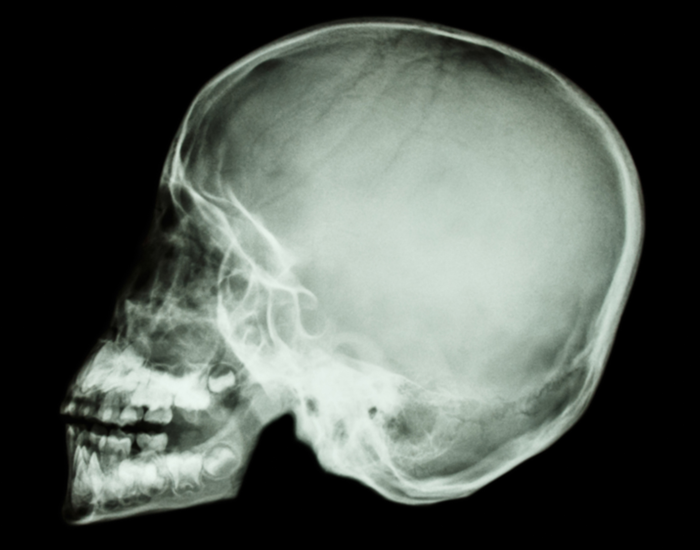
      Almost all the students failed the question, and worse felt like idiots when the answer was revealed: the patient must be dead because the spinal column and the rest of the body are not attached. Compare:

  3. Nov 2021
    1. (the VTA is also part ofthis system, but is too small to image with standard fMRImethods, but see [35] for successful imaging methods).

      All imaging studies face questions of validity and should (and many do) link to comprehensive details on instrumentation, methodology, and interpretation. Apparently, the professional consensus remains that, properly executed and interpreted, fMRI and other functional imaging techniques based on detection of oxygenation can lead to highly valid conclusions. (See Nautil.us article.)

  4. Jul 2021
  5. Feb 2021
  6. Nov 2020
    1. ound discriminationexperiment during the pre-training fMRI scan, and then six weeks later the post-training fMRI scan

      fMRI scans were done prior to exposure to training and then after in order for researchers to view any neural changes in the brain

    2. neuroimagingmethods especially functional magnetic resonance imaging (fMRI

      fMRI is the main tool to collect data in this specific experiment

  7. Aug 2020
  8. May 2020
  9. Apr 2020
  10. Dec 2017
  11. Oct 2016
    1. Previously, intensity-dependent metabolic changes have been found with positron emission tomography and blood oxygen level dependent magnetic resonance imaging after TMS to motor/prefrontal cortex; bilateral motor/prefrontal and auditory activation is induced, which becomes stronger with increasing pulse intensity [Bohning et al.,1999,2000; Fox et al.,1997; Nahas et al.,2001; Siebner et al.,1999; Speer et al.,2003]. However, these results are not directly comparable with our EEG findings. Arising a few seconds poststimulus, metabolic changes reflect relatively long-lasting activity of interconnected neuronal networks, whereas we were interested in the TMS-evoked events that occurred within a fraction of a second.
  12. Jul 2014
    1. 4. Identify Lacunas of Knowledge in Neuroscience Using the NIF Platform Develop a comprehensive web-based interactive meta-analytical map (heat map) of task fMRI activation foci across all human fMRI studies in the literature. Thus create an “activity map” of the human brain based on functional brain imaging by creating a visualization that depicts:

      NIF Grant